저작자표시-비영리-변경금지 2.0 대한민국 이용자는 아래의 조건을 따르는 경우에 한하여 자유롭게 l 이 저작물을 복제, 배포, 전송, 전시, 공연 및 방송할 수 있습니다. 다음과 같은 조건을 따라야 합니다: l 귀하는, 이 저작물의 재이용이나 배포의 경우, 이 저작물에 적용된 이용허락조건 을 명확하게 나타내어야 합니다. l 저작권자로부터 별도의 허가를 받으면 이러한 조건들은 적용되지 않습니다. 저작권법에 따른 이용자의 권리는 위의 내용에 의하여 영향을 받지 않습니다. 이것은 이용허락규약(Legal Code)을 이해하기 쉽게 요약한 것입니다. Disclaimer 저작자표시. 귀하는 원저작자를 표시하여야 합니다. 비영리. 귀하는 이 저작물을 영리 목적으로 이용할 수 없습니다. 변경금지. 귀하는 이 저작물을 개작, 변형 또는 가공할 수 없습니다.
Is craniofacial asymmetry progressive
in the untreated congenital muscular
torticollis?
by
Seungjo Seo
Major in Medicine
Department of Medical Sciences
The Graduate School, Ajou University
Is craniofacial asymmetry progressive
in the untreated congenital muscular
torticollis?
by
Seungjo Seo
A Dissertation Submitted to The Graduate School of
Ajou University in Partial Fulfillment of The
Requirements for The Degree of Master of Medicine
Supervised by
Myong Chul Park, M.D., Ph.D.
Major in Medicine
Department of Medical Sciences
The Graduate School, Ajou University
This certifies that the dissertation
Of Seungjo Seo is approved.
SUPERVISORY COMMITTEE
Myong Chul Park
Kwang Woo Baek
Yun-Hoon Choung
The Graduate School, Ajou University
June, 22
nd, 2012
i
- ABSTRACT-
Is craniofacial asymmetry progressive in the untreated congenital
muscular torticollis?
Although craniofacial asymmetry is frequently involved in the congenital muscular torticollis (CMT) patients who require surgical release, it has not been evaluated appropriately. Therefore, little is still known concerning severity and change of craniofacial asymmetry with aging. The authors analyzed preoperative craniofacial asymmetry objectively and confirmed relationship between craniofacial asymmetry and aging in CMT patients who got surgical release.
The authors retrospectively measured preoperative craniofacial asymmetry using cranial vault asymmetry index (CVAI) and inter-commissural angle (ICA) and reviewed preoperative rotational and flexional deficit of neck movement for 123 CMT patients who underwent complete tight fibrous band release and resection at the Center for Torticollis, Ajou Medical Center from February 2007 to February 2011. The relationship between CVAI, ICA, rotational deficit, and flexional deficit, and age at operation were analyzed. Mean values of CVAI, ICA, rotational deficit, and flexional deficit were compared between the age groups, after grouping the patients by age (Age Group 1, surgically treated before the age of 1 year; Age Group 2, from 1 to 3 years; Age Group 3, from 3 to5 years; Age Group 4, from 5 to 10 years; and Age Group 5, after 10 years).
Mean age at operation was 82.5 months (range, 5-498 months). Seventy-one percent (n=87) of patients had a significant cranial asymmetry and eighty-seven percent (n=107) of patients had a significant facial asymmetry. In correlation analysis, ICA increased proportionally to age (Pearson correlation coefficient r=0.334, p=0.000), especially patients under 3 years old (r=0.42, p =0.001). CVAI was unrelated to age, rotational deficit, and flexional deficit. Rotational deficit decreased proportionally to age (r=-0.229, p=0.032). In ANOVA test, ICA and rotational deficit between the age groups were statistically significantly different (p<0.05)
In congenital muscular torticollis, facial asymmetry is progressive if the contracted SCM muscle is not released although cranial asymmetry is already determined younger than 6 months
ii
old. Early operation should be considered to prevent progression of facial asymmetry in CMT patients if they need surgical release.
iii
TABLE
OF
CONTENTS
ABSTRACT i
TABLE OF CONTENTS iii
LIST OF FIGURES iv
LIST OF TABLES v
I. INTRODUCTION 1
II. MATERIALS AND METHODS 3
A. Method of Assessment 3 B. Assessment of Result 4 III. RESULTS 5 IV. DISCUSSION 12 V. CONCLUSION 16 REFERENCES 17 국문요약 20
iv
LIST
OF
FIGURES
Fig 1. Full supracranial view of 3-D brain CT 9 Fig 2. Standardized anteroposterior photograph before operation 10 Fig 3. The severity of facial asymmetry depends on the age at the operation 11
v
LIST
OF
TABLES
Table 1. Patients’ characteristics 6 Table 2. Correlation analysis between cranial vault asymmetry index, inter-commissural angle, rotational deficit, flexional deficit, and age. 7 Table 3. Comparison in mean ± SD of cranial vault asymmetry index, inter-commissural angle, rotational deficit, and flexional deficit between age groups. 8
1
I.
I
NTRODUCTIONCongenital muscular torticollis (CMT) is a neck deformity involving shortening of the sternocleidomastoid (SCM) muscle.(Canale et al. 1982; Cheng et al. 1994; Cheng et al. 1999) The reported incidence of torticollis has varied from 0.3%-2%.(Cheng et al. 2001) Although various theories such as birth trauma, ischemia, intrauterine malposition, hereditary hypothesis, neurogenic theory, and infection theory have been proposed, the actual cause of torticollis remains uncertain.(Cheng et al. 1994)
The limitation in range of motion of neck and head tilt is typical symptom. The typical lesion is a hard mass within the tight SCM muscle. After reaching its maximum size, the mass gradually recedes within the first year and, in most patients, the muscle tightness subsides after the mass has resolved. In some patients, large portions of the muscle may become fibrotic.(Cheng et al. 2001) Contracture of SCM muscle causes the head to turn towards the affected side and the chin to point to the opposite side. This unilateral contracture of the SCM muscle may induce cranial and facial asymmetry at presentation.(Cheng et al. 2001; Chate 2004; Rogers et al. 2009)
Although craniofacial asymmetry is more frequently involved in the CMT patients who require surgical release,(Cheng et al. 1999) craniofacial asymmetry has not been evaluated appropriately. Most of studies measured craniofacial asymmetry as the categorical value.(Wirth et al. 1992; Bharadwaj 1997; Cheng et al. 1999; Cheng et al. 1999; Stassen et al. 2000; Cheng et al. 2001; Shim et al. 2008) And cranial asymmetry and facial asymmetry were not evaluated separately.(Bharadwaj 1997; Cheng et al. 1999; Cheng et al. 1999; Stassen et al. 2000; Cheng et al. 2001; Shim et al. 2008) Therefore, little is still known concerning how many CMT patients have craniofacial asymmetry and how craniofacial asymmetry change with aging. And craniofacial asymmetry has not been considered as the important factor in determination of surgical timing. However, craniofacial asymmetry will be remained permanently on some patients, although functional problems such rotational and flexional deficit are improved, even after the contracted SCM muscle is released, if surgical release is delayed.
The authors objectively measured frequency and severity of preoperative craniofacial asymmetry of CMT patients who had surgical release at the Center for Torticollis, Ajou Medical
2
Center. And relationship between aging, preoperative limitation in range of motion of neck, and craniofacial asymmetry was analyzed.
3
II.MATERIALS AND METHODS
Surgical release was performed for CMT patients according to the Therapeutic Protocol of the Center for Torticollis, Ajou Medical Center.(Lee et al. 2011) A stretching exercise was
considered for all CMT patients younger than 6 months old. Surgical release was considered around that age. MRI is acquired for patients older than 6 months with limitation of passive range of motions (PROMs) in rotation and lateral flexion of the neck. If they have already been practicing stretching exercise for more than 6 weeks and the focal low signal intensity and/or diffuse low signal intensity in the involved SCM was noted, surgical release was considered. If they did not have previous stretching exercise, additional stretching exercises and/or surgical release are considered, although an abnormal MRI finding is noted.
One hundred seventy one CMT patients underwent complete tight fibrous band release and resection (Lee et al. 2010) at the Center for Torticollis, Ajou Medical Center from February 2007 to February 2011. A retrospective review was carried out of all patients who had surgical release at this center.
Preoperative craniofacial asymmetry was evaluated for only 123 patients using standardized anteroposterior photographs with a digital camera and the three-dimensional (3D) brain computed tomography (CT), because the quality of photographs of other 48 patients were not adequate to evaluate facial asymmetry. Chart reviews were performed to identify age at
operation, sex, presenting symptom, and preoperative PROMs of the rotation and lateral flexion of the neck.
B. Method of Assessment
PROMs of the neck were measured for rotation and lateral flexion under general anesthesia immediately preoperatively because most infants resist attempts to manipulate their head quite vigorously at conscious status. This method provides a more sensitive measure of subtle differences in sternocleidomastoid muscle contraction. Measurement was done with the
arthrodial protractor, with the patient in the supine position, both shoulders stabilized, and head and neck supported by so that the neck was free to rotate and bend laterally. When the limitation occurred during neck movement, further movement was stopped and the PROM was recorded. The limitation in passive rotation and lateral flexion (rotational deficit and flexional deficit)
4 were calculated.
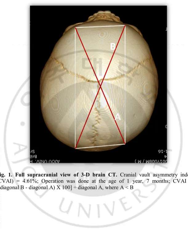
The cranial vault asymmetry index (CVAI) was used to measure the degree of cranial asymmetry.(Loveday et al. 2001) Initial anthropometric parameters for head diagonals were determined from the full supracranial view of the 3-D brain computed tomography (CT). The oblique distances were obtained from the orbitale superius directly above the midpupil to the occipital landmark, which is defined by a parallel line directed posteriorly from the orbitale superius on the opposite side.(Farkas 1994; Mulliken et al. 1999; Schaaf et al. 2010)The CVAI was determined using the following equation:
CVAI = [(diagonal B - diagonal A) X 100] ÷ diagonal A, where A < B (Fig. 1). The CVAI is a meaningful index to describe cranial asymmetry independent of head size.
An inter-commissural angle (ICA) was used to measure the degree of facial asymmetry on standardized anteroposterior photographs, which is the sharp angle between the interexocanthal plane and the intercommissural plane (Fig. 2).(Yu et al. 2009) Variable anthropometric points and angles can be used for measuring facial asymmetry. ICA is the one of parameters easy to measure and understand.(Yu et al. 2009) The CVAI and intercommissural angle were measured twice for each patient and the average was recorded.
C. Assessment of Results
Pearson correlation coefficients were calculated to analyze the relationship between CVAI, ICA, rotational deficit, and flexional deficit, and age at operation.
The sample of patients was divided into five subgroups according to the age at the time of presentation: Age Group 1, surgically treated before the age of 1 year; Age Group 2, from 1 to 3 years; Age Group 3, from 3 to5 years; Age Group 4, from 5 to 10 years; and Age Group 5, after 10 years. CVAI, ICA, rotational deficit, and flexional deficit between the age groups were analyzed for significant differences using one-way analysis of variance (ANOVA).
Measurements of CVAI and ICA were performed on digital photographs with Adobe Photoshop CS3 (Adobe Systems, San Jose, CA), and statistical analysis was performed with PASW Statistics 18.0 (SPSS, Chicago, IL).
5
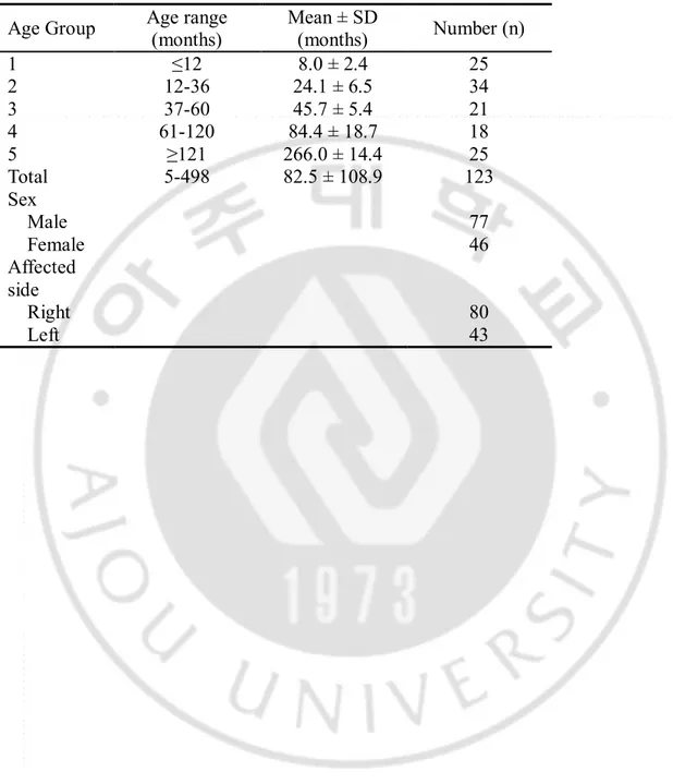
III.RESULT
The data set is comprised of 123 patients (77 males and 46 females). CMT occurred on the right side in 80 cases and on the left in 43 cases. Mean age at operation was 82.5 months (range, 5-498 months).
Among 123 patients, 25 patients were included in Age Group 1, 34 in Age Group 2, 21 in Age Group 3, 18 in Age Group 4, and 25 in Age Group 5 (Table 1).
All but two patients had a variable degree of ipsilateral occipital flattening and frontal bossing. Seventy one percent (n=87) of patients had a significant cranial asymmetry, defined as a CVAI >3.5%.(Loveday et al. 2001) Eighty seven percent (n=107) of patients had a significant facial asymmetry, defined as an ICA >0.5%. There were no patients whose unaffected face was smaller than the affected side.
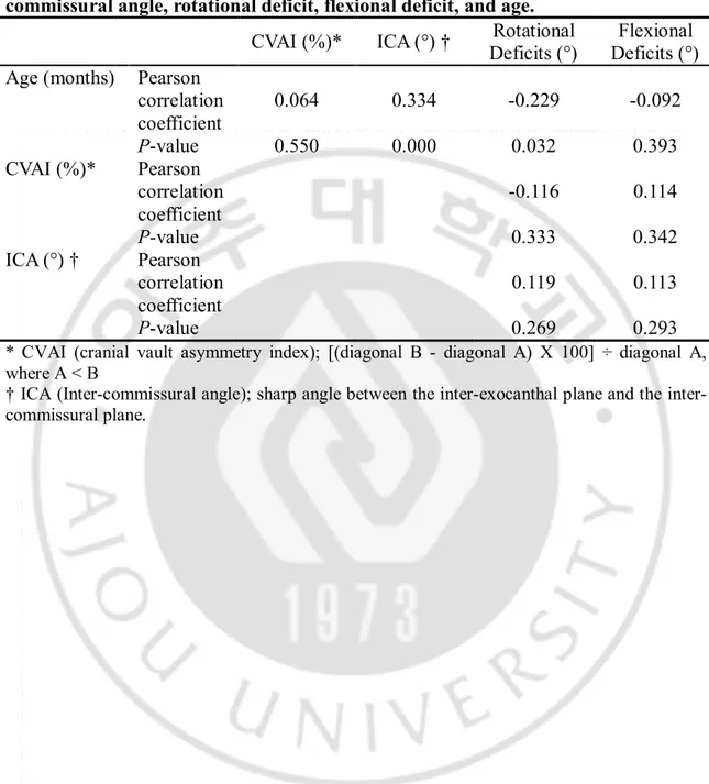
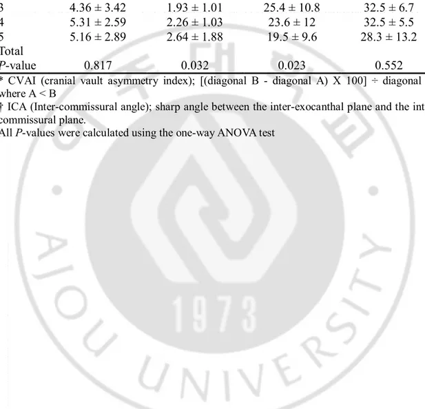
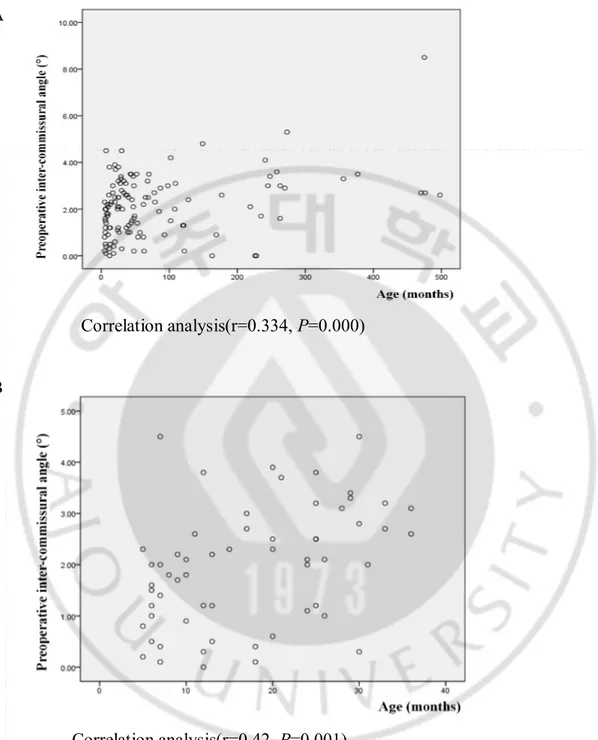
In correlation analysis, ICA increased proportionally to age (Pearson correlation coefficient r=0.334, p=0.000) (Table 2.) (Fig. 3A), especially patients under 3 years old (r=0.42, p =0.001) (Fig. 3B). CVAI was unrelated to age, rotational deficit, and flexional deficit (Table 3).
Rotational deficit decreased proportionally to age (r=-0.229, p=0.032) (Table 2.). In ANOVA test, ICA and rotational deficit between the age groups were statistically significantly different (p<0.05) (Table 3).
6
Table 1. Patients’ characteristics
Age Group
Age range
(months)
Mean ± SD
(months)
Number (n)
1
≤12
8.0 ± 2.4
25
2
12-36
24.1 ± 6.5
34
3
37-60
45.7 ± 5.4
21
4
61-120
84.4 ± 18.7
18
5
≥121
266.0 ± 14.4
25
Total
5-498
82.5 ± 108.9
123
Sex
Male
77
Female
46
Affected
side
Right
80
Left
43
7
Table 2. Correlation analysis between cranial vault asymmetry index,
inter-commissural angle, rotational deficit, flexional deficit, and age.
CVAI (%)*
ICA (°) †
Rotational
Deficits (°)
Flexional
Deficits (°)
Age (months) Pearson
correlation
coefficient
0.064
0.334
-0.229
-0.092
P-value
0.550
0.000
0.032
0.393
CVAI (%)*
Pearson
correlation
coefficient
-0.116
0.114
P-value
0.333
0.342
ICA (°) †
Pearson
correlation
coefficient
0.119
0.113
P-value
0.269
0.293
* CVAI (cranial vault asymmetry index); [(diagonal B - diagonal A) X 100] ÷ diagonal A, where A < B
† ICA (Inter-commissural angle); sharp angle between the exocanthal plane and the inter-commissural plane.
8
Table 3. Comparison in mean ± SD of cranial vault asymmetry index,
inter-commissural angle, rotational deficit, and flexional deficit between age groups.
Age
Group
CVAI (%)*
ICA (°) †
Rotational
Deficits (°)
Flexional
Deficits (°)
1
5.35 ± 2.18
1.52 ± 1.09
32.5 ± 17.5
32.3 ± 7.7
2
5.08 ± 2.1
2.29 ± 1.13
29.3 ± 12.1
29.3 ± 9.8
3
4.36 ± 3.42
1.93 ± 1.01
25.4 ± 10.8
32.5 ± 6.7
4
5.31 ± 2.59
2.26 ± 1.03
23.6 ± 12
32.5 ± 5.5
5
5.16 ± 2.89
2.64 ± 1.88
19.5 ± 9.6
28.3 ± 13.2
Total
P-value
0.817
0.032
0.023
0.552
* CVAI (cranial vault asymmetry index); [(diagonal B - diagonal A) X 100] ÷ diagonal A, where A < B
† ICA (Inter-commissural angle); sharp angle between the exocanthal plane and the inter-commissural plane.
9
Fig. 1. Full supracranial view of 3-D brain CT. Cranial vault asymmetry index
(CVAI) = 4.61%; Operation was done at the age of 1 year, 7 months; CVAI =
[(diagonal B - diagonal A) X 100] ÷ diagonal A, where A < B
10
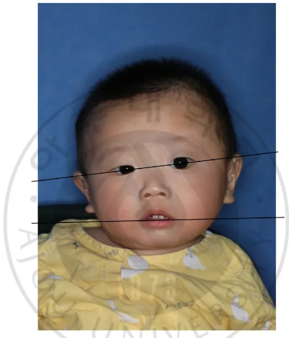
Fig. 2. Standardized anteroposterior photograph before operation.
Inter-commissural angle (ICA) = 4.5°; Operation was done at the age of 7 months ; ICA =
sharp angle between the inter-exocanthal plane and the inter-commissural plane.
11
A
Correlation analysis(r=0.334, P=0.000)
B
Correlation analysis(r=0.42, P=0.001)
Fig. 3. The severity of facial asymmetry depends on the age at the operation. The
scatterplot shows positive linear correlation between age and facial asymmetry.
Panel A displays data acquired at whole age and panel B displays data at an age below
36 months.
12
IV.
D
ISCUSSIONRotational deficit displayed a negative, albeit weak, correlation with age (Table 2.). The limitation in rotation is mainly due to sternomastoid tumors at the affected side of SCM muscle. Tumor is resolved spontaneously as time passes and rotation to the affected side is improved consequently. One study reported that sternomastoid tumors were found only in patients younger than 2 years, with most such patients being under 1 year. After the age of 2 years, all unresolved sternomastoid tumors had changed to a tight fibrotic band.(Cheng et al. 1999) On this study, sternomastoid tumors were found mainly in MRI of patients younger than 1 year and severe fibrotic band in patients over 3 years. Consequently, improvement of rotational deficit in surgical candidates does mean reduction of tumors, not improvement of torticollis.
Craniofacial asymmetry has been noted in 90.1% of patients operated on for torticollis.(Cheng et al. 1999) Torticollis has been associated with 64%-84% of infants born with a normal head shape who develop positional plagiocephaly.(Loveday et al. 2001) In this study, all but two patients had a variable degree of ipsilateral occipital flattening and frontal bossing. Seventy one percent (n=87) of patients had a significant cranial asymmetry, defined as a CVAI >3.5%. However cranial asymmetry was irrelevant to the age after 6 months. This is due to cranial bone growth pattern. A major increase in cranial size occurs in the first 2 years of life. The first 6 months of life are characterized by a period of generalized rapid growth, followed by a slower period of growth from 6 months to 4 years.(Dufresne et al. 2006) And repositioning and physiotherapy for the deformational plagiocephaly is effective until 1-4-months-of-age because cranial remodeling is dependent mainly on the remaining growth potential.(Moss 1997;
Robinson et al. 2009; Kluba et al. 2011) Therefore, cranial shape is most determined during the first 6 months of life. One study showed that the major cause of deformational plagiocephaly is limited head mobility in early infancy secondary to cervical imbalance. And a negative
correlation between age and rotational deficit and a positive correlation between cranial asymmetry and rotational deficit was demonstrated in patients(mean age, 6.1 months) with positional plagiocephaly and torticollis which is resolved spontaneously or by physical therapy.(Rogers et al. 2009) However, in our study, improvement of rotation deficit did not affect cranial asymmetry because most of patients were older than 6 months. Consequently, to
13
correct cranial asymmetry in CMT patients, another therapy such as repositioning and helmet therapy should be considered during physical therapy for CMT before 6 months if cranial asymmetry is severe.
The cluster of findings that comprises the asymmetry associated with torticollis is consistent and has been reported by several authors.(Pirttiniemi et al. 1989; Ferguson 1993) The findings is consistent with a frontal deformational plagiocephaly, with recession of the ipsilateral zygoma and forehead, and a reduction of vertical facial height on the affected side.(Hollier et al. 2000) However, severity of facial asymmetry was not measured as the numerical value(Wirth et al. 1992; Bharadwaj 1997; Cheng et al. 1999; Cheng et al. 1999; Stassen et al. 2000; Cheng et al. 2001; Shim et al. 2008) and was not evaluated separately from cranial asymmetry(Bharadwaj 1997; Cheng et al. 1999; Cheng et al. 1999; Stassen et al. 2000; Cheng et al. 2001; Shim et al. 2008) in most of studies. Although facial asymmetry is measured by objective methods in some studies, younger patient such as infant was not evaluated.(Ferguson 1993; Chate 2004; Chate 2005) Therefore, change in facial asymmetry according to age was not reported. This study measured preoperative facial asymmetry of surgically treated CMT patients from infant to adult as numerical value using ICA and verified that facial asymmetry is progressive with aging unless surgical release is not done appropriately. Chen et al. used four anthropometric angles including midfacial angle, inter-commissural angle, and two chin point angles in a frontal
photograph to measure facial asymmetry of patients who got bimaxillary surgery.(Yu et al. 2009) Although, if all of four angles used, facial asymmetry can be calculated more accurately, ICA is enough to measure severity of facial asymmetry and easy to measure in infant. Some authors used cephalometric values to bony asymmetry.(Ferguson 1993; Chate 2004; Chate 2005) However, cephalometry is hard to be taken in child younger than 5 years and cannot show soft tissue asymmetry.(Hollier et al. 2000)
In this study, preoperative ICA was moderately positively correlated with age, even for patients over 10 years (Table 3). That means that facial asymmetry will progress if the
contracted SCM muscle is not released (Table 3, Fig. 3). Facial bone has a later rapid phase of growth than cranial bone and different rates according to the area of the face.(Dufresne et al. 2006) Therefore, fascial contraction of the involved SCM muscle is responsible for progression of facial asymmetry (Chate 2004; Chate 2005) during long period of facial growth.
14
affected SCM muscle with varying degrees of success, depending on the age of the patient at operation. It is conceded that the cosmetic results of nonsurgical therapy are unsatisfactory if there is facial asymmetry during infancy.(Canale et al. 1982; Ippolito et al. 1985) Candler and Altenberg(Bharadwaj 1997; Cheng et al. 1999) have been the most ardent proponents of surgical correction of the torticollis during infancy. Cheng et al. showed that excellent results can be expected in nearly all cases surgically treated before the age of 3 years.(Cheng et al. 1999) Another study showed that facial asymmetry is persistent and more often observed in children operated on after 3 years.(Maslon et al. 2009) However, Wirth et al. showed that even severe facial asymmetry may resolve completely within a few years if the patient is operated on before 5 years by a long-term follow-up of 55 patients.(Wirth et al. 1992) One case report described that facial asymmetry of a 10-year-old girl with CMT resolved almost completely postoperatively.(Chate 2005)
A drawback of most studies is that facial asymmetry is measured as a categorical value such as mild, moderate, and severe and results were not statistically significant.(Wirth et al. 1992; Bharadwaj 1997; Cheng et al. 1999; Sasaki et al. 2000; Stassen et al. 2000; Swain 2007) Therefore optimal surgical timing to resolve facial asymmetry is hard to determine based on previous reports. However, based on this study, this is clear that an early operation will be helpful to improve facial asymmetry because facial asymmetry in a younger patient is mild and the remodeling potential is higher than in older patients. To confirm optimal surgical timing to get better improvement of facial asymmetry, the authors are following our patients.
This study is the first time that the relationship between craniofacial asymmetry and age were analyzed separately, quantitatively, and objectively in CMT patients. Other authors used categorical variables to measure severity of craniofacial asymmetry, such as mild, moderate, and severe.(Wirth et al. 1992; Bharadwaj 1997; Cheng et al. 1999; Sasaki et al. 2000; Stassen et al. 2000; Swain 2007) In this study, the CVAI and ICA instruments used to measure cranial and facial asymmetry are continuous objective variables. So, the severity of craniofacial asymmetry was measured accurately and the exact association between craniofacial asymmetry and age at operation was analyzed.
Conventional two-dimensional photographs were used to measure ICA. Photography in children is challenging because of patient movement. Accurate positioning for a specific viewpoint is essential to estimate the deformity. The use of a multi-camera system to get a 3D
15
photograph has been described successfully in previous reports. A major advantage of the 3D imaging technique is its quick and easy approach. If this technique is applied for measuring, improved independence of the examiner, unlimited repetition of measurements, and archiving of the results can be attained.
16
V.
C
ONCLUSIONCraniofacial asymmetry is involved frequently in CMT patients, especially patients who require surgical release. Therefore it should be considered as one of important factor in determination of surgical timing. Cranial asymmetry is already determined younger than 6 months. Another therapy such as repositioning and helmet therapy should be considered to correct cranial asymmetry before operation. Facial asymmetry is progressive if the contracted SCM muscle is not released. Early operation should be considered to prevent progression of facial asymmetry in CMT patients.
17
R
EFERENCES1. Bharadwaj, V. K.: Sternomastoid myoplasty: surgical correction of congenital torticollis. J Otolaryngol 26(1): 44-48, 1997.
2. Canale, S. T., D. W. Griffin and C. N. Hubbard: Congenital muscular torticollis. A long-term follow-up. J Bone Joint Surg Am 64(6): 810-816, 1982.
3. Chate, R. A.: Facial scoliosis due to sternocleidomastoid torticollis: a cephalometric analysis. Int J Oral Maxillofac Surg 33(4): 338-343, 2004.
4. Chate, R. A.: Facial scoliosis from sternocleidomastoid torticollis: long-term postoperative evaluation. Br J Oral Maxillofac Surg 43(5): 428-434, 2005.
5. Cheng, J. C. and A. W. Au: Infantile torticollis: a review of 624 cases. J Pediatr Orthop 14(6): 802-808, 1994.
6. Cheng, J. C. and S. P. Tang: Outcome of surgical treatment of congenital muscular torticollis. Clin Orthop Relat Res(362): 190-200, 1999.
7. Cheng, J. C., S. P. Tang and T. M. Chen: Sternocleidomastoid pseudotumor and congenital muscular torticollis in infants: a prospective study of 510 cases. J Pediatr 134(6): 712-716, 1999.
8. Cheng, J. C., M. W. Wong, S. P. Tang, T. M. Chen, S. L. Shum and E. M. Wong: Clinical determinants of the outcome of manual stretching in the treatment of congenital muscular torticollis in infants. A prospective study of eight hundred and twenty-one cases. J Bone Joint Surg Am 83-A(5): 679-687, 2001.
9. Dufresne, C. R. and P. N. Manson. Pediatric Facial Injuries. Plastic Surgery. S. J. Mathes. Philadelphia, PA, SAUNDERS. 3: 381-388.2006
10. Farkas, L. G. Anthropometry of the Head and Face. New York, NY, Raven Press.1994 11. Ferguson, J. W.: Cephalometric interpretation and assessment of facial asymmetry secondary to congenital torticollis. The significance of cranial base reference lines. Int J Oral Maxillofac Surg 22(1): 7-10, 1993.
12. Hollier, L., J. Kim, B. H. Grayson and J. G. McCarthy: Congenital muscular torticollis and the associated craniofacial changes. Plast Reconstr Surg 105(3): 827-835, 2000.
18
tenotomy for idiopathic muscular torticollis. J Bone Joint Surg Am 67(1): 30-38, 1985. 14. Kluba, S., W. Kraut, S. Reinert and M. Krimmel: What is the optimal time to start helmet therapy in positional plagiocephaly? Plast Reconstr Surg 128(2): 492-498, 2011.
15. Lee, I. J., S. Y. Lim, H. S. Song and M. C. Park: Complete tight fibrous band release and resection in congenital muscular torticollis. J Plast Reconstr Aesthet Surg 63(6): 947-953, 2010. 16. Lee, S. J., J. D. Han, H. B. Lee, J. H. Hwang, S. Y. Kim, M. C. Park and S. Y. Yim:
Comparison of clinical severity of congenital muscular torticollis based on the method of child birth. Ann Rehabil Med 35(5): 641-647, 2011.
17. Loveday, B. P. and T. B. de Chalain: Active counterpositioning or orthotic device to treat positional plagiocephaly? J Craniofac Surg 12(4): 308-313, 2001.
18. Maslon, A., R. Lebiedzinski, M. Domzalski, M. Synder and A. Grzegorzewski: [Facial asymmetry in children with congenital muscular torticollis after surgical treatment]. Chir Narzadow Ruchu Ortop Pol 74(1): 31-34, 2009.
19. Moss, S. D.: Nonsurgical, nonorthotic treatment of occipital plagiocephaly: what is the natural history of the misshapen neonatal head? J Neurosurg 87(5): 667-670, 1997.
20. Mulliken, J. B., D. L. Vander Woude, M. Hansen, R. A. LaBrie and R. M. Scott: Analysis of posterior plagiocephaly: deformational versus synostotic. Plast Reconstr Surg 103(2): 371-380, 1999.
21. Pirttiniemi, P., P. Lahtela, J. Huggare and W. Serlo: Head posture and dentofacial
asymmetries in surgically treated muscular torticollis patients. Acta Odontol Scand 47(4): 193-197, 1989.
22. Robinson, S. and M. Proctor: Diagnosis and management of deformational plagiocephaly. J Neurosurg Pediatr 3(4): 284-295, 2009.
23. Rogers, G. F., A. K. Oh and J. B. Mulliken: The role of congenital muscular torticollis in the development of deformational plagiocephaly. Plast Reconstr Surg 123(2): 643-652, 2009. 24. Sasaki, S., Y. Yamamoto, T. Sugihara, K. Kawashima and K. Nohira: Endoscopic tenotomy of the sternocleidomastoid muscle: new method for surgical correction of muscular torticollis. Plast Reconstr Surg 105(5): 1764-1767, 2000.
25. Schaaf, H., C. Y. Malik, P. Streckbein, J. Pons-Kuehnemann, H. P. Howaldt and J. F. Wilbrand: Three-dimensional photographic analysis of outcome after helmet treatment of a nonsynostotic cranial deformity. J Craniofac Surg 21(6): 1677-1682, 2010.
19
26. Shim, J. S. and H. P. Jang: Operative treatment of congenital torticollis. J Bone Joint Surg Br 90(7): 934-939, 2008.
27. Stassen, L. F. and C. J. Kerawala: New surgical technique for the correction of congenital muscular torticollis (wry neck). Br J Oral Maxillofac Surg 38(2): 142-147, 2000.
28. Swain, B.: Transaxillary endoscopic release of restricting bands in congenital muscular torticollis--a novel technique. J Plast Reconstr Aesthet Surg 60(1): 95-98, 2007.
29. Wirth, C. J., F. W. Hagena, N. Wuelker and W. E. Siebert: Biterminal tenotomy for the treatment of congenital muscular torticollis. Long-term results. J Bone Joint Surg Am 74(3): 427-434, 1992.
30. Yu, C. C., L. Bergeron, C. H. Lin, Y. M. Chu and Y. R. Chen: Single-splint technique in orthognathic surgery: intraoperative checkpoints to control facial symmetry. Plast Reconstr Surg 124(3): 879-886, 2009.
20
- 국문요약 -
Is craniofacial asymmetry progressive in the untreated congenital
muscular torticollis?
아주대학교 대학원의학과 서승조 (지도교수 : 박명철) 수술적 치료를 필요로 하는 상당수의 선천성 근성 사경환자들에서 두개안면 비대칭이 동반되지만 이를 적절하게 평가한 연구들은 없었다. 그러므로 아직까지 수술적 치료가 늦어진 선천성 근성 사경환자에 있어서 나이가 듦에 따른 두개안면 비대칭의 심각도 및 그 변화 양상에 대해 정확히 알려져 있지 못하다. 이에 저자들은 수술을 받은 선청성 근성 사경환자들의 술 전 두개안면 비대칭을 객관적으로 분석하여 수술 시 연령과의 관계를 증명하였다. 2007년 2월부터 2011년 2월까지 아주대학교 병원 사경센터에서 수술을 시행받은 123명의 선천성 근성 사경 환자에 대해 후향적 연구를 통해 cranial vault asymmetry index (CVAI) 및 inter-commissural angle (ICA)를 이용하여 술 전 두개안면 비대칭을 측정하였으며 의무기록 조사를 통해 술 전 목의 회전 및 굴곡 운동의 제한을 알아보았다. CVAI, ICA, 회전 제한, 굴곡 제한, 및 수술 시 연령과의 상관관계를 분석하였다. 연령군에 따른 CVAI, ICA, 회전 제한, 및 굴곡 제한의 평균값을 비교하였다 (age group 1, 1세 전에 수술 받은 환자; age group 2, 1-3세; age group 3, 3-5세; age group 4, 5-10세; age group 5, 10세 이상).수술 시 평균 연령은 82.5개월 (5-498개월)이었으며 환자의 71% (n=87)가 상당한 두개 비대칭을, 87% (n=107)가 상당한 안면 비대칭을 가지고 있었다. 상관분석에서 ICA는 연령에 비례하여 증가하였으며 (Pearson correlation coefficient r=0.334, P=0.000), 이는 특히 3세 이하의 환자에서 두드러졌다 (r=0.42, P=0.001). 반면 CVAI는 연령,
21
회전 제한, 및 굴곡 제한과 어떠한 상관성도 보여주지 못했으며 회전 제한은 연령과 반비례하는 양상을 보여주었다 (r=-0.229, P =0.032). ANOVA test에서 ICA와 회전제한은 연령군에 따른 유의한 차이를 보여주었다(P <0.05).
선천성 근성 사경에 있어 두개 비대칭은 6개월 전에 이미 결정될지라도 안면 비대칭은 수축된 흉쇄유돌근을 풀어주지 않으면 진행될 수 있다는 것을 알 수 있다. 그러므로 선천성 근성 사경에서 안면 비대칭의 진행을 예방하기 위해 수술적
치료가 필요할 경우 이른 수술적 치료를 고려해 볼 수 있겠다.