© Korean Powder Metallurgy Institute 1089
-1. Introduction
Ti and its alloys have been used as biomaterials for bone implants [1]. The formation of bone-like apatite layer and increased oseoblast adhesion on Ti and other alloys have been reported by numerous investigaters [1-3]. The apatite formation is generally considered to be the main requirement for bone bonding ability of implant materials. Recently, Zr base amorphous alloys was found to be biocompatible and used as materials in knee-replacement devices [4]. However, the biomedical application of Zr alloys is limited and the biocompatibility of Zr alloys is not as extensively studied as that of Ti. The purpose of this study is to investigate the apatite formation behaviors on the surface of Zr-1Nb with various surface treatment conditions and compare them with those of Ti-6Al-4V in the SBF.
2. Experimental and Results
Rectangular Zr-1Nb and Ti-6Al-4V plates with the size of 10 mm x 10mm were used as a substrate for apatite deposition. The surface of metal plates were polished with abrasive papers (#100, #600, #2400 or 03 µm) to give the various roughness on the surface and some of them were chemically treated with 5mol NaOH for 1 hr. [1]. The SBF was prepared by dissolving chemicals into deionized water in the sequence of 141 mM NaCl, 4.0 mM KCl, 0.5 mM MgSO4, 1.0 mM MgCl2, 4.2 mM NaHCO3, 5.0 mM CaCl2.2H2O, 2.0 mM KH2PO4 and titrated to pH 6.8 at 25oC. The SBF used in this study was 1X modified solution with the concentration of Ca2+ and HPO
42- ions twice as much as those of human blood plasma [4]. Metal plates were immersed in the SBF and kept in an incubator at 37oC. The SBF was refreshed every other day for 10 days in order to
replenish the ion concentration to supersaturated levels. XRD was performed for the structural analyses of the deposited layer. The calcium phosphate deposited specimen was coated with a thin layer of gold and observed with a SEM (Topcon SM-500) for the observation of surface morphology. The deposition rate was examined by measuring the wight gain after the formation of apatite film.
Fig. 1 shows the weight gain of Zr-1Nb and Ti-6Al-4-V. The weigh gain in Zr-1Nb was found to be higher than in Zr-1Nb polished with #100 abrasive paper than in Zr-1Nb polished with #2400 paper, suggesting that the formation of bone-like apatite crystals is enhanced by the increase of roughness. One interesting observation is that the nucleation and growth of hydroxycarbonated apatite (HCA) layer were much faster in Ti-6Al-4V in the early stage, but the nucleation and growth rate of HCA accelerated in Zr-Nb and the weight gains were approximately same for the both alloys.
Fig. 1. Weight gain due to the formation HCA on the surface NaOH-treated Zr-1Nb and Ti-6Al-4V after biomimetic deposition in SBF as a function of time. 2006 POWDER METALLURGY
World Congress
PC05-W-03
Biomimetic Deposition of Apatite on Zr-1Nb and Ti-6Al-4V
J. Kim1, Y. C. Choi1, H.S. Kim1 and S. I. Hong1,a1
Department of Metallurgical Engineering, Chungnam National University, Taedok Science Town, Taejon, 705-364, Korea
a
[email protected] Abstract
Biomimetic apatite deposition behaviors on Zr-1Nb and Ti-6Al-4V plate with various surface conditions were examined. Both alloys were polished with abrasive papers to have different roughness and some of them were treated in NaOH before exposition in simulated body fluid. NaOH treatment was found to enhance the deposition rate of apatite on Ti-6Al-4V significantly. On the other hand, the deposition rate of Zr-1Nb was not influenced by NaOH treatment. Without NaOH treatment, the polished Zr-1Nb with abrasive paper was found to induce more apatite nucleation than the polished Zr-6Al-4V.
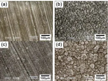
© Korean Powder Metallurgy Institute 1090 -Fig. 2 displays the nodular HCA particles deposited on the surface of NaOH-treated Zr-1Nb (polished with #100 abrasive paper) (a, b) and Ti-6Al-4V (polished with #100 abrasive paper) (c, d) after 2 days (a, c) and 10 days (b, d) deposition. After 2 days of biomimetic deposition, more nodular crystals were formed on Ti-6Al-4V. The linear lines in Fig. 2(a) and 2(c) are scratches intentionally formed by the abrasive paper. After 10 days, whole surface was covred with nodular crystals and no scratches on the surface of Zr-1Nb and Ti-6Al-4V substrates are visible. The nodular particles were observed to be slightly larger in the layer on Ti-6Al-4V.
Fig. 2. Surface morphologies of NaOH-treated Zr-1Nb and Ti-6Al-4V after biomimetic deposition of HCA in SBF.
Fig. 3. Weight gain of Zr-1Nb and Ti-6Al-4V with and without NaOH treatment in SBF.
In order to examine the effect of NaOH treatment, the deposition behaviors on the surface of Zr-1Nb and Ti-6Al-4V with NaOH treatment were compared with those without NaOH treatment. In Fig. 3(a), the weight gain of Zr-1Nb (polished with #600 abrasive paper) with and without NaOH treatment are as a function of deposition days are shown. The wight gain is not greatly influenced by the NaOH treatment in cotradiction to the suggestion of Janasova et al. [1]. The deposition rate of non-NaOH-treated Zr-1Nb was slightly higher than that of NaOH-treated Zr-1Nb in the initial stage. In Ti-6Al-4V(polished with #600 abrasive paper), the deposition rate of HCA increased significantly as shown in Fig. 3(b), consistent with the suggestion of Janasova et al. [1].
3. Summary
Biomimetic apatite deposition behaviors on Zr-1Nb and Ti-6Al-4V plate with various surface conditions were examined. After, 10 days immersion in a SBF, NaOH treated Zr-1Nb and Ti-6Al-4V were completely coated with apatite. The deposition rate of apatite was higher on NaOH-treated Ti-6Al-4V than on NaOH-treated Zr-1Nb initially, but the deposition rate on Zr-1Nb accelerated after 2 days and the total weight gain due to the deposition on Zr-1Nb approached to that of Ti-6Al-4V. NaOH treatment was found to enhance the deposition rate of apatite on Ti-6Al-4V significantly. Whereas the deposition rate of Zr-1Nb was not influenced by NaOH treatment. A rather good compatibility between HCA and Zr-1Nb without lengthy NaOH tretment suggests Zr alloys can be one of the promising biomaterials.
4. References
1. L. Jonasova, F. A. Muller, A. Helebrant, J. Strnad and P. Greil, Biomaterials 25, 1187(2004).
2. T. J. Webster and J. U. Ejiofer, Biomaterials 25, 4731 (2004).
3. R. Godley, D. Starosvetsky and I. Gotman, J. Mater. Sci.: Materials in Medicine 15, 1073(2004).