CASE REPORT
Copyright © 2019 The Korean Society of Neurosonology
This is an Open Access article distributed under the terms of the Creative Commons Attribution Non-Commercial License (http://creativecommons.org/licenses/by-nc/4.0/) which permits unrestricted non-commercial use, distribution, and reproduction in any medium, provided the original work is properly cited.
pISSN 2635-425X eISSN 2635-4357 https://doi.org/10.31728/jnn.2019.00049
Cerebral microbleeds (CMBs) are small (<10 mm), rounded, homogeneous, hypointense lesions revealed on T2*-weighted gradient-echo (GRE) or susceptibil-ity weighted imaging (SWI) sequences, suggestive of extravasation of blood components through patholog-ically fragile small vessels.1 Conventionally, CMBs have
been considered asymptomatic. However, there have been a few recent reports supporting that acutely devel-oping CMBs may cause clinical symptoms mimicking lacunar infarction.2-7 Herein, we present a case of pure
sensory stroke caused by an acute symptomatic CMB with perilesional edema.
CASE REPORT
A 48-year-old woman developed sudden paresthe-sia in the left hemibody 15 hours prior to admission.
Her sensory symptoms started at the left corner of the mouth and the left first two fingers. The oral symptom gradually spread to the face, while the cheiral symptom progressed up to her shoulder over a 60-minute period. A day after symptom onset, she complained of pares-thesia in the left thigh and reduced sensation in the left tongue and gum. The patient had a previous history of hypertension and kidney/pancreas transplantation owing to type 1 diabetes and renal failure. Although her medication included a calcium-channel blocker, her blood pressure was poorly controlled, and the systolic blood pressure occasionally rose to ≥200 mmHg.
Upon neurologic examination, there was no weak-ness, ataxia, or visual field defect. She complained of paresthesia and hypesthesia with symptoms of great-er sevgreat-erity in the regions whgreat-ere the symptoms began while the numbness was milder in the regions where the symptoms developed later. The National Institutes Cerebral microbleeds (CMBs) have conventionally been considered asymptomatic.
However, several reports have suggested that CMBs may cause clinical symptoms that mimic lacunar infarction. Herein, we present a 48-year-old woman who developed pure sensory stroke caused by an acute symptomatic CMB with per-ilesional edema in the right thalamus. Her sensory symptoms started with left cheiro-oral syndrome and then extended to left facio-brachio-crural syndrome over time. The symptoms resolved following the exact reverse order of their exten-sion, suggesting that the clinical symptoms correlated to the extent of perilesion-al edema. The hyperintense rim observed on the previous diffusion-weighted and T2-weighted images disappeared on the follow-up magnetic resonance (MR) imag-es performed 5 months after the symptom onset. Our findings suggimag-est that CMBs can mimic lacunar infarcts and also stroke progression. Early acquisition of MR imaging, including gradient-echo or susceptibility weighted image sequences, may be useful in distinguishing lacunar infarction and CMBs.
J Neurosonol Neuroimag 2019;11(1):96-99
Key Words: Stroke; Cerebral hemorrhage; Thalamus; Magnetic resonance imaging
Pure Sensory Stroke Caused by Acute Symptomatic Thalamic Cerebral
Microbleed with Perilesional Edema
Han Kyu Na*, Taedong Ok*, Bang-Hoon Cho*, Kyung-Yul Lee*,†
Department of Neurology, Gangnam Severance Hospital, Yonsei University College of Medicine*, Seoul; Severance Institute for Vascular and Metabolic Research, Yonsei University College of Medicine†, Seoul, Korea
Address for correspondence: Kyung-Yul Lee
Department of Neurology, Gangnam Severance Hospi-tal, Yonsei University College of Medicine, 211 Eonju-ro, Gangnam-gu, Seoul 06273, Korea Tel: +82-2-2019-3325 Fax: +82-2-3462-5904 E-mail: kylee@yuhs.ac Received: May 27, 2019 Revised: June 13, 2019 Accepted: June 14, 2019
JNN
97 http://www.j-nn.org J Neurosonol Neuroimag 2019;11(1):96-99
Vol. 11 / No. 1 / June 2019 Journal of Neurosonology and Neuroimaging
of Health Stroke Scale score was 1.
Initial non-contrast brain computed tomography did not reveal any acute lesion. Brain magnetic resonance (MR) image performed 19 hours after symptom onset showed no diffusion restriction lesion on the diffu-sion-weighted image (Fig. 1A). However, a CMB was noted on the right inferolateral thalamus on the SWI, which was surrounded by a hyperintense rim on the T2-weighted and fluid attenuated inversion recovery (FLAIR) images (Fig. 1B-D), suggestive of acute CMB with perilesional edema.2 Several other CMBs in the
left basal ganglia, thalamus and pons were also ob-served on the SWI (Fig. 1B), but these lesions did not show adjacent parenchymal edema on the T2-weighted image (Fig. 1C). MR angiography showed no significant intracranial arterial stenosis (Fig. 1E).
The patient was diagnosed with an acute symptomat-ic CMB in the right inferolateral thalamus. Her symp-toms resolved over time following the exact reverse
order of symptom development. Seven days later, she reported mild hypesthesia only in the first two digits of her left hand, which gradually resolved over months. The patient underwent follow-up brain MR imaging 5 months after the symptom onset (Fig. 1F-I). The size of previous symptomatic CMB decreased slightly on the SWI, and the hyperintense rim disappeared on the fol-low-up DWI and T2-weighted images (Fig. 1J).
DISCUSSION
In general, CMBs have been considered asymptom-atic lesions.1 However, a few recent reports reveal that
CMBs can cause clinical symptoms that mimic cerebral ischemia, as in our case.2-7 In a recent study
investigat-ing the incidence of suspected symptomatic CMBs in diffusion-negative stroke patients, seven out of 14 (50%) patients whose symptom persisted for more than 24
Fig. 1. (A) Brain MR image does not show acute infarction on the DWI. (B) A cerebral microbleed in the right inferolateral thalamus is observed on the SWI, and it is surrounded by a perilesional hyperintense rim on the T2-weighted (C) and FLAIR image (D). Another microbleed in the left thal-amus is detected on the SWI, but it is not seen on the T2-weighted or FLAIR image. (E) MR angiography shows no significant stenosis of the in-tracranial arteries. (F-I) Follow-up MR images taken 5 months after the onset of symptoms. (J) The perilesional hyperintense rim observed on the baseline DWI and T2-weighted image (arrowheads) disappeared on the follow-up MR images (arrows). The right thalamus is magnified for better visualization. DWI; diffusion-weighted image, MR; magnetic resonance, SWI; susceptibility-weighted image, FLAIR; fluid attenuated inversion recovery. A F B G C H D I E J
98 http://www.j-nn.org J Neurosonol Neuroimag 2019;11(1):96-99
Han Kyu Na, et al. Acute Symptomatic Cerebral Microbleed
hours showed CMBs surrounded by hyperintense rims on their MR images, implying that a certain propor-tion of diffusion-negative stroke may be explained by
symptomatic CMBs.6 However, ascertaining whether
the symptoms are indeed attributable to the CMBs is challenging as the temporal causal relationship be-tween CMBs and neurological symptoms is difficult to prove.2,6,7 Several reports have claimed that the
exis-tence of hyperintense rim surrounding the CMB could be useful in differentiating acute symptomatic CMBs
from chronic CMBs.2 Contrary to chronic CMBs,
new-ly-developed symptomatic CMBs tend to be surround-ed by a perilesional hyperintense rim on a T2-weightsurround-ed image, considered vasogenic edema due to
microhe-morrhage.2-4 Thus, the CMB-induced acute stroke
syndrome may possibly be explained by perilesional edema.2,8
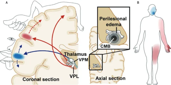
The patient described in this report supports this perspective. It can be inferred that the patient’s CMB developed in the junction of the ventroposterolateral and ventroposteromedial nucleus, causing cheiro-oral symptoms due to close mapping sensory homunculi in the thalamus,9,10 and extension of the sensory
symp-toms to the left hemibody was attributed to gradual ex-pansion of the perilesional edema to a wider portion of thalamic sensory homunculi (Fig. 2). Intriguingly, the symptoms resolved following the reverse order of ini-tial appearance and extension, which may possibly be attributed to shrinkage of perilesional edema over time. This finding holds clinical implication in that CMBs can even mimic stroke progression.
Given that the patient did not undergo MR imaging prior to the onset of symptoms, we cannot assure that the right thalamic CMB observed was indeed an acute symptomatic CMB as it could also be a chronic pre-ex-isting CMB. However, considering that her initial MR images revealed symptom-correlating right thalamic CMB (SWI) surrounded by hyperintense rim (DWI, T2, and FLAIR) and that the hyperintense rim disappeared on the follow-up MR image, it is more likely that the CMB as a newly-developed CMB rather than a chronic one.
Clinicians should be aware that acute stroke syn-drome can also be caused by CMBs, and symptoms can worsen with expansion of perilesional edema. Early MR image acquisition, including the T2*-weighted GRE or
Fig. 2. (A) A CMB developed in the junction of ventroposterolateral and ventroposteromedial nucleus, causing cheiro-oral syndrome. The exten-sion of sensory symptoms (i.e., cheiro-oral pattern of sensory deficit progressing into facio-brachio-crural pattern) may be attributable to expan-sion of perileexpan-sional edema which involves wider regions in the thalamic homunculus. (B) Diagram of the distribution of somatosensory deficits in the patient with an acute symptomatic thalamic CMB. VPM; ventroposteromedial nucleus, VPL; ventrolateromedial nucleus, CMB; cerebral microbleed.
99 http://www.j-nn.org J Neurosonol Neuroimag 2019;11(1):96-99
Vol. 11 / No. 1 / June 2019 Journal of Neurosonology and Neuroimaging
SWI sequences, may be informative in planning treat-ment strategies, especially considering the high risk of hemorrhage in patients with CMBs.
Acknowledgments
We express our gratitude to Jong Shin Park for his work with the illustrations.
Conflicts of Interest
No potential conflicts of interest relevant to this arti-cle was reported.
REFERENCES
1. Greenberg SM, Vernooij MW, Cordonnier C, Viswanathan A, Al-Shahi Salman R, Warach S, et al. Cerebral microbleeds: a guide to detection and interpretation. Lancet Neurol. 2009;8:165-174.
2. Heo SH, Lee D, Lee D, Chang DI. Differentiation of a symptomatic cerebral microbleed from silent microbleeds.
Int J Stroke. 2014;9:E2.
3. Nezu T, Arihiro S, Toyoda K, Minematsu K. Small-vessel
disease in the basal ganglia: lacune or microbleed? J Stroke
Cerebrovasc Dis. 2012;21:905.e5-e6.
4. Teo JTH, Ramadan H, Gregoire SM, Mufti S, Lipman G, Jager HR, et al. Can cerebral microbleeds cause an acute stroke syndrome? Neurol Clin Pract. 2011;1:75-77.
5. Simonsen CZ, Nielsen E. Hypertensive microbleed as a transient ischemic attack mimic. Case Rep Neurol. 2013;5:31-33.
6. Heo SH, Lee D, Kwon YC, Kim BJ, Lee KM, Bushnell CD, et al. Cerebral microbleeds in the patients with acute stroke symptoms. Front Neurol. 2018;9:988.
7. Choi PK, Chung JY, Lee SJ, Kang HG. Recurrent cerebral microbleeds with acute stroke symptoms: a case report.
Medicine (Baltimore). 2018;97:e12480.
8. Renard D. Cerebral microbleeds: a magnetic resonance imaging review of common and less common causes. Eur J
Neurol. 2018;25:441-450.
9. JrK M, Paxinos G. The human nervous system. 3rd ed. Am-sterdam: Elsevier Academic Press, 2012;824-827.
10. Haines DE. Neuroanatomy: an atlas of structures, sections, and systems. 8th ed. Philadelphia: Wolters Kluwer Health/ Lippincott Williams & Wilkins, 2012;178-181.