2004;64:5225-5231.
Cancer Res
Yoo Hong Min, June-Won Cheong, Ji Yeon Kim, et al.
Leukemia
Protein Kinase B and Poor Prognosis in Acute Myelogenous
Associated with Constitutive Phosphorylation of Akt or
Cytoplasmic Mislocalization of p27Kip1 Protein Is
Updated version
http://cancerres.aacrjournals.org/content/64/15/5225
Access the most recent version of this article at:
Cited Articles
http://cancerres.aacrjournals.org/content/64/15/5225.full.html#ref-list-1
This article cites by 49 articles, 25 of which you can access for free at:
Citing articles
http://cancerres.aacrjournals.org/content/64/15/5225.full.html#related-urls
This article has been cited by 19 HighWire-hosted articles. Access the articles at:
E-mail alerts
Sign up to receive free email-alerts
related to this article or journal.
Subscriptions
Reprints and
.
[email protected]
Department at
To order reprints of this article or to subscribe to the journal, contact the AACR Publications
Permissions
.
[email protected]
Department at
[CANCER RESEARCH 64, 5225–5231, August 1, 2004]
Cytoplasmic Mislocalization of p27Kip1 Protein Is Associated with Constitutive
Phosphorylation of Akt or Protein Kinase B and Poor Prognosis in Acute
Myelogenous Leukemia
Yoo Hong Min,
1,2June-Won Cheong,
1Ji Yeon Kim,
3Ju In Eom,
3Seung Tae Lee,
1Jee Sook Hahn,
1Yun Woong Ko,
1and Mark Hong Lee
41Department of Internal Medicine;2Brain Korea 21 Project for Medical Science;3Clinical Research Center, Yonsei University College of Medicine; and4Department of Internal
Medicine, Sungkyunkwan University College of Medicine, Seoul, Korea
ABSTRACT
Cyclin-dependent kinase inhibitor p27Kip1 functions at the nuclear level by binding to cyclin E/cyclin-dependent kinase-2. It was shown that Akt or protein kinase B (Akt/PKB)-dependent phosphorylation of p27Kip1 led to the cytoplasmic mislocalization of p27Kip1, suggesting the potential abrogation of its activity. Here, we evaluated the localization of p27Kip1 protein in leukemic blasts in relation to Akt/PKB phosphoryla-tion and clinical outcomes in acute myelogenous leukemia (AML). West-ern blot analysis of the nuclear and cytoplasmic fractions revealed a heterogenous localization pattern of p27Kip1 in AML. Cytoplasmic mis-localization of p27Kip1 was significantly associated with the constitutive serine473Akt/PKB phosphorylation in AML cells (P< 0.05). Transfection
of U937 cells with an expression construct encoding the constitutively active form of Akt/PKB resulted in a remarkable increase in the levels of cytoplasmic p27Kip1. Whereas the transfection of U937 cells with a construct encoding dominant-negative Akt/PKB resulted in a recovery of nuclear localization of p27Kip1. Both the disease-free survival and overall survival are significantly shorter in AML cases with high cytoplasmic to nuclear ratio of p27Kip1 localization compared with the cases with low cytoplasmic to nuclear ratio (P ⴝ 0.0353, P ⴝ 0.0023, respectively). Multivariate analysis indicated that the cytoplasmic to nuclear ratio of p27Kip1 localization was an independent prognostic variable for both disease-free survival and overall survival (Pⴝ 0.043, P ⴝ 0.008, respec-tively). These findings additionally extend our understanding of the role of p27Kip1 in AML, and buttress the case of p27Kip1 mislocalization as a prognostic indicator and Akt/PKB/p27Kip1 pathway as a ready target for antileukemia therapy.
INTRODUCTION
The cyclin-dependent kinase (CDK) inhibitor p27Kip1 is an im-portant regulator of G1 progression and negatively regulates cell
proliferation. It is expressed highly in G0, where it binds tightly and
inhibits cyclin E/CDK-2 (1– 4). The p27Kip1 protein levels are reg-ulated predominantly by ubiquitin/proteasome-mediated proteolysis (4, 5) in a process that requires its association with cyclin/CDK complexes (6, 7) and phosphorylation of the threonine (Thr)187
of the p27Kip1 protein (6, 8, 9). The expression of S-phase kinase-associ-ated protein 2, a member of the F-box family of the specific substrate-recognition subunit of Skp1/Cull/F-box (SCF) ubiquitin-protein ligase complexes (10) is required for the ubiquitination and subsequent degradation of p27Kip1 (11–15).
Although p27Kip1 is rarely mutated in human cancers, there are considerable evidences that the inactivation of p27Kip1 is a funda-mental step for the development of malignancies (16 –19). The
re-duced expression of p27Kip1, because of increased protein degrada-tion, correlates with the poor prognosis of patients with various types of cancer (16, 17, 19 –23). Paradoxically, some tumors may contain elevated levels of p27Kip1 protein (17, 20, 23), suggesting that they have developed other mechanisms to circumvent p27Kip1 growth inhibition. High levels of p27Kip1 might inhibit the efficacy of chemotherapeutic agents, which rely on the interfering with the cell proliferation to trigger a cytotoxic response (18, 24). This opposing effect of p27Kip1 as a prognostic relevance prompts additional efforts to understand the mechanisms involved in the modulation of the p27Kip1 activity and its consequences on the therapeutic outcomes in specific tumors.
In human tumors, p27Kip1 protein itself, or its activity, appears to be lost by increased cytoplasmic mislocalization or sequestration as well as increased degradation (25). In tumors with abundant p27Kip1 expression, the protein is often mislocalized to the cytoplasm (26 –28). Because the growth-restraining activity of p27Kip1 depends on its nuclear localization, the cytoplasmic mislocalization can effectively inactivate the p27Kip1 inhibitory activity (26, 29 –32). Cytoplasmic p27Kip1 appears to directly correlate with a poor prognosis and advanced tumor grade of esophagus and breast carcinoma (28, 33, 34). These findings suggest that the elucidation of the mechanisms impli-cated in the regulation of p27Kip1 mislocalization can provide the insight into the aberrant p27Kip1 regulatory pathway and its role in tumor cells.
Akt or protein kinase B (Akt/PKB) is a central component of the phosphatidylinositol 3⬘-kinase pathway (35–37), a pathway replete with oncogenetically relevant molecules (38). Akt/PKB can down-regulate the p27Kip1 by increasing its proteolysis (39) or repressing its expression through Akt/PKB-mediated phosphorylation of the forkhead transcription factor (40). Recently, a novel mechanism of Akt/PKB-mediated p27Kip1 regulation was demonstrated (34, 41). The p27Kip1 is phosphorylated at Thr157
by Akt/PKB both in vitro and in vivo (33, 41). This Akt/PKB-dependent phosphorylation results in an impairing of nuclear import of p27Kip1 protein, leading to its cytoplasmic mislocalization and abrogation of its cyclin E/CDK2 inhibitory activity (33, 41). The Akt/PKB phosphorylation has been shown to correlate statistically with the cytosolic mislocalization of p27Kip1 in tumor cells (33, 41). These data indicate that Akt/PKB activation may contribute to the tumor-cell proliferation via phospho-rylation and cytoplasmic retention of p27Kip1, thus, relieving CDK2 from the p27Kip1-induced inhibition.
We demonstrated previously that the constitutive phosphorylation of Akt/PKB was observed in a substantial proportion of acute my-elogenous leukemia (AML) and associated with a poor prognosis (42). However, the cytoplasmic mislocalization of p27Kip1 protein in re-lation to Akt/PKB phosphoryre-lation and clinical outcomes has not been evaluated in AML. In this study, the constitutive phosphorylation of Akt/PKB was correlated highly with the cytoplasmic mislocalization of p27Kip1 protein in AML. The induced overexpression of Akt/PKB activity in leukemia cells resulted in a markedly increased localization
Received 1/19/04; revised 5/5/04; accepted 5/20/04.
Grant support: The 2001 Research Grant of the Institute of Genetic Science provided
by Choongwae (Y. Min).
The costs of publication of this article were defrayed in part by the payment of page charges. This article must therefore be hereby marked advertisement in accordance with 18 U.S.C. Section 1734 solely to indicate this fact.
Requests for reprints: Yoo Hong Min, Department of Internal Medicine, Yonsei
Uni-versity College of Medicine, Seodaemun-ku Shinchon-dong 134, Seoul 120-752, Korea. Phone: 82-2-361-5438; Fax: 82-2-393-6884; E-mail: [email protected].
of p27Kip1 to the cytoplasm. The pronounced cytoplasmic mislocal-ization of p27Kip1 was significantly associated with the reduced survival in AML and remained an independent prognostic factor in a multivariate analysis. These findings additionally extend our under-standing of the role of p27Kip1 in AML and support the case of p27Kip1 mislocalization as a prognostic indicator and the Akt/PKB/ p27Kip1 pathway as a ready target for antileukemia therapy.
MATERIALS AND METHODS
Patients and Treatment. A total of 99 consecutive adults with de novo AML who had not received treatment were included in the study. According to the French-American-British classification, 4 patients had M0, 20 M1, 33 M2, 19 M4, 21 M5, and 2 M6 subtype. Patients with AML (M3) were excluded from the study. Eighty patients received induction chemotherapy, comprising of cytarabine (100 mg/m2/d by continuous infusion for 7 days) and idarubicin
(12 mg/m2/d IV bolus for 3 days). Complete remission was defined as the
normalization of blood counts and bone marrow morphology and the disap-pearance of all signs of leukemia, for at least 4 weeks or longer, in accordance with the recommendations of the National Cancer Institute-sponsored Work-shop (43). All of the patients achieving complete remission then received the same two courses of consolidation chemotherapy consisting of cytarabine (1g/m2/d IV infusion for 2 h every 12 h for 4 days), mitoxantrone (12 mg/m2/d
for 3 days), and VP-16 (100 mg/m2/d for 2 days) as described previously (44).
Isolation of Leukemic Cells. In conjunction with the institutional review board-approved treatment protocol, bone marrow aspirates were prepared prospectively from the patients before the initiation of chemotherapy. Marrows were sedimented on a Ficoll-Hypaque (Pharmacia Biotech, Uppsala, Sweden) density gradient. After washing the mononuclear cells collected from the upper interface, T-cell depletion was performed using a high-gradient magnetic cell separation system/anti-CD3 monoclonal antibody (Miltenyi Biotech, Auburn, CA) according to the manufacturer’s instructions. A morphological evaluation indicated that⬎95% of the isolated cells were leukemic blasts.
Cell Line. The U937 human leukemia cell line was purchased from the American Type Culture Collection (Manassas, VA). The cells were grown in RPMI 1640 (Life Technologies, Inc., Rockville, MD) supplemented with 10% (v/v) heat-inactivated fetal bovine serum (HyClone Laboratories, Logan, UT), 1% penicillin/streptomycin, sodium pyruvate, and 2 mM L-glutamine (Life Technologies, Inc.).
Antibodies and Reagents. The rabbit polyclonal antibodies against total Akt/PKB and phospho-serine (Ser)473Akt/PKB were purchased from Cell
Signaling Technology (Beverly, MA). The mouse monoclonal antibody against p27Kip1 was obtained from Zymed (San Francisco, CA). The rabbit
polyclonal antibody against p27Kip1, HRP-conjugated goat-antimouse IgG and HRP-conjugated goat-antirabbit IgG were obtained from Transduction Laboratories (Lexington, KY). The antihuman␣-tubulin monoclonal antibody was from Cedarlane (Ontario, Canada). The protein A-agarose bead was from Upstate Biotechnologies (Lake Placid, NY). All of the other culture reagents were purchased from Life Technologies, Inc. unless indicated otherwise.
Cytogenetic Analysis. Cytogenetic G-banding analysis was performed on pretreated bone marrow cells. The definition of the cytogenetic clone and descriptions of karyotypes followed the International System for Human Cytogenetic Nomenclature. Patients were divided into three prognostic groups based on their karyotype: (a) prognostic groups were favorable [t(8;21), inv(16)]; (b) intermediate (normal cytogenetics); and (c) unfavorable (all other abnormalities).
Cell Cycle Analysis. Leukemic cells were pelleted, fixed in 70% ethanol on ice for 1 h, and resuspended in 1 ml of cell cycle buffer (0.38 mMsodium
citrate, 0.5 mg/ml RNase A, and 0.01 mg/ml propidium iodide) at a concen-tration of 106cells/ml. Cell cycle analysis was carried out using a
FACSCali-bur flow cytometer equipped with CellQuest software (Becton Dickinson, San Jose, CA).
Transient Transfection. Cells were plated in 12-well plates containing RPMI 1640 supplemented with 10% (v/v) fetal bovine serum and 1% penicil-lin/streptomycin at a density of 2⫻ 106cells/well. The next day, a transfection
mixture was prepared by mixing 5g of DNA (expression vectors) with 100
l of human Nucleofector solution (Amaxa Biosystems, Gaithersburg, MD),
Fig. 1. Western blot analyses of the p27Kip1 protein expression and localization in the representative acute myelogenous leukemia (AML) and normal bone marrrow (BM) samples.
A, the levels of p27Kip1 protein expression were variable according to the samples. The␣-tubulin was used as a control. B, the subcellular localization of the p27Kip1 protein was
analyzed by Western blotting of the nuclear and cytoplasmic fractions of the cell lysates as described in “Materials and Methods.”
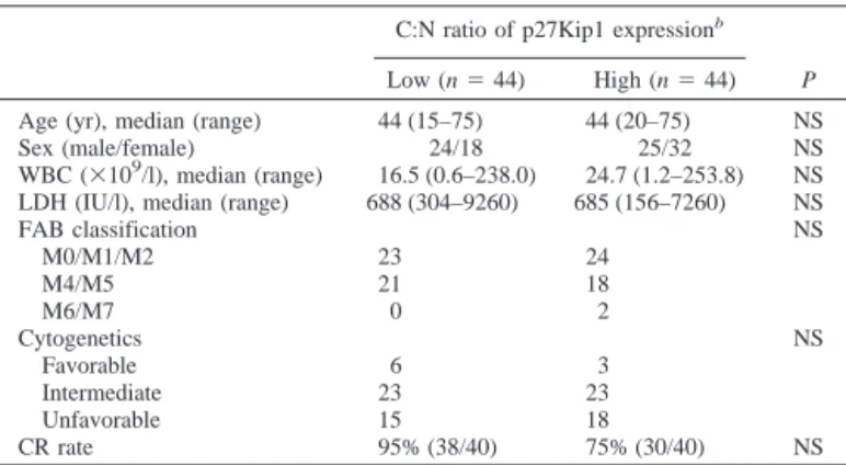
Table 1 Patient characteristics and CRarate according to the C:N ratio of p27Kip1
protein localization
C:N ratio of p27Kip1 expressionb
P
Low (n⫽ 44) High (n⫽ 44) Age (yr), median (range) 44 (15–75) 44 (20–75) NS Sex (male/female) 24/18 25/32 NS WBC (⫻109/l), median (range) 16.5 (0.6–238.0) 24.7 (1.2–253.8) NS LDH (IU/l), median (range) 688 (304–9260) 685 (156–7260) NS
FAB classification NS M0/M1/M2 23 24 M4/M5 21 18 M6/M7 0 2 Cytogenetics NS Favorable 6 3 Intermediate 23 23 Unfavorable 15 18 CR rate 95% (38/40) 75% (30/40) NS
aCR, complete remission; C:N, cytoplasmic to nuclear; NS, not significant; WBC,
white blood cell; LDH, lactic dehydrogenase; FAB, French-American-British.
bC:N ratio of p27Kip1 localization; Low,ⱕ1.97; High, ⬎1.97. 5226
which was then used to transfect 2⫻ 106leukemia cells. Cells were transfected
with expression constructs encoding the constitutively active form of the Akt/PKB (pcDNA3-myristoylated-Akt/PKB) or dominant negative (DN)-Akt/ PKB (pcDNA3-DN-Akt/PKB), which were kindly provided by Dr. Young-Guen Kwon (Yonsei University, Seoul, Korea). The cell suspension was immediately electroporated by a Nucleofector instrument (Amaxa Biosys-tems), according to the manufacturer’s instructions. Immediately after electro-poration, the cells were suspended in the complete medium and incubated in a humidified 37°C/5% CO2 incubator. The cells were harvested 48 h after
transfection and used for the experiments. The experiments were repeated at least three times.
Confocal Microscopy and Image Analysis. Confocal microscopy was used to examine the localization of the p27Kip1 protein in AML cells. Cells were fixed, permeabilized, and blocked with 2% BSA. Cells were incubated with the primary antibody against p27Kip1 (1:100 dilution) for 1 h at 37°C and then incubated with the corresponding FITC-conjugated secondary antibody (1:200 dilution). After the cells had been spun down on a slide, the nuclei were counterstained by 4⬘,6-diamidino-2-phenylindole (Molecular Probes, Eugene, OR) for 1 min. After washing, the slides were mounted in a drop of antifade mounting medium (Vectashield; Vector Laboratories, Burlingame, CA). An-tibody labeling was examined by a Bio-Rad 1024-UV confocal system at-tached to a laser scanning confocal microscope (Leica Lasertechnick GmbH, Heidelberg, Germany). Control experiments, with the omission of the primary antibody, showed negative staining in all of the experiments. Simultaneous FITC or 4⬘,6-diamidino-2-phenylindole images were captured from the same optical section. The captured images were then pseudocolored as follows: (a) blue for 4⬘,6-diamidino-2-phenylindole; and (b) green for FITC. Regions of colocalization appear in cyan, reflecting the additive effect of superimposing the green and blue pixels. Image analysis was performed using the standard system operating software provided with the confocal microscope. All of the illustrations were assembled and processed digitally (Adobe PhotoShop, ver-sion 6.0; Adobe Systems Inc., San Diego, CA).
Preparation of Nuclear and Cytoplasmic Fractions. Cells were subfrac-tionated, as described previously (45), with minor modifications. Briefly, cells were pelleted by centrifugation (5 min, 12,000 rpm, 4°C) and incubated in a hypotonic buffer [10 mMHEPES (pH 7.2), 10 mMKCl, 1.5 mMMgCl2, 0.1 mM
EGTA, 20 mMNaF, 100MNa3VO4, and a 0.1% protease inhibitor cocktail;
Sigma Chemical, St. Louis, MO] for 30 min at 4°C, with rocking. Cells were broken using a Dounce homogenizer (30 strokes), after which the nuclei were pelleted by centrifugation (10 min, 3,500 rpm, 4°C). The nuclei-free superna-tant was subjected to a second 10,000⫻ g centrifugation for 45 min at 4°C to separate the membranes from the cytosolic fractions. The nuclear pellets were resuspended in nuclear lysis buffer [10 nMTris-HCl (pH 7.5), 150 mMNaCl, 5 mMEDTA, and 1% Triton X-100], incubated for 1 min in a sonicating water bath, then incubated for 30 min at 4°C, with rocking. Twentyg of total cytosolic and nuclear proteins were analyzed by Western blotting.
Western Blotting. The cells were dissolved in 100 l of SDS-PAGE sample buffer containing -mercaptoethanol to a final concentration of 3⫻ 106cells. Lysates were sonicated for 15 s with a Vibra Cell Sonicator,
boiled for 10 min, and additionally analyzed by Western blotting. The protein yields were quantified using the Bio-Rad Dc protein assay kit (Bio-Rad, Hercules, CA) and equivalent amounts of protein applied to 15% acrylamide gels. The proteins were separated by SDS-PAGE and transferred to nitrocel-lulose membranes (Amersham Biosciences, Sunnyvale, CA). The membranes were blocked with 3% BSA in Tris-buffered saline-Tween (TBST; 1⫻ Tris-buffered saline, 0.1% Tween 20) for 2 h. After washing twice in TBST, the membranes were incubated with the primary antibodies for 2 h at room temperature. The membranes were then washed four times in TBST and incubated with the relevant horseradish peroxidase-conjugated secondary an-tibodies (1:3000 dilution with 3% BSA in TBST) for 30 min. After washing four times in TBST, the reactive proteins were visualized with an enhanced chemiluminescence detection system (Amersham Biosciences). Densitometry was performed by the Molecular Dynamics Imaging system and ImageQuant 3.3 software (Amersham Biosciences) to quantify relative amounts of protein detected on the Western blots.
Statistical Analysis. The patients were divided into two groups (high and low) in relation to the median value of the cytoplasmic to nuclear ratio (C:N) of the p27Kip1 protein localization. Comparisons among the characteristics of the groups were made using a chi-square test for the binary variables and a Mann-Whitney test for the continuous variables. The disease-free survival (DFS) and overall survival (OS) probabilities were calculated using the Kaplan-Meier method. The log-rank statistic was used to test for the difference in survival times between the groups. In addition to the p27Kip1 protein localization, the WBC count, age, and cytogenetics were analyzed in the
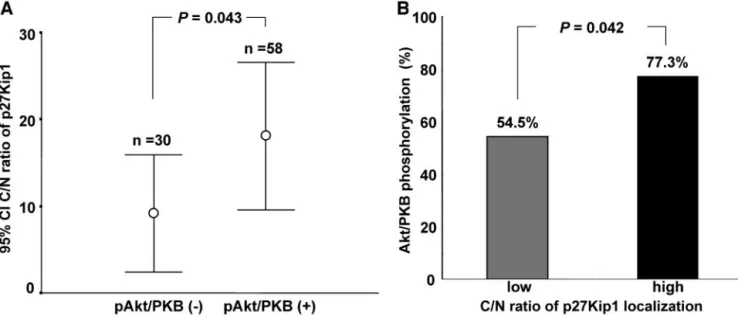
Fig. 2. A, the levels of cytoplasmic to nuclear ratio (C:N) of the p27Kip1 protein localization in relation to the constitutive Akt or protein kinase B (Akt/PKB) phosphorylation in acute myelogenous leukemia cells. The mean value of the C:N ratio of the p27Kip1 protein localization (E) was significantly higher in the pAkt/PKB-positive compared with the pAkt/PKB-negative group (P⫽ 0.043; B) The constitutive Akt/PKB phosphorylation, according to the C:N ratio of p27Kip1 protein localization, in acute myelogenous leukemia cells. The frequency of Akt/PKB phosphorylation was significantly higher in the high C:N ratio group compared with the low C:N ratio group (P⫽ 0.042).
Table 2 Cell cycle distribution according to the C:Naratio of p27Kip1 localization
C:N ratio of p27Kip1 localization
P
Low (n⫽ 44) High (n⫽ 44)
G0/G1 79.4⫾ 27.0% 86.4⫾ 11.9% NS
G2/M 15.9⫾ 27.1% 6.6⫾ 2.5% NS
S 7.8⫾ 12.3% 14.0⫾ 27.7% NS
aC:N, cytoplasmic to nuclear; NS, not significant.
5227
univariate and multivariate analysis. A multivariate analysis was used to test for the independent prognostic significance of the variables using the Cox proportional hazards regression model. Patients alive and still in remission at their last follow-up examination were censored in the analysis. A P⬍ 0.05 was used to indicate statistical significance. All of the calculations were performed using the SPSS software, version 11.0.1 (SPSS Inc., Chicago, IL).
RESULTS
The p27Kip1 Protein Expression in AML Cells. Western blot
analysis demonstrated that the p27Kip1 protein was expressed to a variable degree in 88 (88.9%) of 99 AML cases (Fig. 1A). To perform the quantitative analysis, the expression level of p27Kip1 protein was evaluated in the AML cells (L) by normalization as follows:
Lc ⫽ p27Kip1 (L)/␣-tubulin (L) as determined by Western blot
analysis. In this study, the Lc ranged from 0 to 68.3 (median, 8.08). There was no association between the cellular levels of the p27Kip1 protein and the various clinical parameters such as age, French-American-British classification, WBC count, lactic dehydrogenase level, cell-cycle distribution, cytogenetics, complete remission rate, DFS, and OS (data not shown).
Subcellular Localization of p27Kip1 Protein in AML Cells.
Next, the subcellular localization of the p27Kip1 protein was exam-ined in the leukemia cells obtaexam-ined from 88 AML patients. Western blot analysis of the fractionated cell lysates demonstrated that the subcellular localization of the p27Kip1 protein was different,
accord-ing to the patients (Fig. 1B). To perform the quantitative analysis, the relative levels of the p27Kip1 expression at the subcellular compart-ment were represented as the C:N ratio of the p27Kip1 protein levels as shown in the following data: C:N ⫽ p27Kip1(C)/p27Kip1(N) as determined by the Western blot analysis. In this study, the C:N ratios ranged from 0 to 127.21 (median, 1.97). For the practical evaluations, the median value was used as a cutoff value for the levels of subcel-lular localization of the p27Kip1 in the AML cells. Using this value, the AML cases were classified into two groups; the high and low C:N ratio groups (n⫽ 44 for both; Table 1). Next, the association between the C:N ratio of the p27Kip1 localization and various clinical param-eters in AML were analyzed. As shown in Table 1, the C:N ratio of the p27Kip1 localization was not correlated with age, gender, WBC count, lactic dehydrogenase level, French-American-British classifi-cation, or cytogenetics. Cell cycle analysis demonstrated that the fraction of cells in the G0/G1phase were not different between the
AML cells, showing either a high or low C:N ratio of the p27Kip1 localization (86.4⫾ 11.9 and 79.4 ⫾ 27.0%, respectively; Table 2). Likewise, the C:N ratio of the p27Kip1 localization was not correlated with the fractions of the cells in the G2-M or S-phase (Table 2).
Correlation between Constitutive Akt/PKB Phosphorylation and p27Kip1 Localization. The constitutive phosphorylation of
Ser473
Akt/PKB was demonstrated in 58 (58.6%) of 99 cases (Fig. 2A). Cell cycle analysis showed that neither the fraction of cells in the G0/G1phase, G2-M phase, or S phase were different, according to the Fig. 3. Confocal microscopic analysis of the p27Kip1 protein localization in acute myelogenous leukemia (AML) cells in relation to the constitutive Akt or protein kinase B (Akt/PKB) phosphorylation. A, A representative pAkt/PKB-positive AML specimen was stained for p27Kip1 by FITC (green) as described in “Materials and Methods.” The confocal image demonstrated the preferential localization of the p27Kip1 protein to the cytoplasm. Nuclear counterstain was carried out with 4⬘,6-diamidino-2-phenylindole (DAPI) staining (blue). Cyan represents the merged images. B, nuclear staining of the p27Kip1 protein was evident in the representative AML case, where no Akt/PKB phosphorylation was seen.
5228
phosphorylation of Akt/PKB (data not shown). Next, the association of the phosphorylation of Akt/PKB and localization of the p27Kip1 protein was examined in the AML cells. Akt/PKB phosphorylation was observed more frequently in the high C:N ratio group compared with the low C:N ratio group (77.3% versus 54.5%, P⫽ 0.042; Fig. 2B). The mean value of the C:N ratio of the p27Kip1 localization was significantly higher in the pAkt/PKB-positive AML cases compared with the pAkt/PKB-negative AML cases (18.15 ⫾ 4.2 versus 9.2⫾ 3.3, P ⫽ 0.043; Fig. 2A). The confocal microscopic analysis
revealed the p27Kip1 protein to be preferentially localized to the cytoplasm in a pAkt/PKB-positive AML case (Fig. 3A). In contrast, the nuclear staining of the p27Kip1 protein was evident in a pAkt/ PKB-negative AML case (Fig. 3B).
The p27Kip1 Mislocalization Induced by Akt/PKB Overexpres-sion in Leukemia Cells. To investigate whether the active Akt/
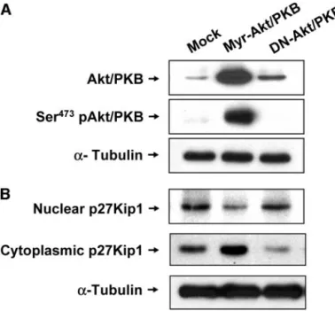
PKB directly affects the localization of the p27Kip1 protein in leukemia cells, the U937 leukemia cells were transfected using the pcDNA3-myristoylated-Akt/PKB or pcDNA3-DN-Akt/PKB ex-pression vectors (Fig. 4A). As shown in Fig. 4B, the exex-pression of the p27Kip1 protein was preferentially localized to the cytoplasm of the myristoylated-Akt/PKB-transfected U937 cells. In contrast, the DN-Akt/PKB-transfected U937 cells revealed a reduced level of the p27Kip1 in the cytoplasm, but increased the p27Kip1 levels in the nuclei.
The p27Kip1 Localization as a Prognostic Variable in AML.
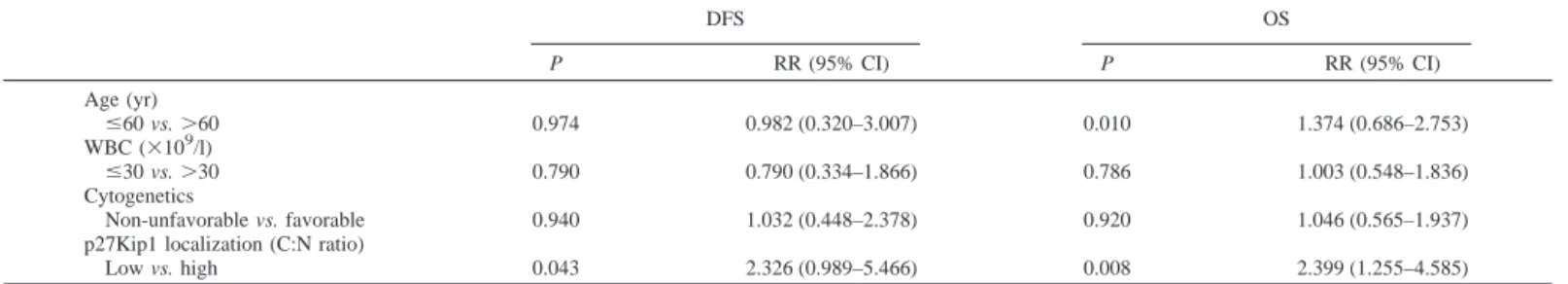
The complete remission rate of the patients with the high C:N ratio of the p27Kip1 localization was lower than that with the low C:N ratio (75% versus 95%), but without statistical significance (P ⫽ 0.116; Table 1). A survival analysis using the Kaplan-Meier method dem-onstrated that the high C:N ratio group had a significantly shorter DFS than the low C:N ratio group (Fig. 5A; P⫽ 0.0353 by log-rank test). The OS rate was also significantly lower in the high C:N ratio group compared with the low C:N ratio group (Fig. 5B; P⫽ 0.0023). The DFS estimates at 5 years for patients with a high or low C:N ratio of p27Kip1 localization were 19.0 (SE ⫽ 14.6%) and 58.2% (SE⫽ 12.4%), respectively (P ⫽ 0.0353). The OS estimates at 5 years for patients with a high or low C:N ratio were 10.8 (SE⫽ 8.4%) and 52.6% (SE ⫽ 11.2%), respectively (P ⫽ 0.0023). The univariate analysis revealed that the C:N ratio of the p27Kip1 localization was a strong prognostic factor both of the DFS and OS (Table 3). A multivariate analysis of the covariates in the Cox regression model demonstrated that the C:N ratio of the p27Kip1 protein localization remained as an independent prognostic factor of the DFS [relative risk (95% confidence interval)⫽ 2.326 (0.989–5.466), P ⫽ 0.043] and the OS [relative risk (95% confidence interval)⫽ 2.399 (1.255–4.585),
P⫽ 0.008] (Table 4). Fig. 4. Effects of myristoylated Akt or protein kinase B (Myr-Akt/PKB) and DN-Akt/
PKB transfection on the subcellular localization of the p27kip protein in the U937 cells. The U937 cells were transiently transfected with Myr-Akt/PKB or DN-Akt/PKB con-structs as described in “Materials and Methods.” A, the total cell lysates were analyzed by Western blotting, using antibodies against total Akt/PKB and Ser473 pAkt/PKB. B,
Myr-Akt/PKB-transfected and DN-Akt/PKB-transfected U937 cells were subjected to the subcellular fractionation. Equal amounts of proteins (20 g) from the nuclear and cytoplasmic fractions were separated by SDS-PAGE and then probed with the anti-p27kip1 antibody. The␣-tubulin was used as a loading control.
Fig. 5. Kaplan-Meier survival curves for the disease-free survival and overall survival rates of patients with acute myelogenous leukemia, according to the cytoplasmic to nuclear ratio (C:N) ratio of the p27Kip1 localization. The acute myelogenous leukemia cases with the high C:N ratio of the p27Kip1 localization had significantly lower disease-free survival (A) and overall survival rates (B) compared with the cases with the low C:N ratio (P⫽ 0.0353 and P ⫽ 0.0023, respectively). The log-rank statistic was used to test for the difference in survival times between the groups.
5229
DISCUSSION
In this study, the cytoplasmic mislocalization of the p27Kip1 pro-tein was demonstrated in a substantial proportion of the AML cases. The p27Kip1 mislocalization to the cytoplasm was highly correlated with the constitutive phosphorylation of Akt/PKB in the leukemic cells and with a poor prognosis in AML. However, the mere cellular levels of the p27Kip1 protein were not correlated with the clinical outcome. This study supports a growing body of evidences suggesting that the cytoplasmic p27Kip1 may play a role as an oncoprotein with antiapoptotic properties (26 –28, 46).
Much less is known about the localization of p27Kip1 protein during the cell cycle progression. To inhibit cyclin E/CDK2, p27Kip1 needs to be imported into the nucleus (3). As cells progress along the cell cycle, the p27Kip1 protein shuttles between the nucleus and cytoplasm (47). The cytoplasmic redistribution of p27Kip1 induced by mitogenic stimulation is dependent on the phosphorylation of the Ser10
residue (47). Recently, it was demonstrated that p27Kip1 phos-phorylation induced by the oncogenically activated kinase Akt/PKB disables the nuclear localization capacity of p27Kip1 (33, 34, 41). Akt/PKB can directly phosphorylate p27Kip1 at a Thr157
residue within the nuclear localization signal both in vitro and in vivo (33, 41). This Akt/PKB-dependent phosphorylation results in an impairing of its nuclear import, leading to cytoplasmic retention of the p27Kip1, abrogation of its CDK2 inhibitory activity, and cell cycle progression (41). The Akt/PKB phosphorylation in primary human breast cancer statistically correlated with the cytoplasmic localization of the p27Kip1 protein (41). An inhibition of the Akt/PKB activation with the phosphatidylinositol 3⬘-kinase inhibitor LY294002 or by the over-expression of DN Akt/PKB allele allows return of the p27Kip1 to the nucleus, resulting in an inhibition of CDK2 activity (33, 34, 41). As with Ser10
, Thr157
-phosphorylated p27Kip1 is detected almost exclu-sively in the cytoplasmic compartment (34, 41). Whereas Ser10
phos-phorylation promotes the nuclear export of the p27Kip1, Thr157
phos-phorylation seems to impair its import into the nuclei (33). The
complex relationship between the p27Kip1 localization and degrada-tion still remains to be addressed. Previous experimental evidence suggests that at least in breast cancer cells, the proteolysis and mislocalization of p27Kip1 occur through separate pathways (33). Although the p27Kip1 phosphorylation at the Ser10
or Thr157
residue was not evaluated in this study, the close correlation between the cytoplasmic mislocalization of p27Kip1 and the constitutive Akt/PKB phosphorylation suggests that an Akt/PKB-mediated Thr157
phospho-rylation mechanism is present in a substantial proportion of AML cases. In this study, the induced Akt/PKB activation resulted in a remarkable increase in the levels of cytoplasmic p27Kip1 in the U937 leukemia cells. Transfection of the U937 cells with a DN Akt/PKB construct resulted in a recovery of the nuclear localization of the p27Kip1 protein. These findings strongly suggest that Akt/PKB acti-vation is one of the crucial mechanisms determining the localization of the p27Kip1 protein in leukemia cells. However, because the Akt/PKB phosphorylation was not observed in a certain proportion of AML cases showing the cytoplasmic mislocalization of p27Kip1 in this study, the contribution of other intracellular oncogenic pathways to the cytoplasmic mislocalization and dysregulation of p27Kip1 protein cannot be excluded.
Whatever the mechanism is, it is important to understand the biological significance of the cytoplasmic mislocalization of p27Kip1 in AML cells. The export of p27Kip1 from the nucleus to the cyto-plasm serves to remove the inhibitory activity of p27Kip1 against cyclin E/CDK2 (34, 41). This allows an activation of an increasing number of cyclin E/CDK2 complexes, which are then free to phos-phorylate the p27Kip1. The transfection studies suggested that the relocalization of the p27Kip1 from the nucleus to the cytoplasm was sufficient to sustain the cellular proliferation (34, 41, 48). Another issue that needs to be addressed is whether the cytoplasmic mislocal-ization of the p27Kip1 observed in cancers merely contributes to abrogate its function or induces the p27Kip1 to acquire new cytoplas-mic functions. The p27Kip1 was demonstrated to inhibit the drug-induced apoptosis (49), although it is uncertain whether the nuclear and cytoplasmic p27Kip1 differ in their efficiencies to protect the cells from apoptosis.
Only recently, the cytoplasmic mislocalization of p27Kip1 in tumor cells has been identified as a mechanism whereby cancer cells pro-mote carcinogenesis in humans (28). Displacement of p27Kip1 into the cytoplasm contributes to the anchorage-independent growth of human transformed fibroblasts by maintaining high cyclin/CDK ac-tivity in the nucleus (31). Cytoplasmic mislocalization of p27Kip1 has been reported for a number of human malignancies (26 –28, 33, 34, 41). It appears that the preferential location of p27Kip1 in the cyto-plasm of tumor cells is predictive of a more aggressive clinical behavior (33). It was shown that the patients with Barrett’s adenocar-cinoma or breast caradenocar-cinoma, which presented the cytoplasmic p27Kip1, showed decreased overall survivals compared with the
Table 3 Univariate analysis of age, WBC,acytogenetics, and the C:N ratio of p27Kip1
localization for CR rate, DES, and OS in AML patients Pb CR DFS OS Age (yr) ⱕ60 vs. ⬎60 0.3300 0.7068 0.1100 WBC (⫻109/l) ⱕ30 vs. ⬎30 0.0200 0.5353 0.9890 Cytogenetics Non-unfavorablecvs. unfavorable 1.000 0.8231 0.8659
p27Kip1 localization (C:N ratio)
Low vs. high 0.116 0.0353 0.0023
aWBC, white blood cell; C:N, cytoplasmic to nuclear; CR, complete remission; DFS,
disease-free survival; OS, overall survival; AML, acute myelogenous leukemia.
bLog-rank test.
cFavorable plus intermediate prognostic group.
Table 4 Multivariate analysis of DFSaand OS in AML patients
DFS OS P RR (95% CI) P RR (95% CI) Age (yr) ⱕ60 vs. ⬎60 0.974 0.982 (0.320–3.007) 0.010 1.374 (0.686–2.753) WBC (⫻109/l) ⱕ30 vs. ⬎30 0.790 0.790 (0.334–1.866) 0.786 1.003 (0.548–1.836) Cytogenetics Non-unfavorable vs. favorable 0.940 1.032 (0.448–2.378) 0.920 1.046 (0.565–1.937) p27Kip1 localization (C:N ratio)
Low vs. high 0.043 2.326 (0.989–5.466) 0.008 2.399 (1.255–4.585)
aDFS, disease-free survival; OS, overall survival; AML, acute myelogenous leukemia; RR, relative risk; CI, confidence interval; WBC, white blood cell; C:N, cytoplasmic to
nuclear.
5230
patients who present with nuclear p27Kip1 (28, 41). In this study, the WBC count or cytogenetics, which had been generally considered as a prognostic factor in adult AML, was not significantly associated with prognosis. These findings were in agreement with the previous report, which revealed that neither WBC nor cytogenetics was an independent prognostic factor in adult AML (50). Instead, we dem-onstrate for the first time that the p27Kip1 mislocalization is the strong prognostic factor in AML cases, whereas the total cellular levels of p27Kip1 do not have any prognostic value. The patients with high C:N ratio of the p27Kip1 localization showed a significantly shorter DFS and OS compared with the patients with low C:N ratio of p27Kip1 localization. The multivariate analysis revealed that the C:N ratio of the p27Kip1 localization remained an independent prognostic factor both the DFS and OS.
In conclusion, the cytoplasmic mislocalization of the p27Kip1 protein was observed in a substantial proportion of the AML cases. The mislo-calization, not the cellular levels, of the p27Kip1 protein was highly associated with the constitutive phosphorylation of Akt/PKB and a poor prognosis in AML. Although the biological functions of cytoplasmic p27Kip1 protein and the genetic alterations that cause its mislocalization to the cytoplasm should be evaluated additionally, these findings suggest that the cytoplasmic mislocalization of p27Kip1 is an independent prog-nostic factor in AML, and the Akt/PKB-p27Kip1 pathway may be a ready target for antileukemia therapy.
ACKNOWLEDGMENTS
We thank Dr. Y. G. Kwon (Yonsei University, Seoul, Korea) for providing the pcDNA3-myristoylated-Akt/PKB and pcDNA3-DN-Akt/PKB expression vectors.
REFERENCES
1. Hengst L, Reed SI. Translational control of p27Kip1accumulation during the cell
cycle. Science (Wash DC) 1996;271:1861– 4.
2. Slingerland J, Pagano M. Regulation of the Cdk inhibitor p27 and its deregulation in cancer. J Cell Physiol 2000;183:10 –7.
3. Reynisdottir I, Massague J. The subcellular locations of p15(Ink4b) and p27(Kip1) coordinate their inhibitory interactions with cdk4 and cdk2. Genes Dev 1997;11:492–503. 4. Pagano M, Tam SW, Theodoras AM, et al. Role of the ubiquitin-proteasome pathway in regulating abundance of the cyclin-dependent kinase inhibitor p27. Science (Wash DC) 1995;269:682–5.
5. Olashaw N, Pledger WJ. Paradigms of growth control: relation to Cdk activation. Sci STKE 2002;134: RE7.
6. Montagnoli A, Fiore F, Eytan E, et al. Ubiquitination of p27 is regulated by Cdk-dependent phosphorylation and trimeric complex formation. Genes Dev 1999; 13:1181–9.
7. Nguyen H, Gitig DM, Koff A. Cell-free degradation of p27(kip1), a G1 cyclin-dependent kinase inhibitor, is cyclin-dependent on CDK2 activity and the proteasome. Mol Cell Biol 1999;19:1190 –201.
8. Sheaff RJ, Groudine M, Gordon M, Roberts JM, Clurman BE. Cyclin E-CDK2 is a regulator of p27Kip1. Genes Dev 1997;11:1464 –78.
9. Vlach J, Hennecke S, Amati B. Phosphorylation-dependent degradation of the cyclin-dependent kinase inhibitor p27. EMBO J 1997;16:5334 – 4.
10. Zhang H, Kobayashi R, Galaktionov K, Beach D. p19Skp1 and p45Skp2 are essential elements of the cyclin A-CDK2 S phase kinase. Cell 1995;82:915–25.
11. Carrano AC, Eytan E, Hershko A, Pagano M. SKP2 is required for ubiquitin-mediated degradation of the CDK inhibitor p27. Nat Cell Biol 1999;1:193–9.
12. Sutterluty H, Chatelain E, Marti A, et al. p45SKP2 promoters p27Kip1 degradation and induces S phase in quiescent cells. Nat Cell Biol 1999;1:207–14.
13. Tsvetkov LM, Yeh KH, Lee SJ, Sun H, Zhang H. p27(Kip1) ubiquitin and degrada-tion is regulated by the SCF (Skp2) complex through phosphorylated Thr187 in p27. Curr Biol 1999;9:661– 4.
14. Ganoth D, Bornstein G, Ko TK, et al. The cell-cycle regulatory protein Cks1 is required for SCF(Skp2)-mediated ubiquitinylation of p27. Nat Cell Biol 2001;3: 321– 4.
15. Spruck C, Strohmaier H, Watson M, et al. A CDK-independent function of mamma-lian Cks1: targeting of SCFSkp2 to the CDK inhibitor p27. Mol Cell 2001;7:639 –50. 16. Esposito V, Baldi A, De Luca A, et al. Prognostic role of the cyclin-dependent kinase
inhibitor p27 in non-small cell lung cancer. Cancer Res 1997;57:3381–5. 17. Loda M, Cukor B, Tam SW, et al. Increased proteasome-dependent degradation of the
cyclin-dependent kinase inhibitor p27 in aggressive colorectal cancers. Nat Med 1997;3:231– 4.
18. Yang HY, Zhou BP, Hung MC, Lee MH. Oncogenic signals of HER-2/neu in regulating the stability of the cyclin-dependent kinase inhibitor p27. J Biol Chem 2000;275:24735–9.
19. Chiarle R, Budel LM, Skolink J. Increased proteasome degradation of cyclin-depend-ent kinase inhibitor p27 is associated with a decreased overall survival in mantle cell lymphoma. Blood 2000;95:619 –26.
20. Catzavelos C, Bhattacharya N, Ung YC, et al. Decreased levels of the cell-cycle inhibitor p27Kip1 protein: prognostic implications in primary breast cancer. Nat Med 1997;3:227–30.
21. Yokozawa T, Towatari M, Lida H, et al. Prognostic significance of the cell cycle inhibitor p27Kip1in acute myeloid leukemia. Leukemia (Baltimore) 2000;14:28 –33.
22. Yang G, Ayala G, Marzo AD, et al. Elevated Skp2 protein expression in human prostate cancer: association with loss of the cyclin-dependent kinase inhibitor p27 and PTEN and with reduced recurrence-free survival. Clin Cancer Res 2002;8:3419 –26. 23. Lloyd RV, Jin L, Qian X, Kulig E. Aberrant p27kip1 expression in endocrine and
other tumors. Am J Pathol 1997;150:401–7.
24. Bales ES, Dietrich C, Bandyopadhyay D, et al. High levels of expression of p27(KIP1) and cyclin E in invasive primary malignant melanomas. J Investig Der-matol 1999;113:1039 – 47.
25. Blain SW, Scher HI, Cordon-Cardo C, Koff A. p27 as a target for cancer therapeutics. Cancer Cell 2003;3:111–5.
26. Baldassarre G, Belletti B, Bruni P, et al. Overexpressed cyclin D3 contributes to retaining the growth inhibitor p27 in the cytoplasm of thyroid tumor cells. J Clin Investig 1999;104:865–74.
27. Ciaparrone M, Yamamoto H, Yao Y, et al. Localization and expression of p27KIP1 in multistage colorectal carcinogenesis. Cancer Res 1998;58:114 –22.
28. Singh SP, Lipman J, Goldman H, et al. Loss or altered subcellular localization of p27 in Barrett’s associated adenocarcinoma. Cancer Res 1998;58:1730 –5.
29. Jiang Y, Zhao RC, Verfaillie CM. Abnormal integrin-mediated regulation of chronic myelogenous leukemia CD34⫹ cell proliferation: BCR/ABL up-regulates the cyclin-dependent kinase inhibitor, p27Kip, which is relocated to the cell cytoplasm and incapable of regulating cdk2 activity. Proc Natl Acad Sci USA 2000;97:10538 – 43. 30. Soucek T, Yeung RS, Hengstschlager M. Inactivation of the cyclin-dependent kinase inhibitor p27 upon loss of the tuberous sclerosis complex gene-2. Proc Natl Acad Sci USA 1998;95:15653– 8.
31. Orend G, Hunter T, Ruoslahti E. Cytoplasmic displacement of cyclin E-cdk2 inhibitors p21Cip1 and p27Kip1 in anchorage-independent cells. Oncogene 1998;16:2575– 83. 32. Yaroslavskiy B, Watkins S, Donnenberg AD, Patton TJ, Steinman RA. Subcellular
and cell-cycle expression profiles of CDK-inhibitors in normal differentiating mye-loid cells. Blood 1999;93:2907–17.
33. Liang J, Zubovitz J, Petrocelli T, et al. PKB/Akt phosphorylates p27, impairs nuclear import of p27 and opposes p27-mediated G1 arrest. Nat Med 2002;8:1153– 60. 34. Viglietto G, Motti ML, Bruni P, et al. Cytoplasmic relocalization and inhibition of the
cyclin-dependent kinase inhibitor p27Kip1 by PKB/Akt-mediated phosphorylation in breast cancer. Nat Med 2002;8:1136 – 43.
35. Datta SR, Dudek H, Tao X, et al. Akt phosphorylation of BAD couples survival signals to the cell-intrinsic death machinery. Cell 1997;91:231– 41.
36. Scheid MP, Woodgett JR. PKB/AKT: functional insights from genetic models. Nat Rev Mol Cell Biol 2001;2:760 – 8.
37. Cantley LC. The phosphoinositide 3-kinase pathway. Science (Wash DC) 2002;296: 1655–7.
38. Testa JR, Bellacosa A. AKT plays a central role in tumorigenesis. Proc Natl Acad Sci USA 2001;98:10983–5.
39. Sun H, Lesche R, Li DM, et al. PTEN modulates cell cycle progression and cell survival by regulating phosphatidylinositol 3,4,5,-trisphosphate and Akt/protein ki-nase B signaling pathway. Proc Natl Acad Sci USA 1999;96:6199 –204. 40. Medema RH, Kops GJ, Bos JL, Burgering BM. AFX-like Forkhead transcription
factors mediate cell-cycle regulation by Ras and PKB through p27kip1. Nature (Lond) 2000;404:782–7.
41. Shin I, Yakes FM, Rojo F, et al. PKB/Akt mediates cell-cycle progression by phosphorylation of p27Kip1 at threonine 157 and modulation of its cellular localiza-tion. Nat Med 2002;8:1145–52.
42. Min YH, Eom JI, Cheong JW, et al. Constitutive phosphorylation of Akt/PKB protein in acute myeloid leukemia: its significance as a prognostic variable. Leukemia (Baltimore) 2003;17:995–7.
43. Cheson BD, Cassileth PA, Head DR, et al. Report of the National Cancer Institute-sponsored workshop on definitions of diagnosis and response in acute myeloid leukemia. J Clin Oncol 1990;8:813–9.
44. Amadori S, Arcese W, Isacchi G, et al. Mitoxantrone, etoposide, and intermediate-dose cytarabine: an effective and tolerable regimen for the treatment of refractory acute myeloid leukemia. J Clin Oncol 1991;9:1210 – 4.
45. Lenferink AE, Busse D, Flanagan WM, Yakes FM, Arteaga CL. ErbB2/neu kinase modulates cellular p27(Kip1) and cyclin D1 through multiple signaling pathways. Cancer Res 2001;61:6583–91.
46. Blagosklonny MV. Are p27 and p21 cytoplasmic oncoproteins? Cell Cycle 2002;1: 391–3.
47. Rodier G, Montagnoli A, Di Marcotullio L, et al. p27 cytoplasmic localization is regulated by phosphorylation of Ser10 and is not a prerequisite for its proteolysis. EMBO J 2001;20:6672– 82.
48. Boehm M, Yoshimoto T, Crook MF, et al. A growth factor-dependent nuclear kinase phosphorylates p27Kip1 and regulates cell cycle progression. EMBO J 2002;21: 3390 – 4001.
49. Eymin B, Haugg M, Droin N, et al. p27Kip1 induces drug resistance by preventing apoptosis upstream of cytochrome c release and procaspase-3 activation in leukemic cells. Oncogene 1999;18:141– 8.
50. Whitman SP, Archer KJ, Feng L, et al. Absence of the wild-type allele predicts poor prognosis in adult de novo acute myeloid leukemia with normal cytogenetics and the internal tandem duplication of FLT3. Cancer Res 2001;61:7233–9.
5231