ABSTRACT
Purpose: With advances in surgical techniques, reduced-port laparoscopic surgery is increasingly being performed for the treatment of gastric carcinoma. Many studies have reported satisfactory short-term outcomes after reduced 3-port laparoscopic gastrectomy (LG). The aim of this study was to investigate the long-term oncological outcomes of 3-port LG in patients with gastric carcinoma.
Materials and Methods: We reviewed the medical records of 1,117 patients who underwent LG for gastric carcinoma in three major institutions between 2012 and 2015. The data showed that 460 patients underwent 3-port LG without assistance, and 657 underwent conventional 5-port LG. We compared the overall and disease-free survival rates between the 2 groups. Results: There were 642 male and 475 female patients with a mean age of 56.1 years. Among them, 1,028 (92.0%) underwent distal gastrectomy and 89 (8.0%) underwent total gastrectomy. In the final pathologic examination, 1,027 patients (91.9%) were stage I, 73 (6.5%) were stage II, and 17 (1.5%) were stage III, and there were no significant difference in the pathologic stage between groups. The 3- and 5-port LG groups showed no significant differences in the 5-year overall survival (94.3% vs. 96.7%, P=0.138) or disease-free survival (94.3% vs. 95.9%, P=0.231). Stratified analyses according to pT and pN stages also showed no significant differences in overall or disease-free survival between the two groups.
Conclusions: Long-term survival after 3- and 5-port LG was comparable in patients with early-stage gastric carcinoma. The 3-port technique requiring limited surgical assistance may be an appropriate surgical option for this patient population.
Keywords: Gastrectomy; Laparoscopy; Reduced port surgery; Stomach neoplasm; Survival
INTRODUCTION
Since its introduction in the early 1990s, laparoscopic gastrectomy (LG) has been rapidly adopted for the treatment of gastric carcinoma [1]. Several studies have demonstrated the
Original Article
Received: Dec 24, 2020 Revised: Mar 23, 2021 Accepted: Mar 23, 2021 Correspondence to Jun Ho LeeDepartment of Surgery, Samsung Medical Center, Sungkyunkwan University School of Medicine, 81 Irwon-ro, Gangnam-gu, Seoul 06351, Korea.
E-mail: [email protected] *Han Hong Lee and Oh Jeong equally contributed to this study.
Copyright © 2021. Korean Gastric Cancer Association
This is an Open Access article distributed under the terms of the Creative Commons Attribution Non-Commercial License (https:// creativecommons.org/licenses/by-nc/4.0) which permits unrestricted noncommercial use, distribution, and reproduction in any medium, provided the original work is properly cited.
ORCID iDs Han Hong Lee
https://orcid.org/0000-0002-7541-8490 Oh Jeong
https://orcid.org/0000-0002-7076-6998 Funding
This study was supported by a grant from the National Research Foundation of Korea (No. 2018R1D1A1B07045486 and 2020R1A2C1012007).
Han Hong Lee 1,*, Oh Jeong 2,*, Ho Seok Seo1, Min Gew Choi3, Seong Yeob Ryu2, Tae Sung Sohn3, Jae Moon Bae3, Sung Kim3, Jun Ho Lee3
1 Division of Gastrointestinal Surgery, Department of Surgery, Seoul St. Mary's Hospital, College of Medicine,
The Catholic University of Korea, Seoul Korea
2Department of Surgery, Chonnam National University Medical School, Gwangju, Korea
3Department of Surgery, Samsung Medical Center, Sungkyunkwan University School of Medicine, Seoul, Korea
Long-Term Oncological Outcomes of
Reduced Three-Port Laparoscopic
Gastrectomy for Early-Stage Gastric
Carcinoma: a Retrospective Large-Scale
Multi-Institutional Study
Author Contributions
Conceptualization: L.H.H., J.O., L.J.H.; Data curation: L.H.H., J.O., L.J.H.; Formal analysis: L.H.H., J.O., L.J.H.; Funding acquisition: L.H.H.; Investigation: L.H.H., J.O., L.J.H.; Methodology: L.H.H., J.O., L.J.H.; Project administration: L.H.H., J.O., L.J.H.; Resources: L.H.H., J.O., S.H.H., C.M.G., R.S.Y., S.T.S., B.J.M., K.S., L.J.H.; Supervision: L.H.H., J.O., L.J.H.; Validation: L.H.H., J.O., L.J.H.; Visualization: L.H.H., J.O.; Writing - original draft: L.H.H., J.O.; Writing - review & editing: L.H.H., J.O., L.J.H.
Conflict of Interest
No potential conflict of interest relevant to this article was reported.
clinical benefits of LG over open surgery, such as faster bowel recovery, shorter hospital stay, and fewer postoperative complications [2,3]. Conventional LG techniques use five or more abdominal ports and require a surgical team that consists of a surgeon, assistant, and scopist. With advances in surgical techniques and instruments, experienced gastric surgeons have developed reduced-port LG, a procedure in which fewer abdominal ports are required, reducing the need for surgical assistance. Previous studies have reported short-term outcomes of reduced-port LG, demonstrating its technical feasibility and safety [4]. Reduced-port LG is also accepted as a useful approach to confront the lack of surgical assistance, especially in small health centers [5].
Single-port LG for gastric cancer was first reported by Omori et al. [6] in 2011. It is performed via a single umbilical incision using a specially designed multichannel port. This technique is demanding even for skilled surgeons because of the operative difficulties caused by handling all instruments through a single channel. Therefore, some surgeons sought to overcome the technical difficulties of single-port LGs by adding additional ports [7,8]. Unlike conventional 5-port LGs, 3-port LGs use two operator ports and one umbilical port. Because this technique uses 2 operator ports and does not require specialized instruments, it can be easily adopted by gastric surgeons who are familiar with conventional LGs [9]. Three-port LG is also called “duet-LG,” emphasizing the fact that it is performed by a surgeon and scopist alone [10]. Several studies have reported the technical feasibility and safety of 3-port LG compared with those of conventional 5-port LG [9-13]. However, the long-term oncological outcomes of 3-port LG have rarely been investigated. In this study, we investigated the long-term oncological outcomes of 3-port LG in a large cohort of patients from multiple institutions.
MATERIALS AND METHODS
Patients
Using institutional gastric cancer databases, we reviewed the records of patients who underwent LG for gastric carcinoma in three major institutions (Samsung Medical Center, Chonnam National University Hwasun Hospital, and Seoul St. Mary's Hospital) in South Korea between January 2012 and December 2015. We excluded patients with other organ malignancies to exclude possible effects on long-term outcomes. A total of 1,117 patients were enrolled, of whom 460 underwent 3-port LG and 657 underwent conventional 5-port LG. The indications for laparoscopic surgery were the same in each institution and included cT1N0 tumors on preoperative staging. The decision regarding 3-port vs. conventional 5-port LG was made by the patient or patient's representative after being informed of the operative procedures. This study was approved by the institutional review boards of each institution, which waived the requirement for informed consent from patients.
Data collection and definition
Clinicopathological data were collected from prospectively constructed databases in each institution. Demographic data included age, sex, body mass index (BMI), and American Society of Anesthesiology (ASA) physical status. Operation data included the extent of gastric resection, reconstruction, operating time, operative blood loss, and need for blood transfusion. Pathological data included tumor size, histological type, resection margin, number of retrieved lymph nodes, and pTNM stage based on the seventh edition of the American Joint Cancer Committee TNM classification [14]. Postoperative outcomes included morbidity, mortality, and hospital stay length. Postoperative morbidity and mortality were defined as any complications
or deaths within 30 days after surgery. The severity of the complications was classified according to the Clavien–Dindo classification of surgical complications [15].
The primary outcomes of this study were overall survival (OS) and disease-free survival (DFS). OS was defined as the time from surgery to death from any cause. DFS was defined as the time from surgery to death or disease recurrence. After surgery, patients were regularly followed up using abdominal computed tomography (CT) and endoscopic evaluations every 6 or 12 months during the subsequent 5 years. Additional work-ups, such as chest CT, liver magnetic resonance imaging, and positron emission tomography/CT, were performed as appropriate. Adjuvant chemotherapy using oral fluoropyrimidine (S-1) or capecitabine plus oxaliplatin was administered to patients with pathologic stage ≥II. The survival status of all patients was ascertained using registration data from the Korea National Statistical Office and medical records. The median follow-up period was 48 months (range, 1–67 months).
Operative techniques
The details of the operative technique of 3-port LG have been described in previous reports [9,10]. Briefly, the patient was placed in the reverse Trendelenburg position. One camera port and two operator ports were made in the umbilicus and on the right and left side of the patient. The operation was performed with no assistance. The operative techniques, including gastric resection and lymph node dissection, were performed as in conventional LG, following the principles of gastric cancer treatment guidelines [16,17]. D1+ lymph node dissection (LND) was performed for cT1N0 tumors. All reconstructions were performed intracorporeally, and the choice of reconstruction was decided at the discretion of the surgeon.
In conventional LG, five abdominal ports, including two operator ports, two assistant ports, and one umbilical port for the laparoscope, were used. The operative techniques, including gastric resection and LND, were the same as those for 3-port LG, and the operation was performed with assistance.
Statistical analyses
Student's t-test was used to compare continuous variables, and the χ2 test or Fisher's exact
test was used to compare categorical variables, as appropriate. Patient survival was analyzed using the Kaplan–Meier method and compared using the log-rank test. Multivariate survival analyses were performed using the Cox proportional hazards model. All statistical analyses were performed using SPSS ver. 19.0 software (SPSS Inc., Chicago, IL, USA), and a P-value <0.05 was considered statistically significant.
RESULTS
Patient characteristics
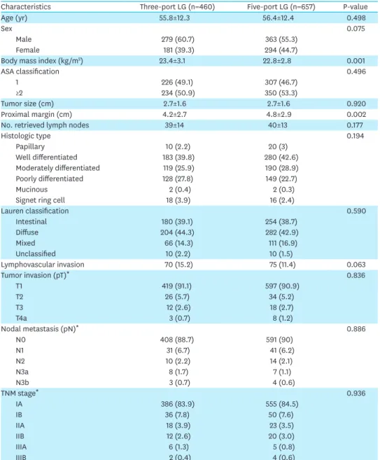
Table 1 presents the clinicopathological characteristics of patients. There were no significant differences in age, sex, or ASA status between the two groups; however, the mean BMI was higher in the 3-port LG group (23.4 vs. 22.8 kg/m2, P=0.001). Tumor size, histologic type, and
Lauren classification did not significantly differ between the two groups. The mean numbers of retrieved lymph nodes in the 3- and 5-port LG groups were 39±14 and 40±13, respectively (P=0.177). In the final pathologic examination, 1027 patients (91.9%) were stage I, 73 patients (6.5%) were stage II, and 17 patients (1.5%) were stage III; there were no significant differences in the final pathologic stage between the two groups.
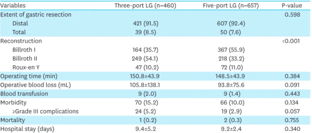
The operative results are shown in Table 2. Among the 1117 patients, 1028 (92.0%) underwent distal gastrectomy and 89 (8.0%) underwent total gastrectomy; there was no significant difference in the extent of gastric resection between the two groups. Billroth II anastomosis was more frequently performed in the 3-port LG group (54.1% vs. 33.2%, P<0.001). All patients underwent laparoscopic surgery without open conversion. No patients in the 3-port LG group required conversion to 5-port surgery. There were no significant differences with respect to operative outcomes, including operating time, operative blood loss, or need for blood transfusion between the two groups. Postoperative morbidity, mortality, and hospital stay did not significantly differ between the two groups.
Table 1. Clinicopathological characteristics
Characteristics Three-port LG (n=460) Five-port LG (n=657) P-value
Age (yr) 55.8±12.3 56.4±12.4 0.498
Sex 0.075
Male 279 (60.7) 363 (55.3)
Female 181 (39.3) 294 (44.7)
Body mass index (kg/m2) 23.4±3.1 22.8±2.8 0.001
ASA classification 0.496
1 226 (49.1) 307 (46.7)
≥2 234 (50.9) 350 (53.3)
Tumor size (cm) 2.7±1.6 2.7±1.6 0.920
Proximal margin (cm) 4.2±2.7 4.8±2.9 0.002
No. retrieved lymph nodes 39±14 40±13 0.177
Histologic type 0.194 Papillary 10 (2.2) 20 (3) Well differentiated 183 (39.8) 280 (42.6) Moderately differentiated 119 (25.9) 190 (28.9) Poorly differentiated 128 (27.8) 149 (22.7) Mucinous 2 (0.4) 2 (0.3)
Signet ring cell 18 (3.9) 16 (2.4)
Lauren classification 0.590 Intestinal 180 (39.1) 254 (38.7) Diffuse 204 (44.3) 282 (42.9) Mixed 66 (14.3) 111 (16.9) Unclassified 10 (2.2) 10 (1.5) Lymphovascular invasion 70 (15.2) 75 (11.4) 0.063 Tumor invasion (pT)* 0.836 T1 419 (91.1) 597 (90.9) T2 26 (5.7) 34 (5.2) T3 12 (2.6) 18 (2.7) T4a 3 (0.7) 8 (1.2) Nodal metastasis (pN)* 0.886 N0 408 (88.7) 591 (90) N1 31 (6.7) 41 (6.2) N2 10 (2.2) 14 (2.1) N3a 8 (1.7) 7 (1.1) N3b 3 (0.7) 4 (0.6) TNM stage* 0.936 IA 386 (83.9) 555 (84.5) IB 36 (7.8) 50 (7.6) IIA 18 (3.9) 23 (3.5) IIB 12 (2.6) 20 (3.0) IIIA 6 (1.3) 5 (0.8) IIIB 2 (0.4) 4 (0.6)
Data are expressed as mean±standard deviation or number (%).
LG = laparoscopic gastrectomy; ASA = American Society of Anesthesiologists.
Long-term survival
Fig. 1 shows the Kaplan–Meier survival curves of the 3- and 5-port LG groups. The 5-year OS rates of the 3- and 5-port LG groups were 94.3% and 96.7%, respectively (P=0.138, Fig. 1A). The hazard ratio of 3-port LG for OS was 1.61 (95% confidence interval [CI] = 0.853.03). The 5-year DFS of the 3- and 5-port LG groups were 94.3% and 95.9%, respectively (P=0.231, Fig. 1B). The hazard ratio of 3-port LG for DFS was 1.42 (95% CI, 0.80–2.52). When adjusting for other clinicopathological factors, including sex, age, ASA classification, extent of gastric resection, histological type, lymphovascular invasion, and pathological stage, the adjusted hazard ratios of 3-port LG for OS and DFS were 1.55 (95% CI, 0.81–2.99) and 1.54 (95% CI, 0.57–4.18), respectively.
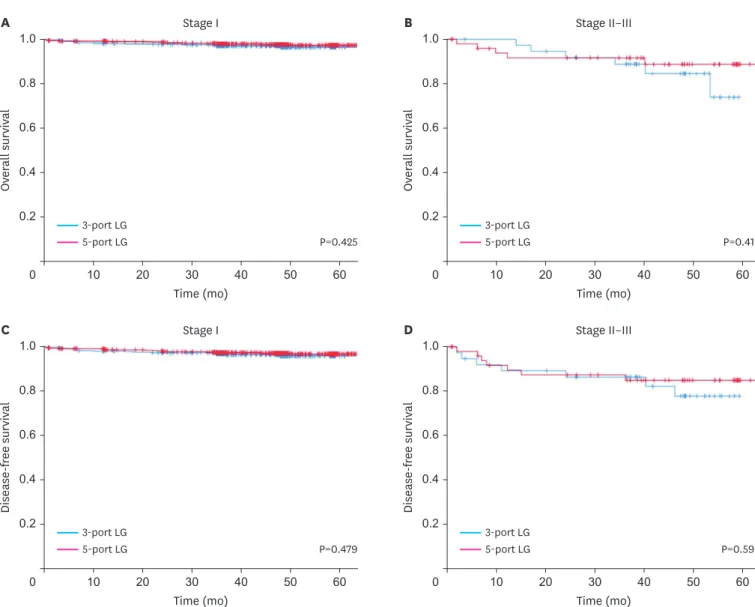
Fig. 2 shows the survival curves of the 3-port and conventional 5-port LG groups at different pathological stages. There were no significant differences in overall survival between the two groups in stage I (P=0.425, Fig. 2A) and stage II–III patients (P=0.419, Fig. 2B). Likewise, DFS did not significantly differ between the two groups for stage I (P=0.479, Fig. 2C) and stage II–III patients (P=0.599, Fig. 2D).
Table 2. Operative outcomes in the two groups
Variables Three-port LG (n=460) Five-port LG (n=657) P-value
Extent of gastric resection 0.598
Distal 421 (91.5) 607 (92.4) Total 39 (8.5) 50 (7.6) Reconstruction <0.001 Billroth I 164 (35.7) 367 (55.9) Billroth II 249 (54.1) 218 (33.2) Roux-en Y 47 (10.2) 72 (11.0)
Operating time (min) 150.8±43.9 148.5±43.9 0.384
Operative blood loss (mL) 105.8±138.1 93.8±75.6 0.091
Blood transfusion 9 (2.0) 9 (1.4) 0.443
Morbidity 70 (15.2) 66 (10.0) 0.134
≥Grade III complications 24 (5.2) 19 (2.9) 0.057
Mortality 1 (0.2) 2 (0.3) 0.755
Hospital stay (days) 9.4±5.2 9.2±2.4 0.340
Data are expressed as mean±standard deviation or number (%). LG = laparoscopic gastrectomy. Time (mo) A Ov er all survival 1.0 0.2 0 0.6 0.8 0.4 60 50 30 40 20 10 3-port LG 5-port LG P=0.138 Time (mo) B Dise ase-free survival 1.0 0.2 0 0.6 0.8 0.4 60 50 30 40 20 10 3-port LG 5-port LG P=0.231
Fig. 1. Kaplan–Meier survival curves for the 3- and 5-port LG groups. (A) Overall survival and (B) disease-free survival. LG = laparoscopic gastrectomy.
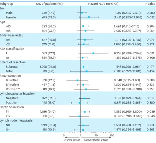
Fig. 3 shows a forest plot of the hazard ratio of the 3-port LG subgroups according to different clinicopathological factors. There were no significant differences in survival between 3- and 5-port LG for all considered subgroups.
DISCUSSION
Following advances in surgical techniques and instruments, gastric surgeons with vast experience in laparoscopy have introduced reduced-port LG for the treatment of gastric cancer. The initial reports mostly demonstrated the technical feasibility and safety of reduced-port LG compared with those of conventional LG [9-13]. However, the long-term oncological outcomes of reduced-port LGs have not been sufficiently investigated. This is the first study to investigate the long-term oncological outcomes of 3-port LG for gastric carcinoma; the results suggest that long-term survival after 3-port LG is comparable to that after conventional 5-port LG in patients with early-stage gastric carcinoma.
Time (mo) A Ov er all survival 1.0 0.2 0 0.6 0.8 0.4 60 50 30 40 20 10 3-port LG 5-port LG P=0.425 Time (mo)
Stage I B Stage II–III
Ov er all survival 1.0 0.2 0 0.6 0.8 0.4 60 50 30 40 20 10 3-port LG 5-port LG P=0.419 Time (mo) C Dise ase-free survival 1.0 0.2 0 0.6 0.8 0.4 60 50 30 40 20 10 3-port LG 5-port LG P=0.479 Time (mo)
Stage I D Stage II–III
Dise ase-free survival 1.0 0.2 0 0.6 0.8 0.4 60 50 30 40 20 10 3-port LG 5-port LG P=0.599
Fig. 2. Kaplan–Meier survival curves for patients with different pathologic stages in the 3- and 5-port LG groups. (A) Overall survival in stage I, (B) overall survival in stage II–III, (C) disease-free survival in stage I, and (D) disease-free survival in stage II–II.
In 2011, Omori et al. [6] first introduced the so-called transumbilical “single-incision” laparoscopic distal gastrectomy. In their technique, they used three working ports through a single 2.5-cm umbilical incision. Later on, various types of multi-channel ports began to be used in this procedure, and “single-port” LG became a common name [18,19]. Some experts have demonstrated the technical feasibility and safety of single-port LG [18-20]. However, single-port LG has not gained wide popularity among gastric surgeons because of the substantial technical difficulties experienced when manipulating multiple instruments through a single umbilical channel. In addition, the merits of single-port LG in improving minimal invasiveness were not significant. In one study that compared single-port and 3-port LG, single-port LG did not improve outcomes in terms of operative blood loss, postoperative pain, morbidity, or hospital stay length [21]. To overcome these technical difficulties, most gastric surgeons agree that additional ports should be inserted if necessary. However, the optimal number of ports in reduced-port LGs remains to be established.
Three-port LG has several advantages over other reduced-port techniques. First, unlike single-port LG, 3-port LG does not require specialized devices, such as a multi-channel port, flexible laparoscope, or curved instruments. Second, by using two operator ports, 3-port LG is free from the operative difficulties of single-port LG, such as loss of triangulation, conflict of instruments, and parallel laparoscopic view. Lastly, the operative techniques of 3-port LG are nearly the same as those of conventional LG, except for some technical tips to overcome
Subgroup No. of patients (%) Hazard ratio (95% CI) P value
Sex Male 642 (57.5) 1.227 (0.595–2.531) 0.580 Female 475 (42.5) 3.451 (0.855–13.926) 0.082 Age ≥65 293 (26.2) 1.494 (0.716–3.115) 0.284 <65 824 (73.8) 2.087 (0.589–7.397) 0.254
Body mass index
≥23 547 (49.0) 1.914 (0.599–6.120) 0.274 <23 570 (51.0) 1.620 (0.758–3.466) 0.213 ASA classification 1 533 (47.7) 6.758 (0.789–57.842) 0.081 ≥2 584 (52.3) 1.339 (0.669–2.678) 0.409 Extent of resection Subtotal 1,028 (92.0) 1.543 (0.798–2.984) 0.197 Total 89 (8.0) 2.503 (0.227–27.611) 0.454 Reconstruction Billroth-I 531 (47.5) 0.648 (0.135–3.123) 0.589 Billroth-II 467 (41.8) 1.502 (0.654–3.447) 0.338 Roux-en-Y 119 (10.7) 2.325 (0.388–13.918) 0.355 Lymphovascular invasion Negative 972 (87.0) 1.861 (0.879–3.944) 0.105 Positive 145 (13.0) 0.871 (0.265–2.866) 0.820 Depth of invasion T1 1,016 (91.0) 1.859 (0.910–3.800) 0.089 ≥T2 101 (9.0) 0.867 (0.206–3.646) 0.846
Lymph node metastasis
N0 999 (89.4) 1.584 (0.768–3.267) 0.213 N+ 118 (10.6) 1.474 (0.396–5.491) 0.563 Conventional better 3-port better 1 0.1 10 100
Fig. 3. Forest plot of subgroup analyses. Bars represent hazard ratios with 95% CI of the effect of 3-port laparoscopic gastrectomy on overall survival.
the lack of surgical assistance. Previous studies have shown that gastric surgeons who are familiar with conventional LG can easily perform 3-port LG without increasing the risk for patients [5,9,22].
In addition to technical issues, the oncological safety of reduced-port LG has been a major concern. To date, only a few studies have reported the long-term outcomes of reduced-port LG for gastric carcinoma. Kunisaki et al. [23] compared the long-term survival of dual-port LG (including distal and total gastrectomy) with that of conventional LG in a propensity score-matched cohort. In their study, most patients had early-stage gastric carcinoma, and the two procedures did not show significant differences in overall or relapse-free survival. Our study investigated the long-term survival of 3-port LG in a large cohort of patients from multiple institutions. We found that the long-term survival of patients with early-stage gastric carcinoma treated with 3-port LG was comparable to that of patients treated with conventional 5-port LG. Furthermore, 3-port LG did not increase operative risks, such as morbidity or mortality.
Although the present study suggests the feasibility of adopting 3-port LG for the treatment of early-stage gastric carcinoma, application of reduced-port LG for advanced gastric carcinoma is questionable. Currently, laparoscopic surgery is only indicated for early-stage gastric carcinomas [17]. Likewise, 3-port LG in this study was indicated for patients with early-stage gastric carcinoma (cT1N0) as identified in preoperative staging. Most previous studies, including ours, have demonstrated the technical feasibility and safety of reduced-port LG when performing limited LND for early-stage gastric carcinoma [9-12]. Therefore, this study does not provide useful information about the advantages of reduced-port LG for advanced gastric carcinoma. Recently, some experts have reported techniques for D2 LND using reduced-port LG [20,24]. However, this may only be applicable to surgeons with sufficient knowledge and experience in LG because of the complexity of the extended LND. Therefore, further studies are needed to determine whether 3-port LG can be applied to patients with stage II/III disease.
Although the effect of reducing the number of ports could be trivial in terms of improving the minimal invasiveness of LG, previous studies have reported decreased operative blood loss [5,20,25], less postoperative pain [20], earlier postoperative oral intake [5,20], and shorter hospital stay [20] when using reduced-port LG. However, these results are all derived from small, single-institution studies and need to be verified in large randomized trials. In fact, one of the main reasons for adopting 3-port LG is to perform LG without assistance, as the lack of a surgical resident and assistant has become a practical problem in many institutions. In the present study, we demonstrated comparable short-term surgical outcomes between the two procedures, despite the lack of a surgical assistant during 3-port LG. The clinical benefits of reduced-port LG include diverse aspects, not just measurable surgical outcomes.
Our study had a few limitations. First, although it was a large multi-institutional study, an inherent selection bias is inevitable because of the retrospective study design. However, patient characteristics, including pathological stage, were comparable between the two groups. Second, this study was performed in large health centers, and all participating surgeons were highly skilled gastric surgeons with substantial LG experience. In other words, the surgeons' learning curve for reduced-port laparoscopic surgery was not considered in our study. This may limit the generalizability of our results. A large multi-institutional clinical trial, including small and large hospitals, is required to solve this problem.
In conclusion, this large multi-institutional study demonstrated that long-term oncological outcomes of 3-port LG were comparable to those of conventional 5-port LG in patients with early-stage gastric carcinoma. We believe that 3-port LG may be an appropriate option to perform LG without a surgical assistant, without increasing the risk for patients. Due to the retrospective nature of this study, large randomized clinical trials are warranted to determine the clinical benefits of 3-port LG in treating gastric carcinoma.
REFERENCES
1. Information Committee of Korean Gastric Cancer Association. Korean Gastric Cancer Association nationwide survey on gastric cancer in 2014. J Gastric Cancer 2016;16:131-140.
PUBMED | CROSSREF
2. Kim W, Kim HH, Han SU, Kim MC, Hyung WJ, Ryu SW, et al. Decreased morbidity of laparoscopic distal gastrectomy compared with open distal gastrectomy for stage i gastric cancer: short-term outcomes from a multicenter randomized controlled trial (KLASS-01). Ann Surg 2016;263:28-35.
PUBMED | CROSSREF
3. Viñuela EF, Gonen M, Brennan MF, Coit DG, Strong VE. Laparoscopic versus open distal gastrectomy for gastric cancer: a meta-analysis of randomized controlled trials and high-quality nonrandomized studies. Ann Surg 2012;255:446-456.
PUBMED | CROSSREF
4. Kim SM, Lee JH. Advancement of single-port and reduced-port laparoscopic gastrectomy for gastric cancer: a systemic review. Ann Laparosc Endosc Surg 2017;2:71.
CROSSREF
5. Kim HG, Kim DY, Jeong O. Transition from conventional to reduced-port laparoscopic gastrectomy to treat gastric carcinoma: a single surgeon's experience from a small-volume center. J Gastric Cancer 2018;18:172-181.
PUBMED | CROSSREF
6. Omori T, Oyama T, Akamatsu H, Tori M, Ueshima S, Nishida T. Transumbilical single-incision laparoscopic distal gastrectomy for early gastric cancer. Surg Endosc 2011;25:2400-2404.
PUBMED | CROSSREF
7. Kunisaki C, Ono HA, Oshima T, Makino H, Akiyama H, Endo I. Relevance of reduced-port laparoscopic distal gastrectomy for gastric cancer: a pilot study. Dig Surg 2012;29:261-268.
PUBMED | CROSSREF
8. Kawamura H, Tanioka T, Kuji M, Tahara M, Takahashi M. The initial experience of dual port laparoscopy-assisted total gastrectomy as a reduced port surgery for total gastrectomy. Gastric Cancer 2013;16:602-608.
PUBMED | CROSSREF
9. Jeong O, Park YK, Ryu SY. Early experience of duet laparoscopic distal gastrectomy (duet-LDG) using three abdominal ports for gastric carcinoma: surgical technique and comparison with conventional laparoscopic distal gastrectomy. Surg Endosc 2016;30:3559-3566.
PUBMED | CROSSREF
10. Kim SM, Ha MH, Seo JE, Kim JE, Choi MG, Sohn TS, et al. Comparison of reduced port totally
laparoscopic distal gastrectomy (Duet TLDG) and conventional laparoscopic-assisted distal gastrectomy. Ann Surg Oncol 2015;22:2567-2572.
PUBMED | CROSSREF
11. Seo HS, Song KY, Jung YJ, Kim JH, Park CH, Lee HH. Right-side approach-duet totally laparoscopic distal gastrectomy (R-Duet TLDG) using a three-port to treat gastric cancer. J Gastrointest Surg 2018;22:578-586.
PUBMED | CROSSREF
12. Kim HB, Kim SM, Ha MH, Seo JE, Choi MG, Sohn TS, et al. Comparison of reduced port totally laparoscopic-assisted total gastrectomy (Duet TLTG) and conventional laparoscopic-assisted total gastrectomy. Surg Laparosc Endosc Percutan Tech 2016;26:e132-e136.
PUBMED | CROSSREF
13. Amin AT, Gabr A, Abbas H. Laparoscopy assisted distal gastrectomy for T1 to T2 stage gastric cancer: a pilot study of three ports technique. Updates Surg 2015;67:69-74.
PUBMED | CROSSREF
14. Washington K. 7th edition of the AJCC cancer staging manual: stomach. Ann Surg Oncol 2010;17:3077-3079.
15. Dindo D, Demartines N, Clavien PA. Classification of surgical complications: a new proposal with evaluation in a cohort of 6336 patients and results of a survey. Ann Surg 2004;240:205-213.
PUBMED | CROSSREF
16. Guideline Committee of the Korean Gastric Cancer Association (KGCA), Development Working Group & Review Panel. Korean Practice Guidelines for gastric cancer 2018: An evidence-based, multi-disciplinary approach. J Gastric Cancer 2019;19:1-48.
PUBMED | CROSSREF
17. Japanese Gastric Cancer Association. Japanese gastric cancer treatment guidelines 2014 (ver. 4). Gastric Cancer 2017;20:1-19.
PUBMED | CROSSREF
18. Kim SM, Lee SH, Ha MH, Seo JE, Kim JE, Choi MG, et al. Techniques of the single-port totally laparoscopic distal gastrectomy. Ann Surg Oncol 2015;22 Suppl 3:S341.
PUBMED | CROSSREF
19. Ozdemir BA, Thomas RL, Soon Y. Single-port laparoscopic subtotal gastrectomy with DIα lymphadenectomy. Surg Innov 2011;18:NP1-NP4.
PUBMED | CROSSREF
20. Omori T, Fujiwara Y, Moon J, Sugimura K, Miyata H, Masuzawa T, et al. Comparison of single-incision and conventional multi-port laparoscopic distal gastrectomy with D2 lymph node dissection for gastric cancer: a propensity score-matched analysis. Ann Surg Oncol 2016;23 Suppl 5:817-824.
PUBMED | CROSSREF
21. Kim SM, Ha MH, Seo JE, Kim JE, Choi MG, Sohn TS, et al. Comparison of single-port and reduced-port totally laparoscopic distal gastrectomy for patients with early gastric cancer. Surg Endosc 2016;30:3950-3957.
PUBMED | CROSSREF
22. Usui S, Tashiro M, Haruki S, Matsumoto A. Triple-incision laparoscopic distal gastrectomy for the resection of gastric cancer: comparison with conventional laparoscopy-assisted distal gastrectomy. Asian J Endosc Surg 2014;7:197-205.
PUBMED | CROSSREF
23. Kunisaki C, Miyamoto H, Sato S, Tanaka Y, Sato K, Izumisawa Y, et al. Surgical outcomes of reduced-port laparoscopic gastrectomy versus conventional laparoscopic gastrectomy for gastric cancer: a propensity-matched retrospective cohort study. Ann Surg Oncol 2018;25:3604-3612.
PUBMED | CROSSREF
24. Lin T, Mou TY, Hu YF, Liu H, Li TJ, Lu YM, et al. Reduced Port laparoscopic distal gastrectomy with D2 lymphadenectomy. Ann Surg Oncol 2018;25:246-246.
PUBMED | CROSSREF
25. Ahn SH, Son SY, Jung DH, Park DJ, Kim HH. Pure single-port laparoscopic distal gastrectomy for early gastric cancer: comparative study with multi-port laparoscopic distal gastrectomy. J Am Coll Surg 2014;219:933-943.