INTRODUCTION
Polyimide (PI) films have been widely used in soft elec-tronics and microelecelec-tronics packages because of their good properties, including excellent thermal and mechanical pro-perties, low moisture absorption, low dielectric constant, and good chemical stability (Kim and Jang 2000; Ramos 2002). For these applications, a good adhesion between a metal and a PI film is essential. However, the adhesion between a metal and a PI film is generally poor because the cohesive energy of metal is two orders of magnitude higher than that of polymers, and because PI films have low wettability (Svorcik et al. 2006). Thus, to improve the adhesion between the metal and a PI film, the surface modification of the PI film has been carried out using various chemical and physi-cal techniques such as wet-chemiphysi-cal treatment, plasma
treat-ment, and ion implantation (Yu et al. 2002).
Among those modification methods, an ion implantation-based technique is attractive for the surface modification of a PI film to improve its adhesion with a deposited metal layer. This type of technique offers several merits: it is a surface-specific method without detrimentally affecting the bulk pro-perties; it is controllable, reproducible, and clean; and it is a low temperature process (Guzman 2002). Thus, ion implan-tation-based surface modification of polymers for the elec-tronic and biological applications has been extensively studied (Jagielski et al. 2007; Zaporojtchenko et al. 2007).
In this research, the surface modification of a PI film by ion implantation is described to improve the interfacial adhesion between the PI and the deposited copper (Cu) layer. The surfaces of the PI films were implanted by energetic ions under various conditions and a Cu layer was then depo-sited. The surface properties of the implanted PI films were characterized in terms of wettability, chemical structure and composition, and morphology. The adhesion strength
bet-─ ─ 335 ──
Ion Beam-based Surface Modification of Polyimide Films for
Adhesion Improvement with Deposited Metal Layer
Hwang-Woo Cho, Chan-Hee Jung, In-Tae Hwang, Jae-Hak Choi* and Young-Chang NhoAdvanced Radiation Technology Institute, Korea Atomic Energy Research Institute, Jeongeup 580-185, Korea
Abstract-- In this study, the surface of polyimide (PI) films was modified using ion implantation to enhance its adhesion to a deposited copper (Cu) layer. The surfaces of the PI films were implanted with 150 keV Xe++ions at fluences varying from 1××1014to 1××1016ions cm--2. The Cu layers were
then deposited on the implanted PI. The surface properties of the implanted PI film were investi-gated based on the contact angle measurements, Fourier transform infrared spectroscopy (FT-IR), X-ray photoelectron spectroscopy (XPS), and atomic force microscopy (AFM). Furthermore, the adhesive strength between the deposited Cu layer and PI film was estimated through a scratch test using a nanoindenter. As a result, the surface environment of the PI film was changed by the ion implantation, which could have a significant effect on the adhesion between the deposited Cu layer and the PI.
Key words : Polyimide, Ion implantation, Surface roughness, Nanoindenter, Adhesion
* Corresponding author: Jae-Hak Choi, Tel. +82-63-570-3062, Fax. +82-63-570-3090, E-mail. [email protected]
ween the deposited Cu layer and the PI film was investigated through scratch testing using a nanoindenter.
MATERIALS AND METHODS
1. Surface modification of polyimide films by ionimplantation
Polyimide (PI) films (Kapton HN, 40 mm in width, 125 μm in thickness) were supplied from Du Pont Chemical Company (Wilmington, DE, USA). PI films were ultrasoni-cally washed in methanol for 20 min and dried in vacuum oven at 50�C for 1 h before use. The resulting films were implanted with 150 keV Xe++ions at fluences ranging from
1×10¹⁴to 1×10¹⁶ions cm-2. The current density of the ion
beam was about 1.0μA cm-2to prevent a detrimental
ther-mal effect on the polyimide films.
2. Copper deposition on the implanted PI films
A copper (Cu) layer was deposited onto the implanted PI films using a DC magnetron sputtering method. The typical deposition parameters used were as follows: an initial vacu-um pressure of 2×10⁻⁶Torr before sputtering and 10 mTorr
at the start of sputtering, argon plasma gas flow of 20 sccm, input DC power of 100 W, and a sputtering time of 25 min. Under these conditions, the thickness of the deposited the Au layers deposited on the implanted PI were around 680 nm.
3. Characterization
The contact angles of the control and implanted PI films were measured by using a contact angle analyzer (Phoenix 300, Surface Electro Optical Company). Changes in the chemical structure of the PI film surfaces after ion implanta-tion were investigated by using an attenuated total reflect-ance Fourier transform infrared spectroscopy (FT-IR, Bruker, Tensor 37). The surface chemical composition of the control and implanted PI films were analyzed by using an X-ray photoelectron spectrometer (XPS, MultiLab 2000, Thermo-Electron Corporation, England) with MgKα X-ray source. The applied power was 14.5 keV and 20 mA, and the base pressure of the analysis chamber was less than 10-9mbar.
All the binding energies were referenced to the C1s neutral carbon peak at 285.0 eV. The surface morphology of the
control and implanted PI films were observed by using an atomic force microscope (AFM, Nanoscope III). All images were collected in air using a tapping mode under a con-stant force (scan size: 2μm, scan rate: 0.5 Hz). The adhesion strength of the deposited Cu layers on the control and im-planted PI films was estimated using a nanoindenter (MTS Systems Corporation). A 2μm diamond tipped stylus was used under a progressive load ranging from 0.3 mN to 15 mN to scratch the deposited Cu layers. The point where the coating fails by cracking is taken as the point failure. Three tests were conducted on a sample in order to determine the exact failure under critical loads.
RESULTS AND DISCUSSION
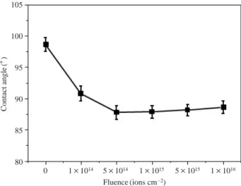
To investigate the changes in the wettability of the PI films by ion implantation, the water contact angles of the control and implanted PI films were measured and the results are present in Fig. 1. In comparison to that of the control PI, 99�, the contact angle of implanted PI films was decreased with an increasing fluence up to 5×10¹⁴ions cm-2, over which it
was increased sightly. This result indicates that dhe PI surface was chemically changed by ion implantation.
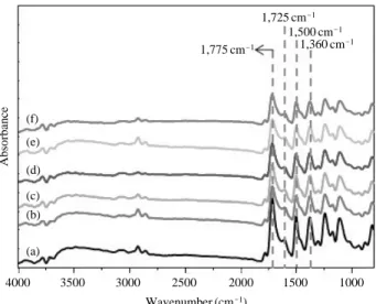
The ATR-FTIR spectra of the control and implanted PI films are shown in Fig. 2. As shown in the control PI spec-trum of Fig. 2(a), the typical bands which are the characteris-tic of PI films appeared at 1,775 and 1,725 (C==O), 1,500 (C==C in aromatic ring), and 1,360 cm-1(C-N-C). In case of
the implanted PI films, the intensities of these characteristic
Fig. 1. The contact angle of PI films as a function of the ion fluence.
Contact angle (� ) 105 100 95 90 85 80 0 1×10¹⁴ 5×10¹⁴ 1×10¹⁵ 5×10¹⁵ 1×10¹⁶ Fluence (ions cm-2)
bands were decreased with an increasing fluence because the imide and aromatic structures were partially destroyed by ion implantation, indicating that the chemical structure of the PI surface was changed by ion implantation.
The C1s spectra for the control and implanted PI films are shown in Fig. 3. In the spectrum of the control PI,
typi-cal peaks corresponding to the chemitypi-cal structure of PI was observed at 284.2 (C==C), 285.0 (C-C), 286.5 (C-N/O), and 288.53 eV (C==O) (Silverman et al. 1986). Upon ion implan-tation, the peak intensities were arbitrarily changed based on the fluence. Moreover, as shown in Fig. 4, the [O]/[C] and [N]/[C] atomic ratios of the implanted PI films were decreased with an increasing fluence in comparison to those of the control PI. These results indicate that the chemical structure of the PI surface was destroyed by ion implantation (Ektessabi et al. 2000).
The morphological changes in the PI surface by ion im-plantation were examined by an AFM observation and the results of which are present in Fig. 5. As shown in Fig. 5, in comparison to that of the control PI film, the RMS value of the implanted PI films, related to their surface roughnesses of PI films, was increased with an increase in fluence up to 5×10¹⁴ions cm-2, beyond which it was deceased. This
morphological changes in the PI surface can be attributed to physical ion bombardment (Kim and Lee 2006).
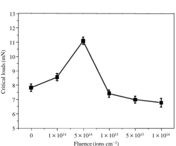
To estimate the adhesion between a deposited Cu layer and PI film, the critical loads for the deposited Cu layers on the control and implanted PI films were measured using a nanoindenter which is a type of the scratch testers (Perry
Fig. 2. FT-IR spectra of the control (a) and implanted PI films films
at fluences of 1×10¹⁴(b), 5×10¹⁴(c), 1×10¹⁵(d), 5×10¹⁵ (e), and 1×10¹⁶ions cm-2(f).
Absorbance 4000 3500 3000 2500 2000 1500 1000 Wavenumber (cm-1) 1,725 cm-1 1,775 cm-1 1,360 cm-1 1,500 cm-1 (f) (e) (d) (c) (b) (a)
Fig. 3. The C1s core-level XPS spectra of the control (a) and implanted PI films films at fluences of 1×10¹⁴(b), 5×10¹⁴(c), 1×10¹⁵(d), 5× 10¹⁵(e), and 1×10¹⁶ions cm-2(f).
Intensity (a.u.) Intensity (a.u.) Intensity (a.u.) Intensity (a.u.) Intensity (a.u.) Intensity (a.u.) 292 290 288 286 284 282 280 292 290 288 286 284 282 280
Binding energy (eV) Binding energy (eV)
292 290 288 286 284 282 280
Binding energy (eV)
292 290 288 286 284 282 280
Binding energy (eV)
292 290 288 286 284 282 280 292 290 288 286 284 282 280
Binding energy (eV) Binding energy (eV)
(a) (b) (c) (d) (e) (f) C==O C==O C==O C==O C==O C==O C==C C==C C==C C==C C==C C==C C-C C-C C-C C-C C-C C-C C-N/O C-N/O C-N/O C-N/O C-N/O C-N/O
1983; Hintermann 1984). Fig. 6 shows the critical loads of the Cu layers deposited on the PI films were measured using a nanoindenter as a function of fluence. For the Cu layer deposited on the control PI film, the critical load was 7.81 mN. On the other hand, for the Cu deposited on the implanted PI films, the critical load was increased up to 11.08 mN with an increase in fluence to 5×10¹⁴ions cm-2
beyond which it was decreased to 6.76 mN. This result revealed that the adhesion strength of the deposited Cu layers was higher than these on the control PI, which was dependant on the fluence. This improvement can be ascribed to the fact that the increase in the surface roughness of the PI film by ion implantation creates a larger surface area for more physical interactions or mechanical interlocking with
Fig. 5. AFM images and root mean square of surface roughness (RMS) for the control (a) and implanted PI films at fluences of 1×10¹⁴(b), 5
×1014(c), 1×10¹⁵(d), 5×10¹⁵(e), and 1×10¹⁶ions cm-2(f).
(a) RMS: 1.13 nm (b) RMS: 1.28 nm (c) RMS: 1.45 nm
(d) RMS: 1.39 nm (e) RMS: 0.78 nm (f) RMS: 0.71 nm
Fig. 4. The [O]/[C] and [N]/[C] ratios of the control PI and implanted PI films measured by a XPS analysis as a function of ion fluence.
[O]/[C] [N]/[C] 0.26 0.24 0.22 0.20 0.18 0.16 0.14 0.12 0.10 0.06 0.05 0.04 0.03 0.02 0.01 0.00 Con. 1×10¹⁴ 5×10¹⁴ 1×10¹⁵ 5×10¹⁵ 1×10¹⁶ Fluence (ions cm-2) Con. 1×10¹⁴ 5×10¹⁴ 1×10¹⁵ 5×10¹⁵ 1×10¹⁶ Fluence (ions cm-2) (a) (b)
the Cu layer, which thereby increases the critical loads.
CONCLUSIONS
The surface modification of a PI film was effectively carried out to improve its adhesion to the deposited Cu layer. The contact angle measurements and XPS analysis results showed that the chemical environment of the PI film was changed by ion implantation. An AFM observation revealed that the surface roughness of the implanted PI was increased in comparison to that of the control PI, which was dependant on the fluence. On the basis of the results of a scratch test using a nanoindenter, the adhesion strength of Cu deposited on the implanted PI film was higher than that on the control PI film, which depended on the fluence. This adhesion im-provement between the Cu layer and the PI film could be attributed to the physiochemical changes on the surface environment of the PI film induced by ion implantation.
ACKNOWLEDGMENTS
This research was supported by the Nuclear R&D program
through the National Research Foundation funded by the Ministry of Education, Science, and Technology, Korea.
REFERENCES
Guzman L, Man BY, Miotello A, Adami M and Ossi PM. 2002. Ion beam induced enhanced adhesion of Au films deposited on polytetrafluoroethylene. Thin Solid Films 420-421:565-570.
Ektessabi AM and Hakamata S. 2000. XPS study of ion beam modified polyimide films. Thin Solid Films. 621:377-378. Hintermann HE. 1984. Adhesion, friction and wear of thin hard
coatings. Wear 100:381-397.
Kim M-H and Lee K-W. 2006. The effect of ion beam treatment on the interfacial adhesion of Cu/polyimide system. IEEE Trans. Comp. Hybrids Manuf. Technol. 12:180-184. Kim H and Jang J. 2000. Corrosion protection and adhesion
promotion for polyimide/copper system using silane-modi-fied polymeric materials. Polymer 41:6553-6561.
Perry AJ. 1983. Scratch adhesion testing of hard coatings. Thin Solid Films. 107:167-180.
Ramos MMD. 2002. Theoretical study of metal-polyimide interfacial properties. Vacuum 64:255-260.
Silverman BD, Bartha JW, Clabes JG, Ho PS and Rossi AR. 1986. Molecular orbital analysis of the XPS spectra of PMDA-ODA polymide and its polyamic acid precursor. J. Polym. Sci. A, Polym. Chem. 24:3325-3333.
Svorcik V, Kotal V, Slepicka P, Blahova O, Sutta P and Hna-towicz V. 2006. Gold coating of polyethylene modified by argon plasma discharge. Polym. Eng. Sci. 46:1326-1332. Yu ZJ, Kang ET and Neoh KG. 2002. Electroless plating of
copper on polyimide films modfied by surface grafting of tertiary and quaternary amine polymers. Polymer 43: 4137-4146.
Zaporojtchenko V, Zekonyte J and Faupel F. 2007. Effect of ion beam treatment on atomic and macroscopic adhesion of copper to different polymer materials. Nucl. Instrum. Methods Phys. Res. Sect. B 265:139-145.
Manuscript Received: November 4, 2010 Revision Accepted: November 12, 2010
Fig. 6. The critical loads of the Cu layers-deposited PI films
mea-sured using a nanoindenteras a function of fluence.
Critical loads (mN) 13 12 11 10 9 8 7 6 5 0 1×10¹⁴ 5×10¹⁴ 1×10¹⁵ 5×10¹⁵ 1×10¹⁶ Fluence (ions cm-2)
![Fig. 4. The [O]/[C] and [N]/[C] ratios of the control PI and implanted PI films measured by a XPS analysis as a function of ion fluence.](https://thumb-ap.123doks.com/thumbv2/123dokinfo/4672035.772/4.892.113.777.470.885/ratios-control-implanted-films-measured-analysis-function-fluence.webp)