1505
ⓒ The Korean Society of Food Science and Technology
In vitro and Cellular Antioxidant Activity of Arginyl-fructose and
Arginyl-fructosyl-glucose
Jung-Sook Lee, Gyo-Nam Kim, Sang-Hyun Lee, Eui-Su Kim1, Kyoung-Soo Ha, Young-In Kwon,
Heon-Sang Jeong2, and Hae-Dong Jang*
Department of Food and Nutrition, Hannam University, Daejeon 305-811, Korea
1Shinwon FI Co., Ltd., Hwaseong, Gyeonggi 445-944, Korea
2Department of Food Science and Technology, Chungbuk National University, Cheongju, Chungbuk 361-763, Korea
Abstract Arginyl-fructose (AF) and arginyl-fructosyl-glucose (AFG) were chemically synthesized and purified. Their in vitro and cellular antioxidant activity was investigated using oxygen radical absorbance capacity (ORAC) and cellular antioxidant activity assay, respectively. The peroxyl radical scavenging activity of AF was much higher than that of AFG, which was in good agreement with their reduction capacity to donate electrons or hydrogen atoms. On the other hand, the hydroxyl radical scavenging activity of AF was weaker than that of AFG, which was consistent with their metal chelating activity, suggesting that AFG-Cu2+ complex may be less redox-active than AF-Cu2+ complex due to 1 glucose molecule
attached. The cellular antioxidant activity of AF and AFG appeared to depend on both their permeability into cell membrane and the scavenging activity on peroxyl or hydroxyl radicals. These results indicate that AF and AFG, Maillard reaction products, may have a high potential as a material for the development of nutraceutical food with antioxidant activity.
Keywords: arginyl-fructose, arginyl-fructosyl-glucose, oxygen radical absorbance capacity (ORAC), cellular antioxidant
activity
Introduction
Oxidative stress is the term referring the imbalance between the generation of reactive oxygen species (ROS) and the activity of the antioxidant defense. Severe oxidative stress has been implicated in aging and such chronic diseases as cancer and coronary heart disease due to the damage of many biological molecules such as lipids, proteins, carbohydrates, and DNA (1). ROS such as superoxide anion radical (O2•−), hydroxyl radical (OH·),
singlet oxygen (O21), and hydrogen peroxide (H2O2) has
been known to be generated as byproducts of normal cellular metabolism or results of exogenous factors including smoking and air pollution (2). A possible scavenger of these ROS may be used as a preventive tool to control oxidative stress-related diseases. Many kinds of natural compounds including polyphenol from plant materials including medicinal herb have shown the antioxidant activity against ROS (3,4).
Amino acid derivatives such as arginyl-fructose (AF) and arginyl-fructosyl-glucose (AFG) shown in Fig. 1 are formed through Amadori rearrangement, the early step of Maillard reaction from arginine and glucose or maltose, respectively, during the processing from raw ginseng to Korean red ginseng (5-7). AF and AFG can contribute to various biological functions of Korean red ginseng including antioxidant and anti-diabetic activity (5,7-9). Their contents are known to be dependent upon the
preparation condition of Korean red ginseng (7,10). However, very limited information is available about theeffect of structural difference between AF and AFG ontheir various biological activities. In this study, we prepared AF and AFG by chemical synthesis, investigated the in vitro and cellular antioxidant activity of AF and AFG, and discussed what kind of the mechanism might be involved in their in vitro and cellular antioxidant activity.
*Corresponding author: Tel: +82-42-629-8795; Fax: +82-42-629-8805 E-mail:[email protected]
Received August 26, 2009; Revised October 1, 2009; Accepted October 7, 2009
Fig. 1. Chemical structure of arginyl-fructose (AF) and arginyl-fructosyl-glucose (AFG).
Materials and Methods
Materials 2,2'-Azobis(2-amidinopropane) dihydrochloride (AAPH) was obtained from Wako Pure Co. (Osaka, Japan). Dulbecco’s modified Eagle’s medium (DMEM), fetal bovine serum (FBS), and Hank’s balanced salt solution (HBSS) were purchased from Gibco BRL. (Calsbad, CA, USA). Arginine, maltose, glucose, 1,10-phenanthroline, Calcein, fluorescein, 2',7'-dichlorofluorescin diacetate (DCFH-DA), 3-(4,5-dimethylthiazol-2-yl)-2,5-diphenyltetrazolium bromide (MTT), phosphate buffered saline (PBS, pH 7.4), Trolox, dimethyl sulfoxide (DMSO), neocuproine, and heat-inactivated fetal bovine serum were purchased from Sigma-Aldrich (St. Louis, MO, USA). HepG2 cell was obtained from the American Type Culture Collection (ATCC, Rockville, MD, USA).
Chemical synthesis of AF and AFG AF and AFG were prepared by modifying a method developed by Matsuura et al. (5). AF was synthesized by dissolving 0.88 g of L
-arginine and 1.80 g of glucose in 10 mL of glacial acetic acid with reflux condenser in water bath at 80oC for 2 hr.
AFG was also synthesized by heating the mixture of maltose (1.80 g) and L-arginine (0.44 g) dissolved in 10
mL of glacial acetic acid with reflux condenser in water bath at 80oC for 1 hr. The reaction mixture was dried and
purified by column chromatography of silica gel (Kieselgel 160, 70-230mesh, Merck, Damstadt, Germany) with butanol-acetic acid-water (2:1:1) solution. Structures of synthesized AFG and AF were identified using liquid chromatography-tandem mass spectrometry (LC-MS/MS) (Applied Biosystems, Foster City, CA, USA) by which purities were confirmed to be over 90%. The mass of AFG and AF was analyzed to be m/z 449 and 337 ([M+H]), respectively, in LC-MS/ MS analysis. The samples were prepared by dissolving at 1 g/mL in 1:1 solution of water to methanol. Mass spectrometric measurements were performed in API 4000TM triple quadrupole LC-MS/MS system equipped with a turbo electrospray ion source. Software version used was Analyst 1.4.2. The sample solutions were directly infused into the electronic spray ionization (ESI) source with a flow rate of 200 L/min. ESI mass spectra were measured using Q1 scan mode at a scan range from m/z 100 to 600 with a spray voltage of 4.5 keV operated in the positive ion mode. As other mass spectrometric parameters, the declustering and entrance potential were 118 and 10 eV, respectively. The curtain gas and ion source gas 1 (GS1) were 10 and 17 psi, respectively.
Determination of AF and AFG The analysis was performed by high performance anion-exchange chromato-graphy (HPAEC) with electrochemical detector (ED-50) (10). The chromatographic separation was carried out with a CarboPac PA1 anion-exchange column (250×4 mm, Dionex, Sunnyvalue, CA, USA) and a CarboPac PA1 guard column (50×4 mm). The column temperature was 30oC. The
elution was performed with water (A) and 400 mM NaOH (B) (10:90, v/v) using binary gradient elution. The flow rate was 0.7 mL/min and injection volume was 20µL. The AFG and AF were detected by electrochemical detector equipped with a gold working electrode and Ag/AgCl reference electrode operating quadruple potential waveform.
Oxygen radical absorbance capacity (ORAC) assay The ORAC assay was carried out on a Tecan GENios multi-functional plate reader (Salzburg, Austria) with fluorescent filters (excitation wavelength: 485 nm and emission filter: 535 nm). In the final assay mixture, fluorescein (40 nM) was used as a target of free radical attack with either AAPH (20 mM) as a peroxyl radical generator in peroxyl radical scavenging capacity (ORACROO·) assay (11) or
H2O2-CuSO4 (H2O2, 0.75%; CuSO4, 5mM) as mainly a
hydroxyl radical generator in hydroxyl radical scavenging capacity (ORACHo·) assay (12). Trolox (1 mM) was used
as a control standard and prepared fresh daily. The analyzer was programmed to record the fluorescence of fluorescein every 2 min after AAPH or H2O2-CuSO4 was added. All
fluorescence measurements were expressed relative to the initial reading.Final results were calculated based on the difference in the area under the fluorescence decay curve between the blank and each sample. ORACROO· and
ORACOH· were expressed as mM of Trolox equivalents
(TE). One ORAC unit is equivalent to the net protection area provided by 1 mM of Trolox.
Determination of reduction capacity The reducing capacity of AF and AFG was determined according to the method of Aruoma et al. (13). The 40µL of different concentrations of AF or AFG in distilled water were mixed with 160µL of the mixture containing 0.5 mM CuCl2 and
0.75 mM neocuproine in 10 mM phosphate buffer, pH 7.4. The absorbance was measured with a Tecan microplate reader at 454 nm for 1 hr. Increased absorbance of the reaction mixture indicates increased reducing power.
Metal chelating activity The metal chelating activity was measured by the competitive binding procedure of Argirova and Ortwerth (14). One-hundred µL of different concentration of AF or AFG were mixed with 100µL of 0.1µM CuSO4. After 100µL of mixture solution was added
to 100µL of 0.1µM calcein, the fluorescence of mixture solution was measured using a Tecan GENios multi-functional plate reader with fluorescent filters (excitation wavelength: 485 nm and emission filter: 535 nm) and compared to the fluorescence intensity of control which contained only calcein.
Cell viability assay The tetrazolium dye colorimetric test 3-(4,5-dimethylthiazol-2,5-diphenyltetrazolium bromide (MTT) test was used to determine the viability of HepG2 cells. The MTT assay is based on the ability of functional mitochondria to catalyze the reduction of MTT to insoluble purple formazan, the concentration of which can be measured spectrophotometrically. HepG2 cells were first cultured in 12-well plates (5×10⁵/mL) for 24 hr, washed twice using
PBS, and pretreated with different concentrations of sample tested. After 4 hr incubation, MTT reagent was added to each well, and the plate was incubated at 37oC for
1 hr. The media was removed, and the plate was washed twice with PBS (pH 7.4). The intracellular insoluble formazan was dissolved in DMSO. The absorbance of each cell was then measured at 570 nm using enzyme-linked immunosorbent assay (ELISA) reader, and the percentage viability was calculated.
Intracellular ROS measurement using DCFH-DA assay Cellular oxidative stress owing to ROS generating by AAPH or Cu2+ was measured by spectrofluorometrically
by the DCFH-DA method (15). DCFH-DA diffuses through the cell membrane and is enzymatically hydrolyzed by intracellular esterase to the non-fluorescent DCFH, which can be rapidly oxidized to the highly fluorescent DCF in the presence of ROS. HepG2 cells were first cultured in 96-well plates (5×10⁵/mL) for 24 hr. After the cell was
incubated with different concentration of AF or AFG for 30 min, media was discarded, and wells were gently washed twice using PBS. Instead of media, Hank’s balanced salt solution (HBSS) which is stable to fluorescence, was added to each well. AAPH was used as an inducer of peroxyl radical oxidative stress, and Cu2+ were used as an
inducer of another oxidative stress. After wells were treated with 100µM AAPH or 20 mM Cu2+ for 30 min,
DCFH-DA was added to the culture plates at a final concentration of 40 mM and incubated for 30 min at 37oC
in the darkness. DCF fluorescence intensity was measured with excitation wavelength at 485 nm and emission wavelength at 535 nm using Tecan GENios fluorometric plate reader.
Statistical analysis All data are presented as mean± standard deviation (SD). Statistical analyses were done using statistical package SPSS (Statistical Package for Social Science, SPSS Inc., Chicago, IL, USA) program, and significance of each group was verified with the analysis of one-way analysis of variance (ANOVA) followed by Student’s t-test for comparison of means.
Results and Discussion
Preparation of AF and AFG AF and AFG were synthesized by refluxing at 80oC for 2 or 1 hr in acidic
condition from L-arginine with glucose or maltose,
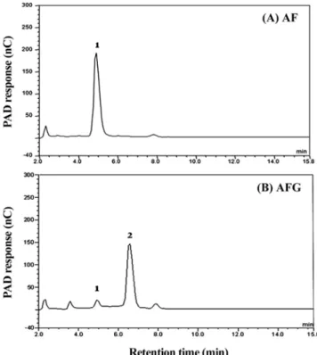
respectively and purified. Finally, 1.40 g of AF was obtained and identified using LC-MS/MS by which the purity was confirmed to be over 90%. Approximately 0.70 g of AFG as an off-white powder was gotten by a freeze-drying of fractionated sample. Their molecular weights were found as 337 and 449 ([M+H]) through LC-MS analysis, respectively, which was same as a value expected theoretically. High performance liquid chromatography (HPLC) analysis showed that AF and AFG were relatively pure (over 90%). In the optimized chromatographic condition, AF and AFG were detected at 4.8 and 6.2 min, respectively (Fig. 2). Purified AF and AFG were used for further in vitro and cellular antioxidant activity assay. In vitro antioxidant activity of AF and AFG The ORAC assay developed by Cao et al. (17)has been proved to be a widely accepted method for evaluating antioxidant capacity of various foods and biological samples (18,19). Antioxidant activity of KRGextracts was investigated for their peroxyl radical scavenging capacity using ORAC assay system, where AAPH, an azo compound, was used as a generator of peroxyl radicals. AAPH can decompose to produce carbon-centered free radicals at constant rate that react with oxygen to yield peroxyl radicals.
Table 1 demonstrates that the scavenging activity of AF
on peroxyl radicals generated from AAPH is much stronger than that of AFG. To determine whether the peroxyl radical scavenging· activityof them could result from their reduction
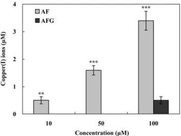
capacity donating electrons or hydrogen to peroxyl radical, the ability of AF and AFG to stimulate the reduction of copper ion was investigated by measuring the concentration of Cu+ formedfrom Cu2+ through the reduction by electrons.
AF showed much higher reduction capacity when compared to AFG, indicating that a glucose molecule attached to AFG caused to decrease its reduction capacity (Fig. 3). This result was in the good agreement with the peroxyl radical scavenging activity of AF and AFG. Therefore, it means that a little difference in composition of sugar moiety between AF and AFG may induce the substantial effect on the reduction capacity to donate electrons or hydrogen atoms to various radicals. It was also suggested that AF and AFG, Maillard reaction products, may contribute to the strong peroxyl radical scavenging activity of Korean red ginseng (20-22).
ORAC assay system has been successfully used to determine the reaction capacity with hydroxyl radical, one of most harmful and reactive oxygen species in biological system. Hydroxyl radicals were generated by H2O2-Cu2+
system. AFG showed much higher level of hydroxyl radical scavenging activity than AF (Table 1). The hydroxyl radical scavenging capacity of antioxidant in ORAC assay system has been known to be dependent on 3 factors such as the chelating activity with transition metal ions, the redox activity of chelator-metal complex and the scavenging activity on hydroxyl radical itself. AF and AFG can block cupric ions from the interaction with H2O2 in ORACassay
Fig. 2. HPLC chromatogram of AF and AFG. Chromatographic conditions 400 mM NaOH/water=90/10; flow rate of mobile phase 0.7 mL/min; column temperature 30oC; injection volume
system by chelating them to inhibit the generation of hydroxyl radicals and then contribute to their hydroxyl radical scavengingactivity. Therefore, the metal chelating activity with Cu2+ was determined by measuring the
inhibition percentage of Calcein-Cu2+ complex formation
because Calcein is a well-known metal chelator giving fluorescence. As a positive control, 1,10-phenanthroline was used since it have been known as a good metal ion chelator permeable through cell membrane. The metal chelating activity of AFG was stronger than that of AF, which was shown in Fig. 4. This was consistent with the result of their hydroxyl radical scavenging activity. There is possible that 2 kinds of chelator-Cu2+ complex,
redox-active or not, may be formed in ORAC assay system. Because the hydroxyl radical scavenging activity of AF and AFG was proportional to their concentration and metal chelating activity, AFG-Cu+2 complex looks like being less
redox-active than AF-Cu2+ complex. These results suggest
that AFG can strongly chelate with Cu2+ to form
AFG-Cu2+ complex which will be less redox active than
AF-Cu2+ complex.
Intracellular antioxidant activity of AF and AFG in HepG2 cells The cytotoxicity of AF and AFG was analyzed using MTT test with Hepatoma HepG2 cells. Cytotoxicity was expressed as a percentage of cell viability relative to that of control. Over the concentrations range tested, neither AF nor AFG had any significant effect on cell viability (Fig. 5A). The exposure of AAPH inducing peroxyl radical did not affect the viability of HepG2 cells. On the addition of Cu2+ as CuSO
4, cell viability was slightly
reduced (91%) only at 40µM compared to control cells (Fig. 5B). From these results, AF and AFG concentrations that corresponded to 87% cell viability were selected for subsequent assays. None of compounds at the selected concentrations significantly affected cell viability compared to control cells.
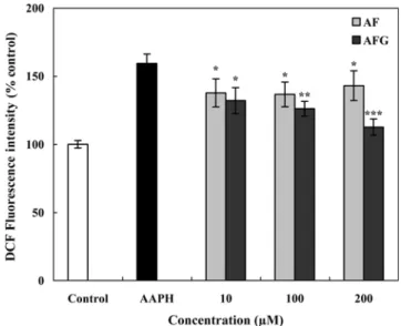
Hepatoma HepG2 cells were pre-incubated with different concentrations of AF or AFG for 30 min. After incubation, cells were exposed to 100 mM AAPH for 30 min and treated with DCFH-DA for 30 min which is a fluorescent probe for detecting ROS to measure intracellular oxidative stress. AF did not show the dose-dependent antioxidant activity against intracellular oxidative stress due to peroxyl radicals, but AFG did (Fig. 6). Even if AF had the higher in vitro peroxyl radical scavenging activity when compared to AFG (Table 1), its cellular antioxidant activity against peroxyl radicals was so weak, suggesting that a small amount of AF may be permeated into HepG2 cell membrane so that it can not efficiently scavenge the peroxyl radicals generated inside of cell. Previous studies showed that the efficiency of quercetin absorption from glycosides was tightly dependent upon the nature of the sugar moiety (23,24). Since quercetin 3-O-glucose was better absorbed
Table 1. The peroxyl radical- and hydroxyl radical scavenging activity of AF and AFG
Concentration (µM) Antioxidant activity
1)
ORACROO•
(TE, µM) ORAC(TE, µM)HO•
10 AFGAG 1,10-Phenanthroline 2)0.09±0.122) 0.00±0.00 -0.00±0.00 1.80±0.29** 11.65±0.24 20 AFGAG 1,10-Phenanthroline -0.06±0.19 3.97±0.21*** 14.51±0.81 50 AFGAG 1,10-Phenanthroline 1.09±0.05** 0.64±0.07 -1.43±0.15 6.61±0.28*** 13.20±0.35 100 AFGAG 1,10-Phenanthroline 2.83±0.05*** 1.34±0.20 -1)ORAC
ROO· and ORACOH· were expressed as mM of Trolox equiva-lents (TE); 1 ORAC unit is equivalent to the net protection area provided by 1 µM of Trolox.
2)Results represent the mean±SD of values obtained from 3 measure-ments. AF vs. AFG; **p<0.01, ***p<0.001.
Fig. 3. The reduction capacity of AF and AFG to reduce Cu2+
ions. Each bar represents mean±SD of 3 measurements. AF vs. AFG; **p<0.01, ***p<0.001.
Fig. 4. The metal chelating activity of AF and AFG against auto-oxidation process. Each bar represents mean±SD of 3 measurements. Sample vs. Cu2+ treated group; *p<0.05, **p<0.01,
than quercetin and quercetin glycosides carrying other sugars (24), the superior permeability of AFG to AF may due to the better recognition of sugar moiety of AFG by transporter molecules existing on the surface of cell membrane when compared to AF.
The intracellular antioxidant activity of AF and AFG against oxidative stress from Cu2+ was shown in Fig. 7. AF
and AFG did not exhibit the dose-dependent antioxidant activity between 10 and 200 mM. However, AFG more efficiently removed Cu2+-induced oxidative stress than AF
at 200 mM, indicating that the former may easily penetrate through cell membrane or have a higher cellular metal chelating activity than the latter. Two important factors such as the permeability into cell membrane and the cellular metal chelating activity may be required for the cellular antioxidant activity against Cu2+-induced oxidative
stress. Therefore, the protection activity of AFG against Cu2+-induced oxidative stress in HepG2 cells could result
from both its good permeability into cell membrane and cellular metal chelating activity. These results obtained from intracellular antioxidant assay of AF and AFG could
be useful for the prediction for their effect as an antioxidant in ex vivo or in vivo model system.
In conclusion, the peroxyl radical scavenging activity of AF was much higher than that of AFG, which was in good agreement with their reduction pacacity to donate electrons or hydrogen atoms. On the other hand, the hydroxyl radical scavenging activity of AF was weaker than that of AFG, which was consistent with their metal chelating activity, suggesting that AFG-Cu2+ complex may be less
redox-active than AF-Cu2+ complex due to 1 glucose molecule
attached. The cellular antioxidant activity of AF and AFG appeared to depend upon both their permeability into cell membrane and the scavenging activity on peroxyl or hydroxyl Fig. 5. Percentage cell viability of HepG2 cells exposed to
increasing concentration of AF and AFG (A), and AAPH and Cu2+ (B). Results are relative to control values and expressed as
mean±SD (n=3) individual experiments.
Fig. 6. Intracellular peroxyl radical scavenging activity of AF and AFG. The results represent the mean±SD (n=3). The
significance of differences between sample and AAPH treated group at p<0.05*, 0.01**, and 0.001*** was determined with
Student’s t-test.
Fig. 7. Intracellular antioxidant activity of AF and AFG against the oxidative stress generated by Cu2+ inHepG2 cells.
The results represent the mean±SD (n=3). The significance of
differences between sample and Cu2+ treated group at **p<0.01
radicals. However, the further study may be needed to investigate the membrane permeability of AF and AFG, and the redox-activity of AF and AFG-Cu2+ complex
Acknowledgments
This study was supported by a research grant from Hannam University, Daejeon, Korea in 2009.
References
1. Halliwell B, Aeschbach R, Löliger J, Aruoma OI. The characterization of antioxidants. Food Chem. Toxicol. 33: 601-617 (1995)
2. Droge W. Free radicals in the physiological control of cell function. Physiol. Rev. 82: 47-95 (2001)
3. Pieta PG. Flavonoid as antioxidants. J. Nat. Prod. 63: 1035-1042 (2000)
4. Moure A, Cruz JM, Franco D, Domíngues JM, Sineiro J, Domíngues H, Núñez MJ, Parajo JC. Natural antioxidants from residual sources. Food Chem. 72: 145-171 (2001)
5. Matsuura Y, Zheng Y, Takaku T, Kameda K, Okuda H. Isolation and physiological activities of a new amino acid derivative from Korean red ginseng. Korean J. Ginseng Sci. 18: 204-211 (1994) 6. Suzuki Y, Choi KJ, Uchida K, Ko SR, Sohn HJ, Park JD.
Arginyl-fructosyl-glucose and arginyl-fructose, compounds related to browning reaction in the model system of steaming and heat-drying processes for the preparation of red ginseng. J. Ginseng Res. 28: 143-148 (2004)
7. Cho EJ, Piao XL, Jang MH, Baek SH, Kim HY, Kang KS, Kwon SW, Park JH. The effect of steaming on the free amino acid contents and antioxidant activity of Panax ginseng. Food Chem. 107: 876-882 (2008)
8. Keum YS, Park KK, Lee JM, Chun KS, Park HP, Lee SK, Kwon HJ, Surh YJ. Antioxidant and anti-tumor promoting activities of the methanol extract of heat-processed ginseng. Cancer Lett. 150: 41-48 (2000)
9. Yoo BC, Park GH, Okuda H, Kim S, Hwang WI. Inhibitory effect of arginine-derivatives from ginseng extract and basic amino acids on protein-arginine N-methyltransferase. Amino Acids 17: 391-400 (1999)
10. Joo KM, Park CW, Jeong HJ, Lee SJ, Chang IS. Simultaneous determination of two Amadori compounds in Korean red ginseng (Panax ginseng) extracts and rat plasma by high-performance anion-exchange chromatography with pulsed amperometric
detection. J. Chromatogr. B 865: 159-166 (2008)
11. Kurihara H, Fukami H, Asami S, Totoda Y, Nakai M, Shibata H, Yao XS. Effects of oolong tea on plasma antioxidative capacity in mice loaded with restraint stress assessed using the oxygen radical absorbance capacity (ORAC) assay. Biol. Pharm. Bull. 27: 1093-1098 (2004)
12. Cao G, Sofic E, Prior RL. Antioxidant and prooxidant behavior of flavonoids: Structure-activity relationships. Free Radical Bio. Med. 22: 749-760 (1997)
13. Aruoma OI, Murcia A, Butler J, Halliwell B. Evaluation of the antioxidant and prooxidant action of gallic acid and its derivatives. J. Agr. Food Chem. 41: 1880-1885 (1993)
14. Argirova AD, Ortwerth BJ. Activation of protein-bound copper ions during early glycation: Study on two proteins. Arch. Biochem. Biophys. 420: 176-184 (2003)
15. Lautraite S, Bigot-Lasserre D, Bars R, Carmichael N. Optimization of cell-based assays for medium through screening of oxidative stress. Toxicol. In Vitro 17: 207-220 (2003)
16. Tenopoulou M, Kurz T, Doulias PT, Galaris D, Brunk UT. Does the Calcein-AM method assay the total cellular ‘labile iron pool’ or only a fraction of it? Biochem. J. 403: 261-266 (2007)
17. Cao G, Alessio HM, Cutler R. Oxygen-radical absorbance capacity assay for antioxidants. Free Radical Bio. Med. 14: 303-311 (1993) 18. Prior R, Hoang H, Gu L, Wu X, Bacchiocca M, Howard L,
Hampsci-Woodill M, Huang D, Ou B, Jacob R. Assays for hydrophilic and lipophilic antioxidant capacity (oxygen radical absorbance capacity (ORACFL) of plasma and other biological and food samples. J. Agr. Food Chem. 51: 3273-3279 (2003)
19. Wu X, Beecher GR, Holden JM, Haytowitz DB, Gebhardt SE, Prior RL. Lipophilic and hydrophilic capacities of common foods in the United States. J. Agr. Food Chem. 52: 4026-4037 (2004)
20. Nam KY. The comparative understanding between red ginseng and white ginsengs, processed ginsengs (Panax ginseng C.A. Meyer). J. Ginseng Res. 29: 1-18 (2005)
21. Kang KS, Kim HY, Pyo JS, Yokozawa T. Increase in the free radical scavenging activity of ginseng by heat-processing. Biol. Pharm. Bull. 29: 750-754 (2006)
22. Kim YK, Guo Q, Packer L. Free radical scavenging activity of red ginseng aqueous extracts. Toxicology 172: 149-156 (2002) 23. Hollman PCH, van Trijp JMP, Buysman MNCP, Gaag MSvd,
Mengelers MJB, de Vries JHM, Katan MB. Relative bioavailability of the antioxidant flavonoid quercetin from various foods in man. FEBS Lett. 418: 152-156 (1997)
24. Morand C, Manach C, Crespy V, Remesy C. Respective bioavailability of quercetin aglycone and its glycosides in a rat model. Biofactors 12: 169-174 (2000)