저작자표시-비영리-변경금지 2.0 대한민국 이용자는 아래의 조건을 따르는 경우에 한하여 자유롭게 l 이 저작물을 복제, 배포, 전송, 전시, 공연 및 방송할 수 있습니다. 다음과 같은 조건을 따라야 합니다: l 귀하는, 이 저작물의 재이용이나 배포의 경우, 이 저작물에 적용된 이용허락조건 을 명확하게 나타내어야 합니다. l 저작권자로부터 별도의 허가를 받으면 이러한 조건들은 적용되지 않습니다. 저작권법에 따른 이용자의 권리는 위의 내용에 의하여 영향을 받지 않습니다. 이것은 이용허락규약(Legal Code)을 이해하기 쉽게 요약한 것입니다. Disclaimer 저작자표시. 귀하는 원저작자를 표시하여야 합니다. 비영리. 귀하는 이 저작물을 영리 목적으로 이용할 수 없습니다. 변경금지. 귀하는 이 저작물을 개작, 변형 또는 가공할 수 없습니다.
The novel physiological function of mitochondrial
E3 ubiquitin ligase, MARCH5 during viral
infection
By
Young Suk Yoo
Major in Cancer Biology
Department of Biomedical Sciences
The Graduate School, Ajou University
The novel physiological function of mitochondrial
E3 ubiquitin ligase, MARCH5 during viral
infection
by
Young Suk Yoo
A Dissertation Submitted to The Graduate School of
Ajou University in Partial Fulfillment of the Requirements for the
Degree of
Ph.D of Biomedical Sciences
Supervised by
Hyeseong Cho, Ph.D.
Major in Cancer Biology
Department of Biomedical Sciences
The Graduate School, Ajou University
This certifies that the dissertation
of Young Suk, Yoo is approved.
SUPERVISORY COMMITTEE
Sun Park
Hyeseong Cho
Myung-Hee Kwon
You Sun Kim
Jong Soo Lee
The Graduate School, Ajou University
18th, December, 2016
i
-ABSTRACT-
Mitochondria have a central role in preserving cellular homeostasis. Ubiquitin proteasome system (UPS) on the mitochondria is one of quality control systems that works as a first line barrier against aggregated or misfolded proteins. In this study, I investigated functional role of mitochondrial E3 ligase, MARCH5, against protein aggregated at the mitochondria membranes. First, the mitochondrial antiviral signaling (MAVS) protein forms aggregates that elicit robust type-I interferon induction on viral infection, but persistent MAVS signaling leads to host immunopathology; it remains unknown how these signaling aggregates are resolved. Here I identify the mitochondria resident E3 ligase, MARCH5, as a negative regulator of MAVS aggregates. March5 +/-mice and MARCH5-deficient immune cells exhibited low viral replication and elevated type-I interferon responses to RNA viruses. MARCH5 bound MAVS only during viral stimulation when MAVS forms aggregates, and these interactions required the RING domain of MARCH5 and the CARD of MAVS. MARCH5, but not its RING mutant of MARCH5H43W, reduced the level of MAVS aggregates. MARCH5 transferred ubiquitin to Lys7 and Lys500 of MAVS and promoted its proteasome-mediated degradation. Second, the hepatitis B virus X (HBx) is non-structural protein encoded by hepatitis B virus (HBV) genome. Highly expressed HBx accumulates at the mitochondria and induces mitochondrial dysfunction that contributes to the pathogenesis of liver cancer. I found that MARCH5 was co-immunoprecipitated with HBx, which was subsequently ubiquitinated through interaction with N-terminal RING domain of MARCH5, whereas MARCH5H43W, which has no ligase activity failed to ubiquitinate HBx. In vitro and in
vivo ubiquitination assays revealed that MARCH5 facilitated polyubiquitin chain
formation at HBx protein and its proteasome-dependent degradation. Taken together, MARCH5 is a crucial mitochondria-resident regulator that plays a protective role against accumulation of aggregated MAVS and HBx.
ii
TABLE OF CONTENTS
ABSTRACT . . . i TABLE OF CONTENTS . . . .. . . ii LIST OF FIGURES . . . .. . .. . . v PART1 I. INTRODUCTION . . . .. . . .. . 1II. MATERIALS AND METHODS . . . .. . .. . 3
A. Mice and in vivo viral infection . . . .. . 3
B. Cell culture and transfections . . . .. . . .. . . 3
C. Plasmid construction . . . .. . . .. . . 3
D. RNA interference . . . .. . . 4
E. Luciferase reporter assays . . . .. . . .. . . 4
F. Virus replication assay . . . .. . 4
G. Type-I interferon and pro-inflammatory cytokines bioassay . . . . . . 5
H. In vivo and in vitro ubiquitination assay . . . 5
I. Immunoblot analysis and immuneprecipitation assay . . . .. . . 5
J. Cycloheximide chase assay . . . .. . . 6
K. Immunofluorescence . . . .. . . 6
iii
II. RESULTSA. March5+/- mice exhibited low viral replication and reduced morbidity in response
to RNA viral infection . . . . .. . . 9
B. MARCH5-deficient immune cells produce elevated levels of type-I interferon and proinflammatory cytokines upon infection with RNA viruses . . . 13
C. The E3 ligase activity of MARCH5 is necessary for inhibition of RLR-mediated antiviral signaling . . . .. . 17
D. MARCH5 suppresses the MAVS-mediated type-I IFN signaling . . . 20
E. MARCH5 binding to MAVS is necessary for the inhibition of type-I IFN responses . . . .. . . 22 F. MARCH5 reduced the MAVS aggregates via the proteasome-mediated degradation pathway . . . 24
G. MARCH5 transfers ubiquitin to Lys7 and Lys500 residues of MAVS and accelerates MAVS degradation . . . .. . . 28
IV. DISCUSSION . . . 32
V. CONCLUSION . . . .. 35
PART2. I. INTRODUCTION . . . .. . . . 37
II. MATERIALS AND METHODS . . . 40
A. Cell culture and transfection . . . 40
iv
C. Immunoprecipitation and immunoblotting . . . .. ... 40
D. Antibodies . . . .. . . 41
E. Immunocytochemistry and confocal microscopy . . . .. . . .. 41
F. Analysis of intracellular ROS levels by flow cytometry . . . 42
G. In vivo and in vitro ubiquitination assay . . . .. . . 42
III. RESULTS A. MARCH5 facilitates the degradation of mitochondrial HBx . . . .. 44
B. MARCH5 interacts and polyubiquitinates mitochondrial HBx . . . .. 49
C. MARCH5 attenuates Cycloocygenase-2 induction by HBx . . . 53
IV. DISCUSSION . . . .. . . 56
V. CONCLUSION . . . .. . . .. . . .. . . ... 59
VI. REFERENCES . . . .. . . .. . . .. . . 60
v
LIST OF FIGURES
PART 1
Fig. 1. Generation of heterozygous MARCH5+/- mice.
Fig. 2. March5+/- mice exhibited low viral replication in response to RNA viral infection. Fig. 3. March5+/- mice reduced morbidity in response to RNA viral infection.
Fig. 4. MARCH5-deficient immune cells produce elevated levels of type-I interferon and proinflammatory cytokines upon infection with RNA viruses.
Fig. 5. MARCH5 depleted cells induced type-I interferon and proinflammatory cytokines by RNA viruses.
Fig. 6. MARCH5 depleted BMDM cells induced interferon and proinflammatory cytokines by RNA viruses.
Fig. 7. The E3 ligase activity of MARCH5 is necessary for inhibition of RLR-mediated antiviral signaling.
Fig. 8. Expression of IFN response genes are elevated in MARCH5-knockdown cells. Fig. 9. MARCH5 suppresses the MAVS-mediated type-I IFN signaling.
Fig. 10. RING of MARCH5 and CARD/TM of MAVS are important for their association. Fig. 11. MARCH5 degradates MAVS through the proteasome-dependent pathway
Fig. 12. The reduction of MAVS aggregates by MARCH5 overexpression. Fig. 13. MARCH5 transfers ubiquitin to MAVS through a K48-linkage. Fig. 14. MARCH5 transfers ubiquitin to Lys7 and Lys500 residues of MAVS.
vi
PART2
Fig. 1. Differential subcellular localization of HBx in a expression time dependent manner.
Fig. 2. MARCH5 degrades HBx in a proteasome-dependent manner.
Fig. 3. MARCH5 degrades mitochondrial localized HBx in a proteasome-dependent manner.
Fig. 4. MARCH5 interact mitochondrial HBx via RING domain containing N-terminus. Fig. 5. MARCH5 ubiquitinates HBx.
1
I. INTRODUCTION
MARCH5 belongs to the MARCH family of membrane bound E3 ubiquitin ligase, which are localized in the plasma membrane and in membranes of intracellular organelles such as endosome, the endoplasmic reticulum, and mitochondria 1,2. As expected most identified
MARCH proteins have shown E3 ligase activity as conferred by their RING-CH domain. The RING-CH domains of viral E3 ligase and cellular MARCH family share the highest level of sequence conservation, which are modulators of immune recognition. And notably, half of the cellular MARCH proteins appear to either directly or indirectly regulates immune functions by affecting surface turnover of immune regulatory molecules on the plasma membrane 3-7. They regulate the expression of cell surface molecules such as major
histocompatibility complex proteins and B7-2, a co-stimulatory molecule important for antigen presentation, thus functioning as immune modulators. To date, however, the specific immune modulatory functions of the MARCH family in organelles remain unknown. MARCH5 (also known as MITOL or RNF 153) is the only member of the MARCH family that on the outer membrane of mitochondria. MARCH5 is a ubiquitin ligase characterized by its four transmembrane and RING-CH domain. MARCH5 is shown to regulate mitochondrial dynamics via ubiquitination of the mitochondria fission and fusion proteins, Drp1 and Mitofusins. Thus, MARCH5 plays a critical role of the balance between fission and fusion protein 8-11. In addition, MARCH5 also contributes to cellular homeostasis by
degrading misfolded or aggregated proteins, as well as from various stress caused by the accumulation and aggregation of misfolded protein. MARCH5 specifically recognizes and binds to mutated superoxide dismutase1 (SOD1) and expanded polyglutamine (polyQ) aggregates that accumulated in mitochondria. MARCH5 targets them for degradation by ubiquitination. Therefore, mSOD and ployQ are good models for investigating the mechanism of mitochondrial quality control regulated by MARCH512,13.
To mount an effective innate immune response against invading viruses, it is critical to sense viral pathogen-associated molecular patterns (PAMPs) and trigger downstream signaling pathways to establish an antiviral state. RIG-I-like receptors (RLRs), RIG-I,
2
MDA5, and LGP2 are cytoplasmic RNA sensors that detect RNA derived from RNA viruses14-16. Although these sensors exhibit different ligand specificities for viral RNA, they
share a common adaptor protein called mitochondrial antiviral signaling (MAVS, also known as VISA, Cardif or IPS-I) that links RLRs to downstream signaling molecules. MAVS forms well-ordered prion-like aggregates17-19. These aggregates are potent activators of NF-kB,
IRF3, and IRF7 cascades, which result in production of type-I interferons (IFNI) and proinflammatory cytokines20-22. The RLR signaling is tightly regulated to ensure a rapid response to pathogens and to limit host immunopathology. As a central element in RLR signaling, MAVS is subjected to diverse regulatory mechanisms including oligomerization, alternative translation initiation, and posttranslational modification. UBXN1-mediated interference with intracellular MAVS oligomerization and miniMAVS, a truncated variant of MAVS generated by alternative translation initiation, attenuate MAVS-mediated immune responses. PCBP2 and Ndfip1 facilitate the degradation of MAVS by recruiting cytosolic E3 ligases, atrophAIP4 and Smurf1, respectively. PSMA7 also induces MAVS degradation, although the molecular mechanism of this process remains to be determined23-26. MAVS is strategically localized in mitochondria that serve as critical platforms for antiviral immunity. Importantly, MAVS is strategically localized to mitochondria, which serve as critical platforms for MAVS activation and propagation. To date, no other MAVS regulators have been proposed to target MAVS aggregates at mitochondria.
Here, we demonstrate that under viral stimulation, mitochondria-resident MARCH5 targets MAVS aggregates and functions as an E3 ubiquitin ligase, promoting K48-linked ubiquitination-mediated degradation of MAVS. This MARCH5–MAVS interaction and degradation of MAVS aggregates at mitochondrial membranes contributes to the cellular homeostasis by preventing host immunopathology.
3
II. Methods
A. Mice and In vivo viral infection. C57BL/6 March5tm1a transgenic mice were purchased
from EMMA. All mice were maintained in a specific pathogen-free animal facility. Offspring were genotyped by PCR with specific primer. The following primer set was used: forward 5’-GTGACACTACTTTTGATGTGAAG-3’, reverse 5’-ATGCTACAGCTCATGTGTAAG-3’ For mice viral infection experiments, 5-6 weeks-old mice were infected with VSV Indiana virus (2 X 10⁸ pfu per mouse) or 200 ug of poly(I:C) via tail intravenous injection. All challenge tests were conducted inside an approved BSL-3+ facility under appropriate conditions.
B. Cell culture and transfections. HEK293T, Raw264.7 cells were cultured in Dulbecco’s
modified Eagle’s medium (DMEM, Invitrogen) supplemented with 10% heat-inactivated fetal bovine serum (FBS) and 1% penicillin-streptomycin (GIBCO BRL) in a 5% CO2
incubator at 37℃. THP-1 cells were cultured in RPMI supplemented with 10% heat-inactivated FBS and 1% penicillin-streptomycin (GIBCO BRL). BMDMs were isolated from tibias and femurs of 5-6 week-old mice. After collection of bone marrow, red blood cells were lysed with ACK lysis buffer (GIBCO BRL) and pellet cells were cultured in 10% L929-cell conditioned medium (LCCM) containing GM-CSF at 37 oC in a 5% CO2 incubator.
MARCH5 knockout HEK293T cells were generated using transcription activator-like effector
nucleases (TALEN) technology. The MARCH5-specific TALEN plasmids were obtained from ToolGen, Inc. (Korea) 27. Plasmid DNA constructs were transfected using polyethylenimine (PEI, Polysciences). Poly(I:C) (InvivoGen) or 5’ppp-dsRNA (InvivoGen) was transfected using Lipofectamine 2000TM (Invitrogen) according to manufacturer’s instructions. Raw264.7 cells stably expressing pIRES, pIRES-MARCH5WT-Flag or pIRES-MARCH5HW-Flag were grown in 1 μg/ml puromycin containing DMEM after selection with 2 μg/ml of puromycin for 2 weeks.
C. Plasmid construction. Myc tagged MARCH5 truncated mutants (△RING, △T234)
4
pIRES-MARCH5-Flag, MARCH5 genes were amplified from template DNA by PCR and cloned into plasmid pEF-Flag-IRES-puro using AflII and XbaI for selection of stable transfectants. Flag-MAVS WT, △CARD, △PR and △TM constructs were kindly provided by Dr. Zhijian J. Chen. Flag-RIG-I and Flag N-RIG-I was kindly provided by Dr. Hui Zhong. Flag-MAVS lysine mutants (K7R, K10R, K461R and K500R) were generated by PCR, using a site directed mutagenesis kit (Intron). For reporter assays, IRES, IRF3, IFN and NF-kB luciferase reporter plasmids were used.
D. RNA interference. Human specific siRNA for MARCH5 was custom synthesized by
Invitrogen. Sequences targeting human MARCH5 were
5’-GGGUGGAAUUGCGUUUGUUTT-3’28. The siRNA targeting mouse March5 was purchased from Thermo Scientific.
E. Luciferase reporter assays. HEK293T cells were transfected with a mixture of luciferase
reporter plasmid, renilla-luciferase (pRL-TK) plasmid, an indicated variety expression plasmid or control (pcDNA3.1) plasmid. Transfected cells were stimulated with PR8-GFP, VSV-GFP viruses or poly(I:C). Luciferase activity was measured at 24-36 hours after transfection using a luminometer (Promega) with a dual-luciferase reporter assay system according to the manufacturer’s instructions (Promega). Data represent relative firefly luciferase activity, normalized to Renilla luciferase activity.
F. Virus replication assay. To analyze viral replication, after cells were infected with
VSV-GFP and PR8-VSV-GFP in serum-free media for 2 hours, extracellular viruses were removed by replacing with complete media. The cultured media of virus-infected cells were collected and used for measurement of viral titer. Viral titer was determined in Vero cells by standard plaque assay. GFP tagged VSV or H1N1 influenza virus (A/PR8/8/34) replication levels were measured with Fluorescence module of GloMax® -Multi Microplate Multimode Reader (Promega). Fluorescence and phase-contrast images were acquired using a Nikon eclipse Ti with a 20X 1.4 NA Plan-Apochromat objective. Images were captured with an iXonEM+897 Electron Multiplying CCD camera and analyzed using NIS elements Ar microscope imaging
5
software.G. Type-I interferon and pro-inflammatory cytokines bioassay. To measure type-I
interferon and pro-inflammatory cytokines, mouse IFN-β (PBL interferon source), mouse IL-6 (BD biosciences), human IFN-β (PBL interferon source) and human IL-IL-6 (BD biosciences) ELISA kits were used. After infection of virus to cells, the levels of secreted cytokines were measured in supernatant of infected-cell. For mice, plasma levels of type-I interferon and pro-inflammatory cytokines were analyzed.
H. In vivo and In vitro ubiquitination assay. Cells were treated with 10 μM of MG132 for
12 hours before harvest. Whole cells were lysed with RIPA buffer (50 mM Tris-HCl [pH7.4], 150 mM NaCl, 1% NP-40, 0.1% SDS, 0.1%s sodium deoxycholate, 5 mM EDTA, 5 mM EGTA) containing complete protease and phosphatase inhibitors. The same amount of protein lysates (700 ~ 1,000 ug) were immunoprecipitated with anti-Flag or anti-MAVS antibody and further incubated with protein G agarose beads 4 oC for 1 hour 30 min. The immune complex was washed extensively 4 times with RIPA buffer and boiled for 5 min with 2X sample buffer. Analysis of ubiquitylation was performed by immunoblotting using anti-Ub antibody. For In vitro ubiquitination assay, immunoprecipitated Myc-MARCH5WT and Flag-MAVSWT were prepared from lysates of HEK293T cells transfected with Myc-MARCH5WT or Flag-MAVSWT, individually. Immunoprecipitates were incubated with 100 ng of E1 (Boston Biochem), 400 ng of E2 (UbcH5b, Boston Biochem), and 2 ug of ubiquitin (Boston Biochem) in reaction buffer (50 mM Tris (pH 7.4), 2 mM MgCl2, 4 mM ATP
(Sigma), 1 mM DTT at 30 oC for 2 hours, and the reaction was terminated by addition of 4X sample buffer.
I. Immunoblot analysis and immuneprecipitation assay. For immunoprecipitation assays,
cells were lysed with RIPA buffer (50 mM Tris-HCl [pH7.4], 150 mM NaCl, 1% NP-40, 0.1% SDS, 0.1%s sodium deoxycholate, 5 mM EDTA, 5 mM EGTA) supplemented with protease and phosphatase inhibitors. Whole cell lysates (500~1,000 ug) were immunoprecipitated with 1 ug of indicated antibodies at 4 oC for 10-12 hours with agitation. After further incubation with protein A-sepharose beads (GE Healthcare Bio-Science AB) for 1 hour 30
6
min, the bead-bound immunoprecipitates were washed with RIPA buffer 4 times, and eluted with 2X SDS sample buffer by boiling for 5 min. For immunoblotting analysis, cell lysates lysed by RIPA buffer were resolved by SDS-PAGE and transferred to the nitrocellulose membrane (Millipore). Immunoblots were visualized by ECL system (Bio-Rad). The following antibodies were used in immunoblotting analysis: Anti-MARCH5 (1:1,000), anti-MAVS (1:1,000) and anti-IRF3 (1:1,000) were from Abcam. Anti-FLAG (M2; 1:2,000) and anti-Actin (1:2,000) were from Sigma. Antibodies for anti-HA (1:1,000), anti-Ub (1:1,000), anti-Tubulin (1:1,000), anti-GAPDH (1:1,000) and anti-c-Myc (1:1,000) were purchased from Santa Cruz Biotechnology. Anti-K48-Ub (1:1,000) and anti-K63-Ub (1:1,000) antibodies were from Millipore. Anti-phospho-IRF3 (Ser396; 1:1,000), anti-NF-κB p65 (1:1,000) and anti-phospho-NF-κB p65 (Ser536; 1:1,000) antibodies were purchased from Cell Signaling Technology.
J. Cycloheximide chase assay. After transfection of plasmids, cells were treated with
cyclohexamide (50 ug/ml) for various times ranging from 0~24 hours. Lysates were lysed and analyzed by immunoblotting with anti-MAVS and GAPDH antibodies.
K. Immunofluorescence. HEK293T cells seeded on coverslips in 6-well plates were
transfected with plasmids and incubated for 36 hours. For visualization of mitochondria, cells were stained with 125 nM MitoTracker RedTM (Molecular Probes) for 30 min before harvest. Cells were fixed with 4% paraformaldehyde in phosphate-buffered saline (PBS) for 10 min at room temperature. Fixed cells were permeabilized with methanol for 20 min at -20
o
C. For immunofluorescence staining, cells were blocked with 1% bovine serum albumin in PBS for 1 hour at room temperature and followed by primary antibody incubation overnight at 4 oC. Cells were washed three times and incubated with fluorescence-conjugated secondary antibody for 1 hour at room temperature. Fluorescence images were captured with an LSM 710 and analyzed using LSM 710 image software (Carl Zeiss) using 40x1.1 NA water immersion objective, controlled by the ZEN2011 program (all from Carl Zeiss).
L. Quantitative real-time PCR and RT-PCR. Total RNA was isolated from cultured cells
7
used to synthesize cDNA with RT-PCR reagent Kit (Enzynomix). Quantitative PCR was performed with gene-specific primer sets and Mygenie 96 thermal block (Bioneer) using the SYBR Green PCR Matster Mix. Real-time PCR was carried out using CFX96 TouchTM Real-Time PCR Detection System (Bio-Rad). Data were normalized to the level of GAPDH expression in each individual sample. The following primer sets for mouse genes were used:
Ifnb1, forward 5'-TCCAAGAAAGGACGAACATTCG-3' reverse 5'-TGCGGACATCTCCCACGTCAA-3'
Ifit1, forward 5'-AGAGAACAGCTACCACCTTT-3' reverse 5'-TGGACCTGCTCTGAGATTCT-3'
Gbp2b, forward 5'-AAAAA CTTCGGGGACAGCTT-3' reverse 5'-CTGAGTCACCTCATAAGCCAAA-3'
Oas1b, forward 5'-GAGGCGGTTGGCTGAAGAGG-3' reverse 5'-GAGGAAGGCTGGCTGTGATTGG-3'
Mx1, forward 5'-ACAAGCACAGGAAACCGTATCAG-3' reverse 5'-AGGCAGTTTGG ACCATCTTAGTG-3'
Eif2ak2, forward 5'-GCCAGATGCACGGAGTAGCC-3'
reverse 5'-GAAAACTTGGCCAAATCCACC-3'
Isg15, forward 5'-CAATGGCCTGGGACCTAAA-3'
reverse 5'-CTTCTTCAGTTCTGACACCGTCAT-3'
Il6, forward 5'-TCC ATC CAG TTG CCT TCT TGG-3' reverse 5'-CCA CGA TTT CCC AGA GAA CAT G-3'
Gapdh, forward 5’-AGGTCATCCCAGAGCTGAACG-3’ reverse 5’-CACCCTGTTGCTGTAGCCGTAT-3’ The following primer sets for human genes were used:
IFNB, forward 5’-ATGAAGATCTCTGCAGCTGCC-3’ reverse 5’-TAGGCAAAGCAGCAGGGAGTG-3’
IRF3, forward 5’-AGAGGCTCGTGATGGTCAAG-3’ reverse 5’-AGGTCCACAGTATTCTCCAGG-3’
IFIT1, forward 5’-CACAGTGATGCTAGTGGTAC-3’ reverse 5’-AGGTTGTGTATTCCCACACTGTA-3’
8
reverse 5’-GCCCTTGTTATTCCTCACCA-3’
TNF, forward 5’-ATGTGCTCCTCACCCACACC-3’ reverse 5’-CCCTTCTCCAGCTGGAAGAC-3’
IRF7, forward 5’-CCGCCCAAACATGCAAGATGGC-3’ reverse 5’-CCGCCCAAACCAGGTGTCACG-3’
IL6, forward 5’-CTACATTGCCGAAGAGCCCTC-3’ reverse 5’-CCACACAGACAGCCACTCACC-3’
GAPDH, forward 5’-ATGGGGAAGGTGAAGGTCG-3’ reverse 5’-GGGGTCATTGATGGCAACAATA-3’
9
II. Result
A. March5+/- mice exhibited low viral replication and reduced morbidity in response to
RNA viral infection
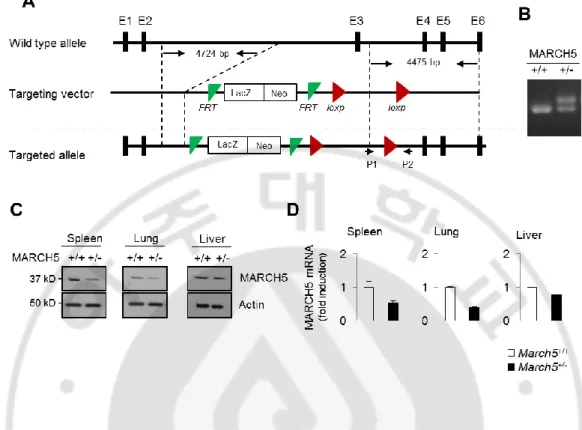
Mitochondria serve as platforms for innate immune responses. Preliminary data obtained by streptavidin-affinity purification of MARCH5 suggested that MAVS is a candidate MARCH5-interacting protein. MAVS is a key molecule in the innate immune response against RNA viruses; hence, we investigated whether MARCH5 exerted any immune modulatory function in innate immunity. We tried to produce MARCH5-deficient mice to identify the functional role of MARCH5 in vivo, but they were all embryonic lethal. Thus, we conducted the experiment with the produced heterozygous March5+/- mice (Fig. 1A). We confirmed targeted disruption of MARCH5 (Fig. 1B). MARCH5 protein and mRNA expression was decreased in spleen, lung and liver from March5+/+ or March5+/- mice (Fig. 1C, D).
10
Fig. 1. Generation of heterozygous MARCH5+/- mice. (A) Generation of heterozygous
MARCH5+/- mice by two-loxp strategy. (B) PCR genotyping of WT and MARCH5+/- mice littermates generated from MARCH5+/- intercrossing. (C) Immunoblot analysis of endogenous MARCH5 expression levels in different tissues of MARCH5+/- mice. (D) Quantitative PCR analysis of MARCH5 mRNA expression in spleen, lung and liver from March5+/+ or March5+/- mice.
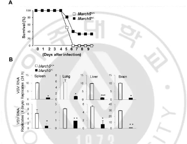
We extended our investigation to an examination of the antiviral effects of MARCH5 in vivo. To accomplish this, we intravenously challenged March5+/+ and March5+/- mice with replication-competent VSV (Indian strain) and assessed infection-related mortality. All March5+/+ mice died within 6 days of VSV infection, whereas 40% of March5+/- mice still survived at 9 days (Fig. 2A). Analysis of VSV RNA levels in various organs (spleen, lung, liver, and brain) from these mice revealed that VSV replication was significantly lower in
11
March5+/- mice than in March5+/+ mice, and remained lower for up to 5 days, indicating that March5+/- mice were more resistant to VSV infection (Fig. 2B).
Fig. 2. March5+/- mice exhibited low viral replication in response to RNA viral infection.
(A) Survival rate (%) of March5+/+ and March5+/- mice (6-7 week of age) were infected with VSV (2x10⁸ pfu per mouse) via tail vein injection. Mortality was monitored for 9 days. (n=15 mice per group). (B) VSV replication analysis by qPCR in March5+/+ and March5+/- mice tissues. March5+/+ and MRCH5+/- mice were infected with VSV for 4 days, tissues from spleen, lung, liver and brain were collected and analyzed. Error bars, mean ± SEM (n=9 mice per group).
12
We next investigated whether the low viral replication in March5+/- mice was due to higher antiviral responses in these animals. Consistent with this increased resistance, March5 +/-mice produced higher levels of serum IFN-β and IL-6 than March5+/+ mice in response to infection with VSV-GFP virus or injection of poly(I:C) (Fig. 3A). We also observed higher expression of IFN-β mRNA and IFN-related genes in the organs of VSV (Indian)-infected
March5+/- mice than in those of March5+/+ mice (Fig. 3B). These results strongly support our findings that MARCH5 plays an important role in antiviral innate response against RNA virus infection by regulating type-I IFN production in vivo.
13
Fig. 3. March5+/- mice reduced morbidity in response to RNA viral infection. (A)
Bioassay of IFN-β or IL-6 production (ELISA) in serum from March5+/+ and March5+/- mice infected with VSV (left, 2x10⁸ pfu per mouse) or poly(I:C, 200 ug) (right) for 12 hours. Error bars, mean ± SEM (n=9 mice per group). (B) Quantitative PCR analysis of Ifnb1, Oas1, Ifit1,
Mx1, Gbp2b and Isg15 mRNA expression in spleen, lung, liver or brain from March5+/+ or
March5+/- mice at 4 days after VSV infection. Graphs represent fold-induction of indicated genes relative to mRNA levels in March5+/+. Error bars, mean ± SEM (n=3 mice per group). *P <0.05, **P <0.01, ***P < 0.001 vs. March5+/+ by Student’s t-test. All data are representative of at least three independent experiments.
B. MARCH5-deficient immune cells produce elevated levels of type-I interferon and proinflammatory cytokines upon infection with RNA viruses
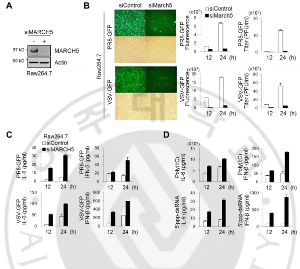
Next, we investigated the modulatory effect of MARCH5 on innate immunity in various cell lines. A MARCH5-specific small interfering RNA (siRNA) efficiently inhibited the expression of endogenous MARCH5 in Raw264.7 macrophage-like cells (Fig. 4A). After siRNA-mediated knockdown of MARCH5 expression, we infected Raw264.7 cells with VSV-fused GFP or PR8 (influenza virus)-fused GFP, and then analyzed GFP expression and virus titer in these cells. Importantly, fluorescence intensities of GFP and virus titer were significantly lower at 24 h after viral infection in cells transfected with MARCH5 siRNA than in control cells transfected with scrambled siRNA (Fig. 4B). Next, we applied four different stimulants of RLR signaling to Raw264.7 cells. We also simultaneously measured IFN-β and the proinflammatory cytokine IL-6, secreted by Raw264.7 cells as a consequence of RLR signaling. Viral infection of VSV-GFP or PR8-GFP (Fig. 4C) and transfection of poly(I:C) or 5’ppp-dsRNA (Fig. 4D), a synthetic ligand for RIG-I, increased secretion of IFN-β and IL-6 from MARCH5-deficient cells.
14
Fig. 4. MARCH5-deficient immune cells produce elevated levels of type-I interferon and proinflammatory cytokines upon infection with RNA viruses. (A) Immunoblot
analysis of MARCH5 expression levels in March5 depleted Raw264.7 cells. (B) Twenty-four hours after infection, PR8-GFP or VSV-GFP virus replication assay by fluorescence microscopy (fluorescence, upper; phase-contrast microscopy, bottom) and virus titration by fluorescence analysis or plaque assay in siControl or siMARCH5 expressing Raw264.7 cells. Error bars, mean ± SEM (n=3). (C-D) Bioassay of IFN-β or IL-6 (ELISA) in supernatants of March5 depleted Raw264.7 (E) infected or transfected with PR8-GFP, VSV-GFP (C), poly(I:C) or 5’ppp-dsRNA (D). After cells were transfected with siControl or siMARCH5 for 24 hours, followed by infection with virus or transfection with poly(I:C) or 5’ppp-dsRNA for indicated times. Error bars, mean ± SEM (n=3). All data are representative of at least three independent experiments.
15
After siRNA-mediated knockdown of MARCH5 expression, we infected 293T cells with VSV-fused GFP or PR8 (influenza virus)-fused GFP, and then analyzed GFP expression and virus titer in these cells. Notably, fluorescence intensities of GFP and virus titer were significantly lower at 24 hours after viral infection in cells transfected with MARCH5 siRNA than in control cells transfected with scrambled siRNA (Fig. 5A). Stronger antiviral effects in MARCH5-deficient cells appeared to be due to elevated production of IFN-β and the proinflammatory cytokine IL-6. Next, we applied four different stimulants of RLR signaling to HEK293T cells. Viral infection of VSV-GFP or PR8-GFP or poly(I:C) (Fig. 5B), a synthetic ligand for RIG-I, increased secretion of IFN-β and IL-6 from MARCH5-deficient cells.
Fig. 5. MARCH5 depleted cells induced type-I interferon and proinflammatory cytokines by RNA viruses. (A) Fluorescence microscopy analysis (left) and viral titer (right)
by fluorescence analysis or plaque-assays after infection for 24 h with VSV-GFP or PR8-GFP in MARCH5 siRNA expressing HEK293T cells. (B) Bioassay of IL-6 or IFN-β (ELISA) in supernatants of MARCH5 siRNA expressing HEK293T cells. Cells were transfected with
16
indicated siRNA, followed by infection of virus (PR8-GFP or VSV-GFP) or transfection with poly(I:C) for 24 h
These findings were verified in bone marrow–derived macrophages (BMDMs) obtained from March5+/+ and March5+/- mice. Viral replication of VSV and PR8 and GFP fluorescence intensity were also consistently reduced in BMDMs obtained from March5+/- mice relative to those from March5+/+ mice (Fig. 6A). Thus, MARCH5 deficiency resulted in stronger antiviral immune responses, consistent with our findings in vivo. Stronger antiviral effects in MARCH5-deficient cells appeared to be due to elevated production of IFN-β and the proinflammatory cytokine IL-6 (Fig. 6B). By contrast, infection of BMDMs from March5+/+ and March5+/- mice with the DNA virus HSV or treatment with poly(dA:dT) had no significant effect on the secretion of either cytokine (Fig. 6C).
17
Fig. 6. MARCH5 depleted BMDM cells induced type-I interferon and proinflammatory cytokines by RNA viruses. (A) Determination of the virus titration by fluorescence analysis
or plaque assay in March5+/+ and March5+/- BMDM cells infected with PR8-GFP or VSV-GFP virus. Error bars, mean ± SEM (n=3). (B) Bioassay of IFN-β or IL-6 (ELISA) in supernatants of March5+/+ and March5+/- BMDM cells infected or transfected with PR8-GFP, VSV-GFP, poly(I:C) or 5’ppp-dsRNA. Error bars, mean ± SEM (n=3). All data are representative of at least three independent experiments. (C) Bioassay of IFN-β (ELISA) in supernatants of March5+/+ and March5+/- derived BMDM cells infected with HSV or transfected with poly(dA:dT) for indicated times.
C. The E3 ligase activity of MARCH5 is necessary for inhibition of RLR-mediated antiviral signaling
Because MARCH5 is a mitochondria-resident E3 ligase, we next addressed whether its E3 ligase activity is required for its ability to inhibit RLR signaling. A previous study predicted that histidine 43 in the RING domain of MARCH5 serves a ubiquitin-transfer function; consequently, the MARCH5H43W mutant, in which His43 is replaced with tryptophan, lacks ubiquitin E3 ligase activity. To determine whether this E3 ligase activity is required for inhibition of MAVS-mediated signaling, we generated a MARCH5-KO HEK293T cell line and examined the MAVS-mediated antiviral response after reconstitution of MARCH5WT or MARCH5H43W. We first found that luciferase activities of IFN-β, IFN-α, ISRE, and IRF3 promoters were all higher in MARCH5-/- HEK293T cells than in MARCH5+/+ cells upon PR8-GFP infection or poly(I:C) treatment (Fig. 7A). This induction of luciferase activity in
MARCH5-/- cells was inhibited by reconstitution of MARCH5WT, but not MARCH5H43W. Expression levels of the corresponding proteins were confirmed by immunoblotting (Fig. 7B). Reconstitution of MARCH5WT in MARCH5-/- cells significantly increased the growth of viruses, which remained low following introduction of MARCH5H43W (Fig. 7C). Consistent with these results, we further found that INF-β and IL-6 secretion upon stimulation with VSV-GFP in MARCH5-/- HEK293T cells was robustly increased compared to that in MARCH5+/+ HEK293T cells; moreover, reconstitution of MARCH5WT into MARCH5-/- HEK293T cells decreased INF-β and IL-6 secretion, whereas reconstitution of
18
MARCH5H43W did not (Fig. 7D), indicating that the E3 ligase activity of MARCH5 is necessary for inhibition of RLR signaling. Together, the data suggest that the E3 ubiquitin ligase function of MARCH5 is necessary for inhibition of RLR-mediated antiviral signaling.
Fig. 7. The E3 ligase activity of MARCH5 is necessary for inhibition of RLR-mediated antiviral signaling. (A) After Myc-MARCH5 WT or H43W expression vectors were
introduced into MARCH5 KO HEK293T cells, cells were infected with PR8-GFP or transfected with poly(I:C) for 24 hours. Promoter activity of β, ISRE,
IFN-Graphs represent fold-induction relative to the luciferase activity in control cells. Error bars, mean ± SEM (n=3). (B) Immunoblot analysis of endogenous or ectopic expression levels of MARCH5 in MARCH5 WT or KO HEK293T cells. (C) Twenty-four hours after infection,
19
VSV-GFP replication assay by fluorescence microscopy analysis. (D) Bioassay of IFN-β or IL-6 production (ELISA). Error bars, mean ± SEM (n=3).
Finally, we confirmed that MAVS-dependent induction of IFN-β and IL-6 genes, as well as IFN stimulated genes, was significantly elevated in MARCH5-knockdown HEK293T cells after challenge with the VSV-GFP virus or 5’ppp-dsRNA (Fig. 8A, B).
20
Fig. 8. Expression of IFN response genes are elevated in MARCH5-knockdown cells.
(A,B) Messenger RNA levels of IFNB, IRF3, IRF7, IRIT1, ISG15, TNF, IL6, GAPDH in control or MARCH5 siRNA expressing HEK293T cells by RT-PCR analysis. Graphs represent fold changes of mRNA levels of indicated genes value were relative to control siRNA expressing cells, and those were normalized by GAPDH. Data represent the average of three independent experiments. Error bars, mean ± s.e.m. (n=3). (A) Control or MARCH5 siRNA expressing cells were infected with VSV-GFP for 24 h, followed by RT-PCR. (B) Control or MARCH5 siRNA expressing HEK293T cells were transfected with 5’ppp-dsRNA for 24 h, followed by RT-PCR.
D. MARCH5 suppresses the MAVS-mediated type-I IFN signaling
After viral infection, various components are involved in the type-I IFN signaling pathway. Next, we investigated whether MARCH5 participates in antiviral signaling by examining the effects of MARCH5 on the various components of the type-I IFN signaling pathway. To this end, we transfected HEK293T cells with an IFN-β-Luc reporter plasmid and expression vectors encoding N-RIG-I, MAVS, the Toll-like receptor adapter TRIF, TRAF-6, TRAF-3, IKK and IRF3 together with increasing amounts of a MARCH5 expression plasmid. MARCH5 inhibited N-RIG-I, MDA5, or MAVS-induced IFN-β-Luc activity in a dose-dependent manner, whereas activation of the IFN-β promoter induced by TRIF was not affected (Fig. 9A). Moreover, the IFN-β-Luc activity induced by TRAF-6, TRAF-3, IKK or IRF3 was unaffected by MARCH5 overexpression, indicating that MARCH5 specifically inhibited RLR-mediated MAVS signaling in mitochondria. Likewise, activation of the IFN-β promoter by viral infection with VSV (Vesicular Stomatitis virus)-GFP or PR8-GFP was inhibited by MARCH5 (Figure 7), and MAVS-stimulated production of IFN-β was diminished by MARCH5 overexpression in a dose-dependent manner (Fig. 9B). In contrast, TRIF-mediated IFN-β production was not affected by MARCH5. The effects of MARCH5 on downstream of MAVS were also examined in HEK293T cells after stimulation with poly(I:C). Transfected poly(I:C) robustly increased IRF3 and p65 phosphorylation in a time-dependent manner; this effect was significantly attenuated in cells transfected with Myc-MARCH5WT (Fig. 9C). These results collectively demonstrate that MARCH5 specifically
21
suppresses MAVS-mediated type-I IFN signaling.Fig. 9. MARCH5 suppresses the MAVS-mediated type-I IFN signaling. (a) IFN-β
promoter activity in N-RIG-I, MDA5, MAVS, TRIF, TRAF6, TRAF3, IKK or IRF3 with Myc-MARCH5WT (0, 50, 100, 200, 400, 800 ng) transfected HEK293T cells for 36 hours. Graphs represent fold-induction of IFN-β relative to the luciferase activity in control cells. Data represent the average of three independent experiments. Error bars, mean ± SEM (n=3). (b) Bioassay of IFN- β (ELISA) in supernatants of HEK293T cells transfected with MAVS or TRIF with Myc-MARCH5WT (0, 100, 200, 400 ng). Error bars, mean ± SEM (n=3). (c) Immunoblotting of p-IRF3, p-p65, total IRF and total p65 in lysates of HEK293T cells. After
22
transfection with control or Myc-MARCH5WT for 24 hours, cells were stimulated by transfection with poly(I:C) for the indicated time.
E. MARCH5 binding to MAVS is necessary for the inhibition of type-I IFN responses
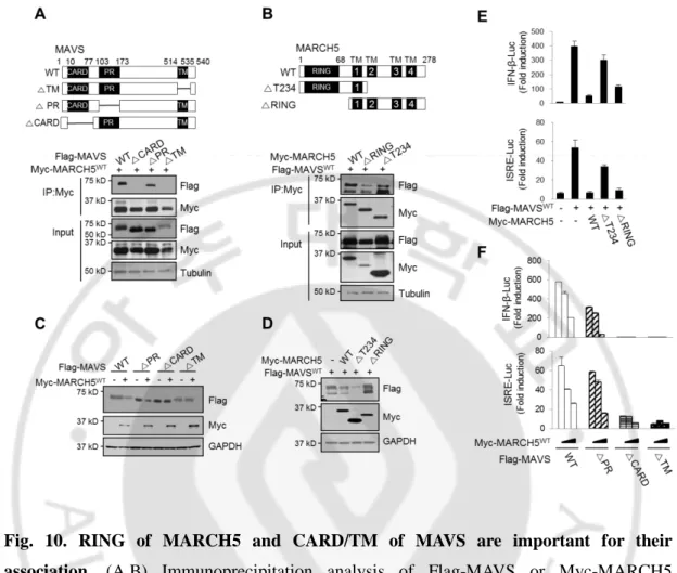
Next, we investigated whether inhibition of MAVS-mediated type-I IFN signaling is mediated by its binding of MARCH5 to MAVS. To study the domains responsible for the interaction between MARCH5 and MAVS, we generated a series of deletion mutants of MARCH5 and MAVS, and then carried out co-immunoprecipitation analyses using these constructs. Both the caspase activation and recruitment domain (CARD) and TM domains of MAVS were required for its interaction with MARCH5 (Fig. 10A), and deletion of the RING domain of MARCH5 significantly reduced MARCH5 binding to MAVS (Fig. 10B). In keeping with these findings, the expression levels of MAVSWT and MAVS∆PR were reduced by MARCH5WT, whereas MAVS∆CARD and MAVS∆TM levels were not affected by MARCH5WT (Fig. 10C). Likewise, FLAG-MAVSWT levels were not affected by overexpression of MARCH5∆RING, but were suppressed by MARCH5WT and MARCH5∆T234 (Fig.10D). To verify the functionality of this interaction, we carried out IFN-β and ISRE luciferase reporter assays in cells co-expressing wild-type or mutant MAVS and MARCH5 proteins. The MAVS-mediated increase in luciferase activity was significantly decreased by MARCH5WT or MARCH5∆T234, whereas luciferase activity remained high upon co-expression of MARCH5∆RING (Fig. 10E, F). Similarly, only the MAVSWT and MAVS∆PR constructs were capable of activating IFN-β and ISRE luciferase reporters; these effects, in turn, were suppressed in a dose-dependent manner by co-expression of MARCH5. The CARD domain of MAVS is necessary for the interaction with RIG-I and its subsequent oligomerization. In the absence of the TM, MAVS hardly positioned to the mitochondria membrane. Thus, the data suggest that binding of the MARCH5 RING domain to the CARD domain of MAVS at the mitochondria is necessary for negative regulation of type-I IFN responses.
23
Fig. 10. RING of MARCH5 and CARD/TM of MAVS are important for their association. (A,B) Immunoprecipitation analysis of Flag-MAVS or Myc-MARCH5
truncated mutants. HEK293T cells were co-transfected with indicated plasmids and interaction was analyzed by immunoblotting with indicated antibodies. Flag-MAVS fragments (WT: full length, △TM; △514-535, △PR; △103-173, △CARD; △10-77) (A). Myc-MARCH5 fragments (WT; full length, △T234; 1-139, △R; 76-278) (B). (C) Immunoblot analysis of Flag-MAVSWT or truncated mutants expression levels in HEK293T cells transfected with or without Myc-MARCH5WT. (D) Immunoblotting of Flag-MAVS expression levels in HEK293T cells transfected with Myc-MARCH5WT or truncated mutants. (E) IFN-β and ISRE promoter activity in Flag-MAVS expressing HEK293T cells transfected with Myc-MARCH5 fragments. Graphs represent fold-induction relative to the luciferase activity in control cells. Error bars, mean ± SEM (n=3). (F) Promoter activity of IFN-β and ISRE in HEK293T cells transfected with Myc-MARCH5WT and Flag-MAVS fragments
24
constructs. Data represent the average of three independent experiments. Error bars, mean ± s.e.m. (n=3).
F. MARCH5reduced the MAVS aggregates via the proteasome-mediated degradation
pathway
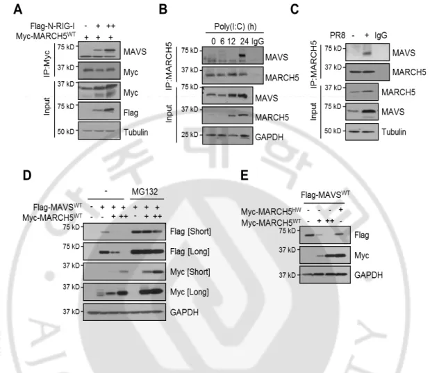
MAVS is strategically localized to mitochondria, where it is necessary for RIG-I signaling. Because MARCH5 also localizes to the mitochondria outer membrane. We next addressed how the MARCH5 and MAVS interaction exerted the observed antiviral effect. Co-immunoprecipitation revealed that Myc-MARCH5 overexpressed in HEK293T bound endogenous MAVS only when RIG-I was activated by overexpression of N-RIG-I (Fig. 11A). Similarly, endogenous MARCH5 bound to MAVS only after poly(I:C) transfection (Fig. 11B) or PR8 infection (Fig. 11C). Notably, the amount of endogenous MAVS bound to Myc-MARCH5WT occurred 24h after poly(I:C) treatment. Thus, the MARCH5 binding to MAVS was induced only under the RIG-I stimulation. Next, we addressed whether the MARCH5 binding of MAVS promoted proteasomal degradation of MAVS. We found that exogenously expressed MARCH5WT induced a dose-dependent decrease in FLAG-MAVSWT protein levels in HEK293T cells; this reduction was absent in the presence of the proteasome inhibitor MG132 (Fig. 11D). By contrast, introduction of MARCH5H43W did not reduce the level of MAVS protein (Fig. 11E), suggesting that MARCH5 E3 ligase promotes MAVS degradation through a proteasome-dependent pathway.
25
Fig. 11. MARCH5 degradates MAVS through the proteasome-dependent pathway. (A,B)
Interaction between MARCH5 and MAVS in HEK293T cells. Cells were transfected with Flag-N-RIG-I (A) or poly(I:C) (B), interaction was determined by immunoblotting with anti-MAVS antibody after lysates were immunopricipitated with anti-MARCH5 or anti-Myc antibody. (C) After cells were infected with PR8 virus for 24h, cell lysates were immunoprecipitated with anti-MARCH5 antibody or control IgG. Interaction was evaluated using anti-MAVS antibody. (D) Immunoblot analysis of Flag-MAVS expression levels in HEK293T cells. Cells were transfected with Flag-MAVSWT and Myc-MARCH5WT, with or without treatment of MG132 (10uM). (E) Immunoblot analysis of Flag-MAVS expression levels in HEK293T cells. Cells were co-transfected with Flag-MAVS and Myc-MARCH5 WT or H43W.
26
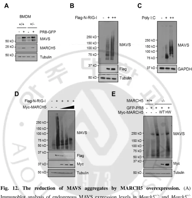
Under Virus stimulation, we observed higher MAVS accumulation in MARCH5+/- mice than MARCH5+/+ mice on PR8-GFP infection (Fig. 12A). MAVS forms prion-like aggregates that potently propagate downstream signaling. MARCH5 bound MAVS only under conditions of RIG-I stimulation, in which MAVS forms aggregates. In semi-denaturing detergent agarose gel electrophoresis (SDD-AGE), we also observed a smear of SDS-resistant high-molecular weight MAVS aggregates 24h after N-RIG-I or poly(I:C) transfection (Figs. 12B, C). Likewise, the MAVS aggregates stimulated by N-RIG-I were significantly reduced by overexpression of MARCH5 in a dose-dependent manner (Fig. 12D). Importantly, formation of the MAVS aggregates induced by viral infection was intensified in MARCH5 KO cells; re-expression of MARCH5WT, but not MARCH5H43W, significantly reduced the levels of aggregates (Fig. 12E). These results suggest that MARCH5 may not interact with monomeric MAVS at the mitochondria membrane. Under RIG-I stimulation, MARCH5 recognizes MAVS aggregates and targets them for proteasome-mediated degradation. Together, these data suggest that MARCH5reduces the level MAVS aggregates through the proteasome-mediated degradation pathway. To our knowledge, mitochondria-resident MARCH5 is the first negative regulator shown to target MAVS aggregates.
27
Fig. 12. The reduction of MAVS aggregates by MARCH5 overexpression. (A)
Immunoblot analysis of endogenous MAVS expression levels in March5+/+ and March5 +/-BMDM cells infected with PR8-GFP. (B, C) Cells were transfected with Flag-N-RIG-I or poly(I:C) for 36h. Lysates were mixed with or without 6X native sample buffer, followed by SDS-PAGE. (D) Immunoblot analysis of oligomeric MAVS in HEK293T cells. Cells were transfected with Flag-N-RIG-I with Myc-MARCH5WT dose dependent manner. Lysates were mixed with or without 6X native sample buffer, followed by SDS-PAGE. (E) After Myc-MARCH5 WT or H43W expression vectors was reintroduced into MARCH5 KO HEK293T cells, cells were infected with PR8-GFP. The lysates were mixed with or without 6X native sample buffer, followed by SDS-PAGE.
28
G. MARCH5 transfers ubiquitin to Lys7 and Lys500 residues of MAVS and accelerates MAVS degradation
Next, we characterized the patterns of MARCH5-mediated ubiquitination of MAVS. The poly(I:C) transfection into HEK293T cells significantly increased MAVS polyubiquitin complexes at 24h (Fig. 13A). Notably, the polyubiquitin chains in MARCHWT-expressing cells contained lysine (K)48-linked ubiquitin, but not K63-linked ubiquitin (Fig. 13B). MARCH5-induced ubiquitination was not observed in MARCH5H43W expressing cells (Fig. 13C). Moreover, challenging cells by overexpressing N-RIG-I increased MAVS ubiquitination in MARCH5+/+ HEK293T cells, but not in MARCH5-/- HEK293T cells (Fig. 13D). Finally, in vitro ubiquitination assays verified that MARCH5, in combination with an E1 ubiquitin-activating enzyme and the E2 ubiquitin-conjugating enzyme UbcH5b, effectively transferred ubiquitin to MAVS (Fig. 13E). In the absence of MARCH5, no ubiquitination occurred, and in the absence of MAVS, MARCH5 was auto-ubiquitinated. Thus, the data indicate that MARCH5 transfers the K48-linked ubiquitin to MAVS under RIG-I stimulation.
29
Fig. 13. MARCH5 transfers ubiquitin to MAVS through a K48-linkage. (A)
Ubiquitination assay of endogenous MAVS by using anti-MAVS antibody in poly(I:C) transfected HEK293T cells. Levels of ubiquitination were determined by anti-Ub antibody. (B, C) Ubiquitination assays in Flag-MAVSWT and Myc-MARCH5WT expressing HEK293T cells. (B) After cells were transfected with Flag-MAVSWT and control or Myc-MARCH5WT, ubiquitination levels of Flag-MAVS were determined by immunoblotting with anti-Ub, K48-Ub or K63-K48-Ub specific antibodies. (C) Cells were co-transfected with Flag-N-RIG-I and Myc-MARCH5-WT or H43W. Endogenous ubiquitination levels of MAVS were determined by anti-Ub antibody after IP with endogenous anti-MAVS antibody. (D) Ubiquitination of endogenous MAVS in MARCH5+/+ or MARCH5-/- HEK293T cells transfected with
Flag-N-30
RIG-I. (E) In vitro ubiquitination assay of Flag-MAVS. Myc-MARCH5WT and Flag-MAVSWT were incubated with a reaction mixture with ubiquitin, E1, UbcH5b as E2, and ubiquitnation levels were determined by immunoblotting with anti-Ub antibody.
Next, to identify the lysine (K) residues of MAVS to which ubiquitin is attached, we first performed an in silico search for ubiquitin-binding sites on MAVS using the UbPred program and PhosphoSitePlus (http://www.phosphosite.org). This analysis revealed four putative lysine target residues: K7, K10, K461, and K500. We then generated FLAG-MAVS mutants in which each of these lysine residues was replaced with arginine (R). The steady-state levels of these point-substitution mutant proteins were comparable, but they responded differently to MARCH5. MAVSK10R was as sensitive to MARCH5 as MAVSWT, whereas neither MAVSK7R nor MAVSK500R was affected by MARCH5WT overexpression (Fig. 14A).
MAVSK461R protein levels were decreased by MARCH5WT, but to a lesser extent than
MAVSK10R or MAVSK500R. In vivo ubiquitination assays verified a substantial decrease in ubiquitin conjugation in the MAVSK7R and MAVSK500R mutants (Fig. 14B). To address the functional importance of ubiquitination on these Lys sites in the type-I IFN responses, HEK293T cells were transfected with these ubiquitin conjugation-defective MAVS mutants and Myc-MARCH5WT together with IFN-β, ISRE, or NF-B luciferase reporter constructs, MAVSK7R and MAVSK500R fully retained the ability to increase luciferase activity from all promoters tested, even in the presence of MARCH5WT (Fig. 14C). However, MAVSWT as well as the MAVSK10R and MAVSK461R mutants only weakly activated these promoters when co-expressed with MARCH5WT. Taken together, these data demonstrate that MARCH5 transfers ubiquitin to Lys7 and Lys500 of MAVS through a K48 linkage and accelerates MAVS degradation.
31
Fig. 14. MARCH5 transfers ubiquitin to Lys7 and Lys500 residues of MAVS. (A)
Expression of Flag-MAVS lysine mutants by Myc-MARCH5WT in HEK293T cells. (B) Ubiquitination assay of Flag-MAVS lysine mutants by Myc-MARCH5WT. Ubiquitination of MAVS lysine mutants was assessed by immunoblotting with anti-Ub antibody. (C) Promoter assay of IFN-B, ISRE or NF-KB in HEK293T cells transfected with Myc-MARCH5WT and Flag-MAVSWT or lysine mutants for 36h. Graphs represent fold-induction relative to the luciferase activity in control cells. Error bars, mean ± SEM (n=3). All data are representative of at least three independent experiments.
32
IV. DISCUSSION
On the mitochondria membrane, MAVS acts as a critical adaptor protein of RLR signaling pathway that links upstream viral RNA recognition to downstream signal activation to control viral replication. A remarkable feature of MAVS is the formation of prion-like aggregates that are not only functional, but also potently propagate downstream signaling. The persistence of MAVS signaling, however, results in an excessive immune response, leading to detrimental effects on the host. Understanding the mechanism by which MAVS aggregation is initiated, and how it is subsequently resolved, is crucial for a complete understanding of innate immunity.
In this study, we provided several lines of evidence that MARCH5 is a potent negative regulator of MAVS-mediated antiviral innate immunity, and that it is a unique mitochondria-resident E3 ubiquitin ligase. First, March5+/- mice exhibited reduced morbidity upon VSV infection due to low viral replication. Second, knockdown/knockout of endogenous MARCH5 in immune cells resulted in reduced viral replication that correlated well with high type-I IFN responses. Third, MARCH5 bound MAVS at the mitochondria membrane only under conditions of RIG-I activation. Fourth, MARCH5 could reduce the level of MAVS aggregates via proteasome-mediated degradation. Thus, MARCH5 is the crucial mitochondria-resident regulator that coordinates the MAVS signaling via timely degradation of the aggregated MAVS. This MARCH5-mediated quality control provides an efficient way to prevent excessive host immunity.
The MARCH proteins were initially identified as a family of viral ubiquitin ligases; to date, 11 mammalian homologues have been identified. Both viral E3 ligases and mammalian homologues have been proposed to acts as immune modulators in viral immune evasion strategies. Most mammalian MARCH proteins contain two or more transmembrane domains that target them to intracellular organelles or the plasma membrane. So far, the reported functions of mammalian MARCH proteins have been limited to down-regulation of immunomodulatory surface proteins of immune cells. MARCH4 and MARCH9 decrease the surface expression of MHC-I and/or ICAM1 in T- and B-cells. In addition, MARCH8 triggers degradation of IL-1 receptor accessory protein (IL1RAP). MARCH5 is the first MARCH family member shown to be involved in TLR7 (Toll-like receptor 7) signaling.
33
MARCH5 catalyzes poly-ubiquitination of TANK (TRAF family member-associated NF-B activator) through a K63-Ub linkage that leads to activation of NF-κB. In this study, we found that MARCH5 facilitated the degradation of MAVS after RIG-I mediated recognition of cytosolic viral dsRNA, thereby controlling excessive production of type-I interferon. To our knowledge, this is the first demonstration of a negative modulatory function for a MARCH family protein in antiviral innate immunity. Unlike other cytosolic E3 ligases that negatively regulate RLR signaling pathways, MARCH5 is a mitochondrial protein that directly binds and degrades activated MAVS oligomers following viral infection. AIP4 E3 ligase–mediated degradation of MAVS requires a scaffolding protein, poly(rC) binding protein 2 (PCBP2). Similarly, Ndfip1 leads to the recruitment of Smurf1 E3 ligase, triggering proteasome-mediated degradation of MAVS. Other E3 ligases, such as cytosolic TRIM2530 and RNF5 in the endoplasmic reticulum, have also been shown to accelerate MAVS degradation after viral infection.
In non-activated cells, MAVS is anchored to the mitochondrial outer membrane in its monomeric form. During viral infection, RIG-I and MAVS proteins undergo conformational changes that are essential for antiviral signaling. Of particular note, MAVS forms well-ordered, functional aggregates, and these MAVS fibrils behave like prions. In this study, we showed that MARCH5 did not interact with MAVS in unstimulated cells, and that its binding to MAVS was induced by stimulation with virus, N-RIG-I, or poly(I:C) treatment. MARCH5 binding to MAVS occurred 12–24h after viral infection or mimetic stimulation, suggesting selective and timely interaction between MARCH5 and the activated MAVS oligomer. These results strongly suggest that MARCH5 is likely to bind only to the activated MAVS oligomers, but not to the non-activated monomeric MAVS. In fact, we observed that the level of high–molecular weight MAVS aggregates induced by influenza virus infection was significantly reduced by MARCH5. At present, it remains unclear how MARCH5 distinguishes the monomeric MAVS from the MAVS oligomer. However, previous work showed that MARCH5 selectively facilitates the degradation of protein aggregates and misfolded SOD1 accumulated at mitochondria. Thus, MARCH5 plays an important role in protein quality control and maintenance of mitochondrial homeostasis. Our deletion analysis showed that the N-terminal CARD domain of MAVS was necessary for MARCH5 binding. Because the CARD domain is required for MAVS polymerization and RIG-I–dependent
34
IRF3 activation, the inability of the MAVS∆CARD mutant to bind MARCH5 may be due to a failure of MAVS polymerization. Furthermore, we identified amino acids Lys7 and Lys500 of MAVS as critical for MARCH5-mediated ubiquitination. Paz et al. reported that K63-linked ubiquitination at Lys500 of MAVS leads to recruitment of inhibitor of κB kinase ε. In this study, we demonstrated that Lys500 residue of MAVS is also a target for K48-linked ubiquitination by MARCH5.
35
V. Conclusion
In this study, we identified the E3 ubiquitin ligase MARCH5 as a novel mitochondrial regulator of the immune response against RNA virus infection. MARCH5 targets the activated MAVS oligomer and attenuates MAVS-mediated IFN signaling through K48-linked ubiquitination and degradation of MAVS. Thus, MARCH5 is a crucial mitochondria-resident regulator that coordinates the MAVS signaling via timely degradation of activated MAVS oligomers, and this MARCH5-mediated quality control provides an efficient way of preventing excessive host immunity. Improved understanding of MARCH protein–mediat ed modulation of RLR signaling may facilitate development of new strategies for targ eting the RLR pathway to achieve therapeutic control of viral infection and enhance ment of the innate immune response.
36
PART II
Mitochondrial E3 Ligase, MARCH5 degrades
Hepatitis B Virus X protein on Mitochondria
37
I. Introduction
Hepatocellular carcinoma (HCC) is a leading cause of death worldwide. HCC is a common type of liver cancer. It is strongly associated with cirrhosis, from viral etiologies. One of the primary etiological factors for the development of liver cancer is chronic infection with hepatitis B virus (HBV). Hepatitis B virus (HBV) infection is one of the most prevalent global health problems and attack the liver of more than 2 billion people worldwide. Among them, more than 350 million chronic carriers lead to chronic liver disease and cirrhosis of the liver and liver cancer, hepatocellular carcinoma (HCC), the most common malignancy of the liver. The incidence of hepatocellular carcinoma is highest in Asia, where the endemic high prevalence of hepatitis B and C strongly predisposes to the development of chronic liver disease. HCC are typically diagnosed in adults in late middle age. Surgical resection and liver transplantation are major curative modalities, but they are limited to patients with early HCC.
Human hepatitis B virus (HBV) is one of the causative pathogens of acute hepatitis, chronic hepatitis and hepatocellular carcinoma. HBV is composed of a partially double-stranded circular DNA, 3.2 kb genome that contains 4 overlapping genes: S (HBsAg, Hepatitis surface antigen), C (HBcAg, core antigen) P (DNA polymerase), and X (HBx). Among these four proteins, HBx, an essential factor for viral replication, contributes to HBV-induced hepatocarcinogenesis. Forced expression of HBx protein enhanced the survival and proliferative potential of liver cells whereas knockdown of HBx reduces the tumorigenicity of HCC cells. HBx has been implicated in HBV pathogenesis and cancer development through its ability to deregulate cell cycle check points, activate the expression of oncogenes and inflammatory cytokines, and to induce liver tumors in transgenic mouse. Several studies on HBx transgenic mice have investigated characteristics of malignant transformation. In order to viral replication, HBx exerts transactivation function to activate transcription factors such as nuclear factor kB (NF-kB), activator protein-1 (AP-1) and activation transcription factor (ATF)/cAMP responsive element binding transcription factor (CREB) in cytoplasm and nucleus. HBx has been known to be involved in multiple functions in nucleus and cytosol, and recently reports have focused on mitochondria, which play an important role in apoptosis. Mitochondrial function contributes to cancer through alteration
38
in glucose metabolism, the production of reactive oxygen species (ROS). High level of HBx expression leads to an abnormal mitochondrial aggregation at the perinuclear which was caused by the HBx mediated microtubule network alteration. HBx is multifunctional protein and regulates mitochondrial activities. Mitochondrial HBx decreased mitochondrial membrane potential, cellular ATP level and oxidative stress, inducing the development of HCC and liver inflammation through COX2 activation. HBx also is involved in the apoptosis during viral infection. It has been reported that HBx interacts with various cellular signaling factors to enhance apoptosis. Mitochondria undergo major changes in membrane integrity due to apoptotic signals, and these changes cause the redistribution of numerous apoptogenic factors such as cytochrome c and Bax in the intermembrane space. High expression level of HBx is crucial in the pathogenesis of HCC mediated by mitochondrial dysfunction. Therefore, negative regulatory mechanism of HBx is required for cellular homeostasis. In general, HBx was maintained at a low intracellular level since rapid degradation by the ubiquitin-proteasome degradation. It has been known to be deregulated by 26S proteasome complex, heat shock protein 40, p53, Siah-1, MDM2 and ID-1. However, quality control system for HBx accumulation in mitochondrial has not been addressed.
Mitochondria have a central role in preserving cellular homeostasis and they have quality control system to regulate cell life and death. Ubiquitin proteasome system (UPS) is molecular quality control system as a first line barrier on the mitochondrial outer membrane. Additionally, severely damaged mitochondria is removed by mitophagy or apoptosis by the release of pro-apoptotic proteins in organellar or cellular quality control system, respectively. In mammals, a mitochondrial E3 ubiquitin ligase, membrane-associated RING-CH5 (MARCH5, also known as MITOL), localizes to the mitochondrial outer membrane and has four transmembrane domains and contains a RING-finger domain that has ubiquitin ligase activity. MARCH5 regulates mitochondrial dynamics. Mitochondria is a highly dynamic organelle via the control of the balance between mitochondrial fusion proteins Mfn1/2 and OPA1 and fission protein hFis1 and DRP1. Thus, MARCH5 regulates the balance between mitochondrial fusion and fission proteins via ubiquitination. Recent reports describe that MARCH5 degrades the accumulation and aggregation of misfolding proteins localized to mitochondria. This quality control protect cells from the cellular toxicity of misfolding
39
proteins. Mitochondrial E3 ubiquitin ligase MARCH5 has know to be involved in protection against mitochondrial accumulation of Mutant SOD and PolyQ and S-nitrosylated microtubule-associated protein 1B-light chain 1 (LC1) by the ubiquitin-proteasome pathway. MARCH5 attenuates the cytotoxicity of mutant SOD or PolyQ by ubiquitination of mitochondrial misfolding proteins and promoting its degradation. The mechanism of mitochondrial quality control regulated by MARCH5. Therefore, MARCH5 has a potent role to maintain mitochondrial outer membrane as a first line barrier quality controller.
We found that MARCH5 as a potent regulator of mitochondrial HBx facilitate its ubiquitination and subsequent proteasomal degradation. It has been reported that HBx is important for ICC development. Therefore, our findings suggest that MARCH5 mediated mitochondrial HBx degradation is important to suppress the progression of hepatocellular carcinoma. Finally, we discuss the possibility of a new gene therapy for HBx induced HCC development through the activation of a mitochondrial quality control system by MARCH5.
40
II. Materials and Methods
A. Cell culture and transfection
Chang, Huh7 and HEK293T cells were cultured in Dulbecco’s modified Eagle’s medium (DMEM; Invitrogen) supplement with 10% heat-inactivated fetal bovine serum (FBS, Invirogen) and 1% penicillin-streptomycin (Invitrogen) in a 5% CO2 incubator at 37°C.
MARCH5 knockout HEK293T cells were generated using transcription activator-like effector nuclease (TALEN) technology. The MARCH5-specific TALEN plasmids were obtained from ToolGen, Inc. Plasmid DNA transfections were carried out using polyethylenimine (PEI; Polysciences) according to manufacturer’s instructions. For RNA interference, siRNA was transfected using Lipofectamine 2000TM (Invitrogen) according to manufacturer’s instruction.
B. siRNA and Plasmids
Human specific siRNA for MARCH5 was custom synthesized by Invitrogen. Sequences targeting human MARCH5 were 5’-GGGUGGAAUUGCGUUUGUUTT-3’. GFP siRNA was used as a negative control. Flag-HBx, Flag-HBx-MTS and Flag-HBx △MTS were established as previously described. Myc tagged MARCH5-WT and MARCH5-H43W were established as previously described. For reporter assays, NF-kB luciferase reporter plasmids were used.
C. Immunoprecipitation and Immunoblotting
For immunoprecipitation assay, cells were lysed with E1A buffer (50 mM Tris-HCl [pH7.4], 150 mM NaCl, 0.1% NP-40, 1 mM DTT, 5 mM EDTA). Cell extracts (500 ~ 1,000ug) were immunoprecipitated with 1 ug of indicated antibody at 4°C for 12 h. After incubation with Protein A-sepharose beads (GE healthcare Bioscience AB) for 1 h 30 min at 4°C,