INTRODUCTION
The p53 tumor suppressor gene has a critical role for regula-tion of cell cycle, cellular growth, and maintenance of genomic integrity (1). And it is well known that the inactivating muta-tion of p53 is the most common genetic alteramuta-tion in human cancers including hepatocellular carcinoma (HCC) (2). The mutational spectrum of p53 has been reported to differ in HCCs from different geographic regions. For example, the G to T transversion at the third position of codon 249 has been detected very frequently, ranging from 30 to 58% of HCCs, in southern Africa and southern China in which aflatoxin B1 is contaminated highly in food and hepatitis B virus (HBV) infection is endemic (3-5). In contrast, few or none of the muta-tions occur at codon 249 in low aflatoxin B1 exposure areas (6-9). In Korea, HCC is one of the most common cancers, HBV-related HCC constitutes about 65-70% of HCC, and dietary exposure of aflatoxin B1is low (10, 11). There are a few reports, however, on the status of p53 mutation at exons 3-9 or 5-8 in HCC patients in Korea (12-14).
The missense mutations of p53 may result in biologically altered proteins with increased stability that is easily detectable by immunohistochemical method. However, deletion or
trun-cated proteins resulted from nonsense or frameshift mutations are usually not detectable by immunohistochemical method, because these proteins are very unstable in the cell, even using antibody containing the corresponding N-terminal epitope (2). Therefore, immunohistochemistry (IHC) will be less sensitive in detecting mutations in tumors with high proportion of these non-missense mutations. p53 overexpression is detected at a higher incidence than gene mutation in human cancers (44% vs 36%) (2).
In p53 IHC, the proportion of cells that show immunore-activity is very important (15). Occurrence of just occasional strongly immunoreactive cells in a tumor does not seem to correlate with obvious molecular abnormality of p53 (16). They may rather represent the normal working of the p53 system in which the wild-type protein accumulate in response to spontaneous genetic errors occurring at a higher frequency in the tumor than in the normal surrounding tissue. In p53 immunohistochemical study on HCC, the threshold value above which the result is regarded positive has not been well established. p53 overexpression in HCC is not always depen-dent on p53 mutation (17). And the degree of correlation between sequencing and IHC for detecting mutations of p53 has not been well established in human HCCs.
Shi Nae Lee�
, Cheol Keun Park, Chang Ohk Sung, Jong Sun Choi, Young Lyun Oh, Jae Won Cho*, Byung Chul Yoo�
Departments of Pathology, General Surgery*and Internal Medicine�
, Samsung Medical Center, Sungkyunkwan University School of Medicine, Seoul; Department of Pathology, Ewha Womans University College of Medicine�
, Seoul, Korea
Address for correspondence Cheol Keun Park, M.D.
Department of Diagnostic Pathology, Samsung Medical Center, Sungkyunkwan University School of Medicine, 50 Ilwon-dong, Kangnam-gu, Seoul 135-710, Korea
Tel : +82.2-3410-2766, Fax : +82.2-3410-0025 E-mail : [email protected]
*This work was supported by the Samsung grant, #SBRI C-A0-033-2.
801
Correlation of Mutation and Immunohistochemistry of p53 in
Hepatocellular Carcinomas in Korean People
The degree of correlation between sequencing and immunohistochemisty (IHC) for detecting mutations of p53 has not been well established in human hepatocellular carcinoma (HCC). We analyzed 36 HCCs from Korean people for p53 mutation at exons 4-10 by PCR-SSCP and sequencing, and compared the results with the IHC positivity. p53 mutations were identified in 7 out of 36 HCCs (19.4%). These mutations were found widely throughout exons 4-8. No mutation was detected in codon 249. Among the 7 mutations, 6 missense mutations were detected in 15 HCCs with ≥5% immunoreactive tumor cells and one nonsense mutation was in 21 HCCs with <5% immunoreactive tumor cells. The sensitivity for p53 mutation was 85.7% (6/7), the specificity 69.0% (20/29), the predictive value of positive IHC 40.0% (6/15), and the predictive value of negative IHC 95.2% (20/21). Two mis-sense mutations were detected in 25 cases with <10% immunoreactive tumor cells. Predictive values of both positive IHC and negative IHC were higher in ≥5% over-expression group than in ≥10% overexpression group or >0% overexpression group. This study suggests that 5% immunoreactivity is a reliable immunohisto-chemical threshold value to detect p53 mutations in HCCs and the spectrum of
p53 mutations in HCCs in Korean people is different from that of high aflatoxin B1
exposure areas.
Key Words : Carcinoma, Hepatocellular; Genes, p53; Immunohistochemistry; Mutation
Received : 15 July 2002 Accepted : 11 September 2002
In this study, we determined what proportion of cells that show immunoreactivity is a reliable immunohistochemical threshold value to detect p53 mutations in HCCs and evaluat-ed the spectrum of p53 mutations at exons 4-10 in 36 HCCs from Korean people.
MATERIALS AND METHODS
Tumor samples
Samples of 36 HCCs were obtained from 34 Korean patients at the time of curative surgical resection at Samsung Medical Center, Seoul, Korea, between 1999 and 2000. None of the patients had any preoperative chemotherapy. All of these were primary HCC tumors: 33 were from patients with a single HCC nodule and 3 were from 1 patient with three HCC nod-ules of Edmondson & Steiner grade I. Informed consent was obtained from each patient. The age of patients ranged from 26 to 89 yr, with a mean age of 51.8 yr. The male-to-female ratio was 27:7. All samples included both tumors and non-tumorous tissues. Halves of the samples were snap-frozen in liquid nitrogen and stored at -80℃until DNA extraction. The other halves were fixed in 10% formalin and embedded in paraffin. The longest diameter of a tumor ranged from 9 to 110 mm, with a mean of 38.6 mm. The degrees of differenti-ation of tumor cells were determined according to the Edmond-son & Steiner’s grading system (18). There were 6 cases of Edmondson & Steiner grade I, 11 grade II, and 19 grade III. The nontumorous liver showed cirrhosis in 23 (67.6%) patients and chronic hepatitis in 11 (32.4%). The etiologies of liver disease were HBV in 27 patients, hepatitis C virus (HCV) in 3, and cryptogenic in 4.
Immunohistochemistry
Formalin-fixed, paraffin-embedded sections of 4 m con-taining both HCC and nontumorous liver were prepared. Immunohistochemical study was performed using the strep-tavidin-biotin complex method and TechMateTM 1,000 auto-mated staining system (DakoChemmate, Glostrup, Denmark). Primary monoclonal antibody against p53 (clone BP53-12) was purchased from Zymed Lab Inc. (San Francisco, CA, U.S.A.) and used at 1:80 dilution. Deparaffinized sections were processed in 0.05 M sodium citrate buffer (pH 6.0) and heated in a microwave oven for 10 min for antigen retrieval. Sections were then incubated with the primary antibody for 60 min at room temperature. DAB (3,3′-diaminobenzidine tetrahydrochloride) was used as chromogen. Negative controls were run simultaneously with an omission of primary anti-body. Blocks of normal liver were prepared from 10 patients with metastatic colonic carcinoma of the liver as control cases. For assessment of the positivity of immunostaining for each section, only nuclear staining was regarded as positive. We
counted tumor cells with clearly brown reaction products in nuclei by monitoring at least 1,000 tumor cells from more than five high power fields where positive cells were present at a relatively uniform density. Three observers evaluated stain-ing results independently and differences in interpretation were resolved by consensus.
DNA extraction
Frozen tissue samples were pulverized to a powder using a mortar and pestle precooled with liquid nitrogen, supend-ed in lysis buffer, and treatsupend-ed with proteinase K. DNA was extracted by phenol-chloroform-isoamyl alcohol as described elsewhere (19).
PCR-SSCP and DNA sequencing
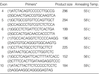
Each of p53 exons 4-10 was amplified by PCR. The primers and the cycling conditions for each exon are listed in Table 1. Each PCR reaction was generally performed under standard conditions in a 10 L reaction mixture containing 1 L of tem-plate DNA, 0.4 M of each primer, 125 M of each dNTP, 1.5 mM MgCl2, 0.4 unit of Taq polymerase, 0.5 Ci of [32P] dCTP (Amersham, Buckinghamshire, U.K.), and 1 L of 10 ×buffer. The reaction mixture was predenatured for 12 min at 95℃and amplified for 35 cycles (denaturing for 30 sec at 95℃, annealing for 30 sec at 55-59℃, and extending for 30 sec at 72℃). Final extension was continued for 5 min at 72℃. PCR products were denatured for 5 min at 95℃at a 1:1 dilu-tion of formamide loading dye containing 95% formamide, 20 mM/L EDTA, 0.05% bromophenol blue, and 0.05% xylene cyanol and were loaded onto a MDE gel (AT Biochem, Mal-vern, PA, U.S.A.) with 10% glycerol. After electrophoresis, the gels were transferred to 3-mm Whatman paper and autora-diography was performed using Kodak X-OMAT film (East-man Kodak, Rochester, NY, U.S.A.). For the detection of
4 (1)ATCTACAGTCCCCCTTGCCG 296 55℃ (2)GCAACTGACCGTGCAAGTCA 5 (1)GCTGCCGTGTTCCAGTTGCT 294 58℃ (2)CCAGCCCTGTCGTCTCTCCA 6 (1)GGCCTCTGATTCCTCACTGA 199 55℃ (2)GCCACTGACAACCACCCTTA 7 (1)TGCCACAGGTCTCCCCAAGG 196 56℃ (2)AGTGTGCAGGGTGGCAAGTG 8 (1)CCTTACTGCCTCTTGCTTCT 225 55℃ (2)ATAACTGCACCCTTGGTCTC 9 (1)GCCTCAGATTCACTTTTATCACC 152 56℃ (2)CTTTCCACTTGATAAGAGGTCCC 10 (1)ATACTTACTTCTCCCCCTCCTC 184 59℃ (2)AGGAAGGCAGGGGAGTAG
*(1), sense primer; (2), antisense primer.
Exon Primers* Product size Annealing Temp.
mutations, DNAs showing mobility shifts were cut out from the dried gels and reamplified for 30 cycles using the same primer set. Sequencing of the PCR products was carried out using the cyclic sequencing kit (Perkin-Elmer, Foster City, CA, U.S.A.) according to the manufacturer’s recommenda-tions. All mutations were verified through triplicated exper-iments including PCR-SSCP and sequencing analysis, which ensured the specificity of the results.
Statistical analysis
Chi-square test was used for the statistical analysis. Two-sided p-value of less than 0.05 was considered as statistically significant.
The sensitivity of p53 mutation was the percentage of IHC-positive tumors among tumors with identified mutation. The specificity was the percentage of IHC-negative tumors among tumors without p53 mutation. The predictive value of positive IHC was the percentage of IHC-positive tumors, which had p53 mutation. The predictive value of negative IHC was the percentage of IHC-negative tumors, which lacked p53 muta-tion. Accuracy was the percentage of tumors correctly classified for p53 mutation by IHC.
RESULTS
Immunohistochemical analysis of p53
Of the 36 HCC cases, 3 had ≥50% of tumor cells stained for p53, 8 had 10-49% of tumor cells stained for p53, 4 had 5-9% of tumor cells stained for p53, 10 had 1-4% of tumor cells stained for p53, and 11 did not stained for p53. No nucle-ar staining was found in nontumorous liver tissues or normal controls.
Fifteen out of 36 HCCs (41.7%) had ≥5% of tumor cells stained for p53: 13 out of 27 HBsAg seropositive cases (48.1%), 1 out of 5 anti-HCV antibody seropositive cases (20%), and 1 out of 4 both HBsAg and anti-HCV antibody seronegative cases (25%). The p53 was overexpressed more frequently in HCCs with poorer histological grade of differentiation
(Ed-mondson I, 0%; Ed(Ed-mondson II, 36.3%; Ed(Ed-mondson III, 57.9%; p=0.039).
DNA sequencing analysis
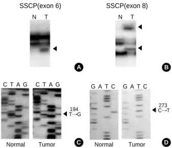
We detected 7 somatic mutations out of 36 HCCs (19.4%) consisting of 6 missense and 1 nonsense mutations (Table 2). These mutations were found widely throughout exons 4-8 without any mutational hot spot. No mutation was detected in codon 249. Two representative cases with aberrant bands and mutations are shown in Fig. 1. Nontumorous liver tissues from all 36 cases showed no mutation.
Mutations were found in 5 HBsAg seropositive cases, 1 HCV antibody seropositive case, and 1 both HBsAg and anti-HCV antibody seronegative case. Mutations were frequently found in HCCs with poorer histological grade of differentiation (Edmondson I, 0%; Edmondson II, 18.2%; Edmondson III 26.3%; p>0.05).
Correlation of p53 mutation and p53 overexpression Among the 7 mutations, 6 missense mutations were detect-ed in 15 HCCs with ≥5% immunoreactive tumor cells and one nonsense mutation was in 21 HCCs with <5% immunore-active tumor cells (40% vs 4.8%, p=0.027). The sensitivity for p53 mutation was 85.7% (6/7), the specificity 69.0% (20/29), the predictive value of positive IHC 40.0% (6/15), the predictive value of negative IHC 95.2% (20/21), and the accuracy 72.2% (26/36).
No missense mutation was detected in all 21 cases with <5% immunoreactive tumor cells but 2 missense mutations were detected in 25 cases with <10% immunoreactive tumor cells.
10 4 111 CTG→CGG Leu→Arg Transversion HCV 11 6 194 CTT→CGT Leu→Arg Transversion -12 5 175 CGC→CTC Arg→Leu Transversion HBV 22 5 158 CGC→CAC Arg→His Transition HBV 30 8 280 AGA→TGA Arg→Stop Transversion HBV 33 8 273 CGT→TGT Arg→Cys Transition HBV 35 6 214 CAT→CGT His→Arg Transition HBV
Exon Codon Case No. Nucleotide change Amino acid change Type of mutation Hepatitis* virus
Table 2.Summary of p53 mutations in 36 cases of hepatocel-lular carcinoma
*HBV, HBsAg(+); HCV, anti-HCV antibody(+).
Fig. 1.PCR-SSCP of exon 6 and 8 of the p53 shows abnormally shifted bands (arrowheads) in A(case 11) and B(case 33) (N, normal; T, tumor). Sequencing reveals CTT→CGT at codon 194 in case 11 (C) and CGT→TGT at codon 273 in case 33 (D).
A B C D ◀ ◀ ◀ ◀ SSCP(exon 6) Normal Tumor 194 T→G ◀ 273 C→T Tumor Normal SSCP(exon 8) N T C T A G C T A G G A T C G A T C N T
If we regarded the p53 IHC as positive when ≥10% of tumor cells were stained, the sensitivity for p53 mutation was 57.1% (4/7), the specificity 75.9% (22/29), the predictive value of positive IHC 36.4% (4/11), the predictive value of negative IHC 88.0% (22/25), and the accuracy 72.2% (26/36). If we regarded the p53 IHC as positive based on the presence of any immunoreactive tumor cells, the sensitivity for p53 mutation was 85.7% (6/7), the specificity 34.5% (10/29), the predictive value of positive IHC 24.0% (6/25), the predictive value of negative IHC 90.9% (10/11), and the accuracy 44.4% (16/36).
DISCUSSION
The p53 consists of 11 exons. Greenblatt et al. (2) had iden-tified 50 studies in which sequencing of the entire coding region of p53 in human cancers was reported. Of the 560 mutations reported in these papers, 87% were in exons 5-8 and most of the others were in exon 4 (8%) and exon 10 (4%). Mutations outside exons 5-8 were most frequent in urinary bladder carcinoma (28%) and HCC (24%). There have been six studies in which sequencing of the entire coding region of p53 in HCCs was reported (6, 17, 20-23). All of the 68 mutations reported in these papers were in exons 4-10. There-fore, we analyzed nucleotide sequences of the p53 in exons 4-10.
The frequency of p53 mutations has been reported to differ in HCCs from various geographic areas: 50-58% in southern China and southern Africa, 15-32% in Japan, and 15.4% in Germany (3, 5, 6, 8, 24). In this study, we showed that p53 mutation in HCCs in Korean people, at a frequency of 19.4%, is not uncommon. The not uncommon frequency of p53 mutations might represent only one of the multiple steps involved in hepatocarcinogenesis, as in other human cancers (1). In addition, no mutation was detected at codon 249 in the present study. This is consistent with the low prevalence of mutations at codon 249 reported in geographic areas where aflatoxin B1is not detectable at high levels in the diet, such as U.S.A., Germany, France, Britain, Taiwan, Australia, Japan, and Korea (6-9, 12-14, 25-27). This finding suggests that factors other than aflatoxin B1may be responsible for the p53 abnormalities in HCCs. The mutations in HCCs in low afla-toxin exposure areas often show a wide distribution over the highly conserved region covering exons 4-10 (6). The findings of the present study are also in line with this observation, with mutations scattered over exons 4-8.
We have found that overexpression of p53 in HCCs was correlated with the mutation of the p53 (p=0.027). However, there was substantial discrepancy between molecular genetic alterations in p53 and overexpression of the protein. In p53 immunohistochemical studies on HCCs, there have been different threshold values above which the result is regarded positive: presence of any labeled cells, 2%, 5%, 10%, or 50% immunoreactivity (28-32). Volkmann et al. (33) regarded the
p53 IHC as positive based on the presence of any active cells and reported that one HCC with a few immunore-active cells had wild-type p53. Volkmann et al. (34) regarded the p53 IHC as positive based on the presence of any immu-noreactive cells and reported that one out of eight HCCs with 1-9% immunoreactive cells showed missense mutation of p53. Hsia et al. (35) regarded 10% immunoreactivity as the thresh-old value and reported that one out of four HCCs with 1-9% immunoreactive cells showed missense mutation of p53.
Our findings showed that HCCs with ≥5% immunore-active tumor cells are the ones in which p53 mutations are most frequently present. No missense mutation was detected in all 21 cases with <5% immunoreactive tumor cells but 2 missense mutations were detected in 25 cases with <10% immunoreactive tumor cells. Predictive values of both positive IHC and negative IHC were higher in ≥5% overexpression group than in ≥10% overexpression group or >0% over-expression group. The specificity for p53 mutation in >0% overexpression group was very low. These findings suggest that 5% immunoreactivity is a reliable immunohistochemical threshold value to detect p53 mutations in HCCs. It should be noted that our findings might not be applicable to other tumor types in which discordance between IHC and molecular genetic analysis of p53 is more frequent.
These results suggest that 5% immunoreactivity is a reli-able immunohistochemical threshold value to detect p53 mutations in HCCs and the spectrum of p53 mutations in HCCs in Korean people is different from that of high aflatoxin B1exposure areas.
REFERENCES
1. Levine AJ, Momand J, Finlay CA. The p53 tumour suppressor gene.
Nature 1991; 351: 453-6.
2. Greenblatt MS, Bennett WP, Hollstein M, Harris CC. Mutations in the
p53 tumor suppressor gene: clues to cancer etiology and molecular pathogenesis. Cancer Res 1994; 54: 4855-78.
3. Bressac B, Kew M, Wands J, Ozturk M. Selective G to T mutations of
p53 gene in hepatocellular carcinoma from southern Africa. Nature 1991; 350: 429-31.
4. Hsu IC, Metcalf RA, Sun T, Welsh JA, Wang NJ, Harris CC.
Muta-tional hotspot in the p53 gene in human hepatocellular carcinomas. Nature 1991; 350: 427-8.
5. Scorsone KA, Zhou YZ, Butel JS, Slagle BL. p53 mutations cluster
at codon 249 in hepatitis B virus-positive hepatocellular carcinomas from China. Cancer Res 1992; 52: 1635-8.
6. Nishida N, Fukuda Y, Kokuryu H, Toguchida J, Yandell DW, Ikenega M, Imura H, Ishizaki K. Role and mutational heterogeneity of the p53
gene in hepatocellular carcinoma. Cancer Res 1993; 53: 368-72.
7. Buetow KH, Sheffield VC, Zhu M, Zhou T, Shen FM, Hino O, Smith M, McMahon BJ, Lanier AP, London WT, Redeker AG, Govindarajan S. Low frequency of p53 mutations observed in a diverse collection of
89: 9622-6.
8. Kress S, Jahn UR, Buchmann A, Bannasch P, Schwarz M. p53
muta-tions in human hepatocellular carcinomas from Germany. Cancer Res 1992; 52: 3220-3.
9. Debuire B, Paterlini P, Pontisso P, Basso G, May E. Analysis of the p53
gene in European hepatocellular carcinomas and hepatoblastomas. Oncogene 1993; 8: 2303-6.
10. Moon HY, Moon YM, Han KH, Chun JY, Kang JK, Park IS. Clinical
aspect and prognosis according to the infection type of hepatitis virus in primary liver cancer. Korean J Med 1994; 47(Suppl): 33.
11. Nam KW, Kim SJ. Clinical studies on 148 cases of primary
hepatocel-lular carcinoma. Korean J Gastroenterol 1987; 19: 184-93.
12. Park YM, Yoo YD, Paik SY, Kim BS, Tabor E. Mutation of tumor
suppressor gene p53 in hepatocellular carcinomas from Korea. Exp Mol Med 1996; 28: 173-9.
13. Jeong JH, Lee KS, Cho YH. A study of the p53 tumor suppressor gene
mutations in hepatocellular carcinomas in Korean patients. J Korean Surg Soc 1998; 55: 726-36.
14. Park NH, Chung YH, Youn KH, Song BC, Yang SH, Kim JA, Lee HC, Yu E, Lee YS, Lee SG, Kim KW, Suh DJ. Close correlation of p53
mutation and microvascular invasion in hepatocellular carcinoma. J Clin Gastroenterol 2001; 33: 397-401.
15. Hall PA, Lane DP. p53 in tumor pathology: can we trust
immunohis-tochemistry?-Revisited! J Pathol 1994; 172: 1-4.
16. Baas IO, Mulder JW, Offerhaus GJ, Vogelstein B, Hamilton SR. An
evaluation of six antibodies for immunohistochemistry of mutant p53 gene product in archival colorectal neoplasms. J Pathol 1994; 172: 5-12.
17. Bourdon JC, D’Errico A, Paterlini P, Grigioni W, May E, Debuire B.
p53 protein accumulation in European hepatocellular carcinoma is not always dependent on p53 gene mutation. Gastroenterology 1995; 108: 1176-82.
18. Edmondson HA, Steiner PE. Primary carcinoma of the liver. A study
of 100 cases among 48900 necropsies. Cancer 1954; 7: 462-503.
19. Sato T, Tanigami A, Yamakawa K, Akiyama F, Kasumi F, Sakamoto G, Nakamura Y. Allelotype of breast cancer: cumulative allele losses
promote tumor progression in primary breast cancer. Cancer Res 1990; 50: 7184-9.
20. Kazachkov Y, Khaoustov V, Yoffe B, Solomon H, Klintmalm GB, Tabor E. p53 abnormalities in hepatocellular carcinoma from United
States patients: analysis of all 11 exons. Carcinogenesis 1996; 17: 2207-12.
21. Honda K, Sbisa E, Tullo A, Papeo PA, Saccone C, Poole S, Pignatelli M, Mitry RR, Ding S, Isla A, Davies A, Habib NA. p53 mutation is a
poor prognostic indicator for survival in patients with hepatocellular carcinoma undergoing surgical tumour ablation. Br J Cancer 1998; 77: 776-82.
22. Karachristos A, Liloglou T, Field JK, Deligiorgi E, Kouskouni E, Spandidos DA. Microsatellite instability and p53 mutations in
hepa-tocellular carcinoma. Mol Cell Biol Res Commun 1999; 2: 155-61.
23. Wong N, Lai P, Pang E, Fung LF, Sheng Z, Wong V, Wang W, Hayashi Y, Perlman E, Yuna S, Lau JW, Johnson PJ. Genomic
aber-rations in human hepatocellular carcinomas of differing etiologies. Clin Cancer Res 2000; 6: 4000-9.
24. Nose H, Imazeki F, Ohto M, Omata M. p53 gene mutations and 17p
allelic deletions in hepatocellular carcinoma from Japan. Cancer 1993; 72: 355-60.
25. Challen C, Lunec J, Warren W, Collier J, Margaret F, Bassedine MF.
Analysis of the p53 tumour suppressor gene in hepatocellular carci-nomas from Britain. Hepatology 1992; 16: 1362-6.
26. Hosono S, Chou MJ, Lee CS, Shih C. Infrequent mutation of p53
gene in hepatitis B virus positive primary hepatocellular carcinomas. Oncogene 1993; 8: 491-6.
27. Vesey DA, Hayward NK, Cooksley WG. p53 gene in hepatocellular
carcinomas from Australia. Cancer Detect Prev 1994; 18: 123-30.
28. Zhao M, Zhang NX, Laissue JA, Zimmermann A.
Immunohistochem-ical analysis of p53 protein overexpression in liver cell dysplasia and in hepatocellular carcinoma. Virchows Arch 1994; 424: 613-21.
29. Cohen C, DeRose PB. Immunohistochemical p53 in hepatocellular
carcinoma and liver cell dysplasia. Mod Pathol 1994; 7: 536-9.
30. Lunn RM, Zhang YJ, Wang LY, Chen CJ, Lee PH, Lee CS, Tsai WY, Santella RM. p53 mutations, chronic hepatitis B virus infection, and
aflatoxin exposure in hepatocellular carcinoma in Taiwan. Cancer Res 1997; 57: 3471-7.
31. Mise K, Tashiro S, Yogita S, Wada D, Harada M, Fukuda Y, Miyake H, Isikawa M, Izumi K, Sano N. Assessment of the biological
malig-nancy of hepatocellular carcinoma: relationship to clinicopathologi-cal factors and prognosis. Clin Cancer Res 1998; 4: 1475-82.
32. Caruso ML, Valentini AM. Overexpression of p53 in a large series of
patients with hepatocellular carcinoma: a clinicopathological corre-lation. Anticancer Res 1999; 19: 3853-6.
33. Volkmann M, Hofmann WJ, Muller M, Rath U, Otto G, Zentgraf H, Galle PR. p53 overexpression is frequent in European hepatocellular
carcinoma and largely independent of the codon 249 hot spot mutation. Oncogene 1994; 9: 195-204.
34. Volkmann M, Schiff JH, Hajjar Y, Otto G, Stilgenbauer F, Fiehn W, Galle PR, Hofmann WJ. Loss of CD95 expression is linked to most but
not all p53 mutants in European hepatocellular carcinoma. J Mol Med 2001; 79: 594-600.
35. Hsia CC, Nakashima Y, Thorgeirsson SS, Harris CC, Minemura M, Momosaki S, Wang NJ, Tabor E. Correlation of immunohistochemical
staining and mutations of p53 in human hepatocellular carcinoma. Oncol Rep 2000; 7: 353-6.