INTRODUCTION
Neonatal hypoxic-ischemic brain injury (HI) remains one of the most common causes of death and severe neurologic morbidity in infants and children, resulting in chronic cogni-tive, motor, and sensory deficits.1,2 However, currently, there is
no therapy to repair the HI except therapeutic hypothermia, which offers very limited functional recovery.3,4 Consequently,
the development of supplementary or alternative therapies, that act synergistically with hypothermia or are effective when used alone, are needed to reduce the mortality and morbidity in
Cellular Response of Ventricular-Subventricular
Neural Progenitor/Stem Cells to Neonatal
Hypoxic-Ischemic Brain Injury and
Their Enhanced Neurogenesis
Jeong Eun Shin
1, Haejin Lee
2, Kwangsoo Jung
1, Miri Kim
2, Kyujin Hwang
3,
Jungho Han
1, Joohee Lim
1, Il-Sun Kim
1, Kwang-Il Lim
4, and Kook In Park
1,2,31Division of Neonatology, Severance Children’s Hospital, Department of Pediatrics, Yonsei University College of Medicine, Seoul; 2Yonsei Biomedical Research Institute, Yonsei University College of Medicine, Seoul;
3Brain Korea 21 Plus Project for Medical Science, Yonsei University College of Medicine, Seoul; 4Department of Chemical and Biological Engineering, Sookmyung Women’s University, Seoul, Korea.
Purpose: To elucidate the brain’s intrinsic response to injury, we tracked the response of neural stem/progenitor cells (NSPCs) located in ventricular-subventricular zone (V-SVZ) to hypoxic-ischemic brain injury (HI). We also evaluated whether transduc-tion of V-SVZ NSPCs with neurogenic factor NeuroD1 could enhance their neurogenesis in HI.
Materials and Methods: Unilateral HI was induced in ICR neonatal mice. To label proliferative V-SVZ NSPCs in response to HI, bromodeoxyuridine (BrdU) and retroviral particles encoding LacZ or NeuroD1/GFP were injected. The cellular responses of NSPCs were analyzed by immunohistochemistry.
Results: Unilateral HI increased the number of BrdU+ newly-born cells in the V-SVZ ipsilateral to the lesion while injury reduced
the number of newly-born cells reaching the ipsilateral olfactory bulb, which is the programmed destination of migratory V-SVZ NSPCs in the intact brain. These newly-born cells were directed from this pathway towards the lesions. HI significantly increased the number of newly-born cells in the cortex and striatum by the altered migration of V-SVZ cells. Many of these newly-born cells differ-entiated into active neurons and glia. LacZ-expressing V-SVZ NSPCs also showed extensive migration towards the non-neurogenic regions ipsilateral to the lesion, and expressed the neuronal marker NeuN. NeuroD1+/GFP+ V-SVZ NSPCs almost differentiated into
neurons in the peri-infarct regions.
Conclusion: HI promotes the establishment of a substantial number of new neurons in non-neurogenic regions, suggesting in-trinsic repair mechanisms of the brain, by controlling the behavior of endogenous NSPCs. The activation of NeuroD1 expression may improve the therapeutic potential of endogenous NSPCs by increasing their neuronal differentiation in HI.
Key Words: Neural stem cells, hypoxia-ischemia, brain, cell proliferation, cell movement, cell differentiation
pISSN: 0513-5796 · eISSN: 1976-2437
Received: February 20, 2020 Revised: April 10, 2020 Accepted: April 18, 2020
Corresponding author: Kook In Park, MD, PhD, Division of Neonatology, Sever-ance Children’s Hospital, Department of Pediatrics, Yonsei University College of Medicine, 50-1 Yonsei-ro, Seodaemun-gu, Seoul 03722, Korea.
Tel: 82-2-2228-2059, Fax: 82-2-393-9118, E-mail: [email protected] •The authors have no potential conflicts of interest to disclose. © Copyright: Yonsei University College of Medicine 2020
This is an Open Access article distributed under the terms of the Creative Com-mons Attribution Non-Commercial License (https://creativecomCom-mons.org/licenses/ by-nc/4.0) which permits unrestricted non-commercial use, distribution, and repro-duction in any medium, provided the original work is properly cited.
Yonsei Med J 2020 Jun;61(6):492-505 https://doi.org/10.3349/ymj.2020.61.6.492
newborn infants with HI.
It is conventionally accepted that neuronal generation does not take place beyond fetal life, and therefore, neuronal regen-eration does not occur in the vast majority of the post-develop-mental “non-neurogenic” central nervous system (CNS) after injury or disease.5 However, neurogenesis from neural
stem/pro-genitor cells (NSPCs) in two restricted neurogenic niches—ven-tricular-subventricular zone (V-SVZ) near the lateral ventricles and hippocampal subgranular zone—of the mammalian CNS has great implications for the growing field of research on en-dogenous NSPCs in the mature nervous system.6-16
Manipula-tion of these actively dividing or relatively quiescent NSPCs could supply cell population with immense neuroregenerative potential. Stimulated endogenous NSPCs could be a source of new neurons or glial cells that replace lost neural cells in CNS diseases. Notably, the V-SVZ peaks in size during the first week of postnatal development in rodents and at approximately the 35th week of gestation in humans as a result of extensive cell proliferation.17 The human neonatal V-SVZ has a remarkable
neurogenic capacity,18,19 suggesting that NSPCs in V-SVZ may
provide an endogenous cell population for neural regeneration after neonatal brain injury.
As a model for HI, we employed the Rice-Vannucci model20
which has been most well-accepted as rendering one of the experimentally destructive injuries: permanently ligating uni-lateral common carotid artery on postnatal day 7 (P7) followed by hypoxia (8–10% O2). This procedure results in extensive HI
that severely damages the ipsilateral cerebral hemisphere, in-cluding non-neurogenic regions such as the sensorimotor cor-tex, subcorcor-tex, and striatum, while preserving the contralateral hemisphere as an intact control. We believed that such a model would support an ideal opportunity to estimate the response of endogenous NSPCs to a devastating, clinically-relevant se-vere HI.
In this study, we tracked the response of endogenous V-SVZ NSPCs to a severe clinically relevant neonatal HI to investigate the brain’s intrinsic repair mechanism to injury, by using ret-roviral vectors transducing the LacZ reporter gene and by bromodeoxyuridine (BrdU) to label proliferative NSPCs. In addition, we also evaluated whether a retroviral transduction of V-SVZ NSPCs with neurogenic gene NeuroD1 could en-hance their neurogenesis in HI, which may enen-hance therapeu-tic potential of endogenous NSPCs in CNS disorders.
MATERIALS AND METHODS
Induction of unilateral HI brain injuryThrough a ventral midline neck incision, the right common carotid artery of anesthetized ICR (CD1) mice on postnatal day 7 (P7) was permanently ligated with the surgical suture silk. After the incision was closed, the animals were kept warm until awake and returned to their dams for 3 h. The pups were
then placed in an acrylic chamber with hypoxic atmosphere of 92% N2 and 8% O2 for 2.2 h. The animals’ body temperature
was maintained at 37°C. BrdU labeling and detection
Two hours after the induction of unilateral HI, the mice were intraperitoneally administered 50 mg/kg bromodeoxyuridine (BrdU; Sigma, St. Louis, MO, USA) every 4 h for the subse-quent 12 h (four administrations total). The animals were sac-rificed 24 h (n=3), 48 h (n=3), 1 wk (n=7), 2 wk (n=5), and 3 wk (n=5) after the final BrdU “pulse” and processed for BrdU de-tection. In some experiments designed to assess the long-term in vivo survival and permanence of BrdU-intercalated cells over months, daily “pulses” of BrdU for a week post-HI were performed. At the pre-determined timepoints for analysis, the animals were deeply anesthetized and perfused transcardially with 0.1 M PIPES buffer (pH 6.9; Sigma) containing 4% para-formaldehyde. The brain was subsequently post-fixed in the perfusing solution overnight at 4°C, cryoprotected overnight in phosphate-buffered saline (PBS) containing 30% sucrose at 4°C, and then embedded in O.C.T compound medium (Sakura Finetek, Torrance, CA, USA). Serial brain coronal cryostat sec-tions (15-μm thick) were treated initially with 2N HCl for 30 min at 37°C to denature cellular DNA and then reacted with a Fluorescein isothiocyanate (FITC)-conjugated mouse anti-BrdU antibody and/or a mouse anti-anti-BrdU antibody (Boeh-ringer Mannheim, Indianapolis, IN, USA) followed by a bi-otinylated-horse anti-mouse secondary antibody (Vector Laboratories, Burlingame, CA, USA), in order to identify Br-dU-labeled cells.
Injection of retrovirus and detection
Retrovirus vector particles encoding lacZ were collected from the supernatants of confluent ψ2 BAG helper virus-free pack-aging cells for 48 h at 37°C, filtered through 0.45 μm pores, ali-quoted, and stored at -80°C.21 The BAG virus stock (8×10⁷
G418-resistant CFU/mL) was mixed with 8 μg/mL polybrene and trypan blue prior to gently expelling 2 μL through a finely drawn glass micropipette into each lateral ventricle of mice 2 h before the induction of HI. The brains were processed via standard methods described below and elsewhere for immu-nohistochemisty.22 Expression of the lacZ gene product, E. coli
β-galactosidase (βgal), was detected by rabbit βgal anti-body (1:1000; Cappel Laboratories, Cochranville, PA, USA) fol-lowed by Texas red (TR)-conjugated goat anti-rabbit IgG (1:200; Vector Laboratories) antibodies.
The pCLPIT GFP plasmid was used to produce the murine leukemia virus (MLV)-based retroviral vector expressing GFP.23
The pCAG-NeuroD1-IRES-GFP sequence was subcloned from CAG-NeuroD1-IRES-GFP plasmid (Addgene, Watertown, MA, USA) and replaced the PuroR-IRES-tTA/TRE-GFP sequence in the pCLPIT GFP plasmid to generate the mouse NeuroD1/GFP-expressing retroviral vector. Retrovirus vector particles
encod-ing GFP or NeuroD1/GFP were collected from the superna-tants of human embryonic kidney 293T cells and concentrated to about 108 TU/mL by ultracentrifugation.24 The virus stock
solution was injected into each lateral ventricle of mice being subjected to HI, and the brains were processed as described above. The expressions of GFP and NeuroD1 gene products were detected by rabbit or mouse anti-GFP (1:100; Thermo Fish-er Scientific, Waltham, MA, USA) and mouse anti-NeuroD1 (1:500; Abcam, Cambridge, MA, USA) antibodies, respectively. Species-specific secondary antibodies, conjugated with FITC (1:200; Vector Laboratories) or TR (1:200; Vector Laborato-ries), were used to detect the binding of primary antibodies. Immunohistochemistry
Immunohistochemistry (IHC) analysis25 employed the
anti-BrdU, anti-βgal, anti-GFP, and/or anti-NeuroD1 antibodies de-scribed above. Dual staining was performed with primary anti-bodies against the following immature, undifferentiated NSPC and established differentiated neural cell type-specific gens: NeuN (1:100; Chemicon, Billerica, MA, USA), anti-neurofilament (NF) (1:500; Sternberger, San Diego, CA, USA), Doublecortin (DCX) (1:50, Santa Cruz, CA, USA), anti-Olig2 (1:200; Millipore, Billerica, MA, USA), anti-CNP’ase (1:500, Sternberger), anti-glial fibrillary acidic protein (GFAP) (1:1000; Dako, Glostrup, Denmark), anti-nestin (1:500; PharMingen, San Diego, CA, USA). Nuclear c-fos and synaptic connection with neighboring neurons were detected by a rabbit polyclonal anti-c-fos (1:10000; Santa Cruz) and anti-synapsin 1 (1:500; Ther-mo Fisher Scientific) antibodies, respectively. Species-specific secondary biotinylated (Vector Laboratories), streptavidin TR-conjugated (1:200; Jackson, Cambridge, UK), FITC-TR-conjugated, or TR-conjugated antibodies were used to detect the binding of primary antibodies. Specimens were mounted using the Vecta-shield mounting medium with 4,6-diamino-2-phenylindole (DAPI; Vector Laboratories), and were analyzed by an immuno-fluorescence microscopy (BX51; Olympus, Center Valley, PA, USA) or a confocal laser scanning microscopy (LSM 700; Carl Zeiss, Oberkochen, Germany).
Quantification
Structures were sampled by selecting predetermined areas on each coronal section as described.26 Every 6th section was
se-lected from a coronal series through the striatum between the following points relative to bregma: +0.98 mm (genu of the cor-pus callosum) and +0.14 mm (anterior commissure crossing). Proliferating cells were ascertained by their BrdU incorporation, and their quantification was based on counts of BrdU-immuno-reactive cells using the optical dissector stereological method.27
BrdU+ cells were counted in three predetermined ventral,
lat-eral, and dorsal squares (50×50 μm) of the lateral ventricle wall on all selected sections. A rectangular area of the striatum (300× 600 μm) was selected at a 50-μm distance from the lateral ventricle wall and analyzed on each section. All BrdU+ nuclei
in these areas were counted. Every 12th section from a coro-nal series [spanning from the olfactory bulb (OB) and frontal cortex rostrally and progressing caudally through the entire tel-encephalon] was selected and processed for BrdU IHC. As de-picted,26 four predetermined areas (50×50 μm) in the rostral
migratory stream (RMS) and granule cell layer of the OB were analyzed on each section. All BrdU+ nuclei in these areas were
counted.
Statistical analysis
All statistical analyses were performed using SPSS version 23 (IBM Corp., Armonk, NY, USA). For statistical comparison of newly-born BrdU+ cell numbers, we performed two-way
ANO-VA to account for the effect of HI (non-injured vs. injured mice) and/or hemispheres (contralateral vs. ipsilateral hemi-sphere). Bonferroni post hoc analysis was sequentially per-formed for pairwise comparisons of groups at each time-points where appropriate. Sample sizes were calculated using power analyses with an α-level of 0.05 and a power of 0.8. Data are presented as the means±SEM. p<0.05 was considered sta-tistically significant.
RESULTS
Proliferation and migration patterns of BrdU-labeled endogenous NSPCs in response to unilateral HI Following unilateral HI, the proliferation and migration pat-terns of endogenous NSPCs in the periventricular regions of both hemispheres was assessed using the serial BrdU injec-tion method. BrdU was selectively and permanently incorpo-rated into the nuclear genomic material of all proliferating cells entering S-phase.10-14 HI induced a significantly increased
pro-liferation of BrdU-labeled V-SVZ NSPC population ipsilateral to the lesioned right side (Figs. 1 and 2). Furthermore, HI ap-peared to induce a significant redistribution of proliferative NSPCs ipsilateral to the lesioned right side (Fig. 1B, D, F, and H) compared to the grossly intact contralateral left side (Fig. 1A, C, E, and G) and uninjured control group (n=5) (Fig. 2). The accumulation of BrdU+ cells was actually most pronounced in
the dorsolateral wall of the lateral ventricles, particularly the anterior one-third of the V-SVZ (Fig. 1B, D, F, and H), adjacent to the infarction cavity (IC) (Fig. 1B, D, and F); and a relatively dense “stream” of newly-born cells (arrowhead in Fig. 1B) ori-ented towards and into the extensively injured cortex was now apparent (arrows in Fig. 1B, D, F, H, and F’). The actual num-ber of newly-born cells was, indeed, increased at the site of HI and its penumbra in the neocortex as well as in adjacent pa-renchymal areas, particularly in the striatum (Fig. 1D, F, F’, and Fig. 2). About 2–4 times as many BrdU+ cells were seen in
the ipsilateral cortex and striatum 24–48 h after BrdU pulse com-pared to uninjured controls [e.g., cortex: 1964.2±246.5 (mean± SEM) vs. 495.6±78.8, p<0.05; striatum: 2950.8± 331.8 vs. 657.6±
78.5, p<0.05, respectively] (Fig. 2).
As the migratory route from V-SVZ to OB via the RMS is so well-characterized developmentally programmed destination of migratory V-SVZ NSPCs in the intact brain,6,8,9,28 examining
it in the context of unilateral HI provides an ideal opportunity to assess the role HI might play in “shifting” migratory pat-terns. The normal fate for most of the cells born in V-SVZ, par-ticularly the anterior portion (“c-d” in the schematic in Fig. 1K and displayed in Fig. 1A-D), is to migrate rostrally along the RMS into the OB (“a-b” in the schematic in Fig. 1K and dis-played in Fig. 1I and J), where they differentiate into the
neu-rons.8,9,28-30 Such typical developmental program is evident in
the intact left hemisphere [e.g., few BrdU+ cells surrounding
the left lateral ventricle (LV) in Fig. 1A and C; many BrdU+ cells
in left OB (LOB) in Fig. 1I and J].
However, although more cells were actually born in the right V-SVZ ipsilateral to the lesion in response to HI (Fig. 1B and Fig. 2), significantly fewer BrdU+ cells (about 53% of
unin-jured controls) were present in the right RMS (Fig. 2) 1 wk after BrdU injection. In addition, the number of newly-born cells that actually reached the right OB (ROB) was reduced significantly (about 28% of uninjured controls and 51% of that observed
con-A B C D I E F H G J F‘ K a b c d e OB SVZ RMS
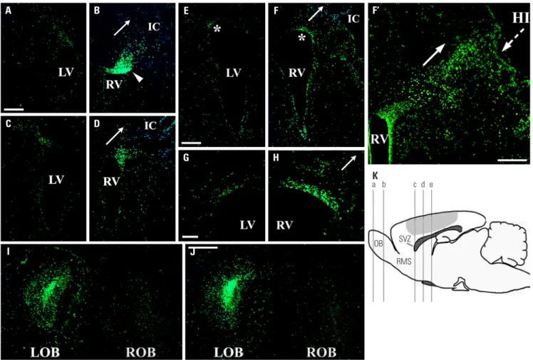
Fig. 1. Proliferation and migration patterns of BrdU-labeled endogenous NSPCs in response to unilateral HI. The schematic (K) shows the level of coronal section represented by each of following immunostained panels: the section through “a” is seen in (I); section through “b” is seen in (J); sections through “c” are seen in (A and B); sections through “d” are seen in (C and D); sections through “e” are seen in (E, F, and F’). The marked (*) areas in (E and F) are magnified in (G and H), respectively. The shaded region in (K) delineates the approximate area of injury in the right hemisphere [the side ipsilateral to (B, D, F, H, F’ and “ROB” in I and J)]. The normal fate of V-SVZ NSPCs is to migrate rostrally to the OB via the RMS. Accordingly, few of BrdU+ (green) V-SVZ
NSPCs (particularly the anterior portion) on the intact left side (e.g., A and C) remain there; rather most have appropriately left the V-SVZ and migrated out rostrally to and become integrated in the LOB in (I and J). Strikingly, on the side ipsilateral to the injury on the right, BrdU+ cells in the V-SVZ [e.g.,
arrow-head in (B)] appear to have been directed from migrating out rostrally to the ROB in (I and J) (where the number of BrdU+ cells is significantly diminished
compare with adjacent LOB), but rather have remained near the injured brain in and around the V-SVZ and adjacent cortex and striatum. Arrows in (B, D, F, H, and F’) points to the extensive adjacent areas of degeneration emanating from the IC. HI induced increased accumulation of BrdU+ V-SVZ NSPCs
ip-silateral to the lesion (B, D, F, H, and F’) compared to the grossly intact contralateral side (A, C, E, and G) and to the non-injured control group (not shown, see Fig. 2). BrdU+ cells were more pronounced in the lateral [particularly dorsolateral anterior one-third, e.g., (B and D)] wall of the ventricles adjacent to
and oriented (arrows) towards the IC. The density of BrdU+ cells entering the damaged cortex (epicenter of HI injury and its penumbra) appeared to be
in-creased [e.g., (B) compared to (A)]; there was also an increase in other adjacent areas: striatum [e.g., (D and F) compared to (C and E)] and cortex [e.g., (F’)]. Scale bars: 200 μm in (A and E); 100 μm in (G); 500 μm in (F’); 300 μm in (J). NSPCs, neural stem/progenitor cells; HI, hypoxic-ischemic brain injury; OB, olfactory bulb; ROB, right OB; RMS, rostral migratory stream; V-SVZ, ventricular-subventricular zone; LOV, left OB; IC, infarction cavity;LV, left lateral ven-tricle; RV, right lateral ventricle.
tralateral to the lesion, e.g., ipsilateral ROB compared to contra-lateral LOB in Fig. 1I, J, and Fig. 2), although the migration route remained anatomically intact. Interestingly, the number of newly-born cells that reached the RMS and OB from the V-SVZ contralateral to the lesion, though certainly much greater than that ipsilateral to the lesion, was also significantly reduced com-pared to the uninjured control mice (Fig. 2). This suggested that injury may draw cells from even distant regions, affecting the migratory pattern of V-SVZ-derived cells even in the grossly in-tact contralateral hemisphere. Indeed, the greater number of BrdU+ cells in the injured ipsilateral cortex contrasted with, and
may also come at the expense of, about 76% fewer BrdU+ cells
noted in the contralateral cortex compared to the age-matched uninjured controls (843.8±77.7 vs. 1117.0±121.9, p<0.05) (Fig. 2), suggesting that NSPCs which normally would have taken up residence in their contiguous cortex31 may be diverted
to-wards the lesion as the SVZ→RMS→OB axis was effected.
HI promotes the maintenance of cerebral nestin expression
Consistent with the view that HI may have a broader effect throughout the brain was the pattern of cerebral nestin ex-pression at various times—1 day (n=5), 1 wk (n=7), and 3 wk (n=5)—following unilateral HI, both at the site of injury and in the contralateral intact hemisphere. We analyzed the expres-sion of an intermediate filament protein nestin via IHC, which is known as NSPC and radial glia (embryonic NSPC during CNS development) markers.32-34 In contrast to the restricted
nestin-immunoreactivity (IR) bordering both lateral ventricles in the intact brain of age-matched control mice on P8 (Fig. 3F), robust nestin-IR was evident not only in the periventricu-lar regions of both lateral ventricles, but also in the septum, striatum, corpus callosum, external capsule, and cerebral cor-tices of both hemispheres, particularly at the right cortical in-farction site, 24 h after HI (Fig. 3A and B). In addition, the den-sity of nestin+ radial glia fibers with long processes appeared to
4000 3000 2000 1000 0 4000 3000 2000 1000 0 4000 3000 2000 1000 0 V-SVZ RMS OB Striatum Cortex V-SVZ RMS OB Striatum V-SVZ RMS OB Striatum Cortex 24 hours after BrdU injection
1 week after Brdu injection
48 hours after BrdU injection Ipsilateral side, non-injured control
Contralateral side, non-injured control Ipsilateral to injury
Contralateral to injury
Ipsilateral side, non-injured control Contralateral side, non-injured control Ipsilateral to injury
Contralateral to injury
Ipsilateral side, non-injured control Contralateral side, non-injured control Ipsilateral to injury Contralateral to injury BrdU + cells BrdU + cells BrdU + cells A C B *† *† *† † * * * *† *† *† *† *† * * * * * * *
Fig. 2. Density of BrdU-positive newly-born cells following unilateral hypoxic-ischemic brain injury. BrdU+ cells of V-SVZ, RMS, OB, striatum, and
cor-tex in non-injured control and injured animal at 24 h (A), 48 h (B), and 1 week after the final BrdU pulse (C). The number of BrdU+ cells was not
avail-able to count at the cortex at 1 week after BrdU injection. Data represent the means±SEM. *p<0.05 vs. the corresponding regions in non-injured con-trols, †p<0.05 vs. the contralateral side to the lesion in an injured animal. V-SVZ, ventricular-subventricular zone; RMS, rostral migratory stream; OB,
be highly increased in both hemispheres (arrowheads in Fig. 3A and B). The robust nestin-IR was maintained in HI site and cortical penumbral area of ipsilateral hemispheres 1 wk (Fig. 3D) and even 3 wk (data not shown) after HI. However, the nestin-IR in the contralateral hemisphere decreased, except the external capsule, 1 wk after HI (Fig. 3C) and almost disap-peared, except the dorsolateral SVZ adjacent to the LV, 3 wk af-ter HI (Fig. 3E). These results suggest that neonatal HI promotes
the maintenance of cerebral nestin expression and has the potential to maintain radial glia fibers after injury.
The hypothesis that such appearance of nestin+ NSPCs or
radial glia fibers (even in regions distant from the lesion) might reflect the rapid mobilization or recruitment of NSPCs to re-gions of cell loss was supported by a further observation of BrdU+/nestin+ cells around both SVZs (Fig. 4A-C) and within
both cortices (Fig. 4D-F). In addition, robust nestin-IR with
A B C D E F J I H G
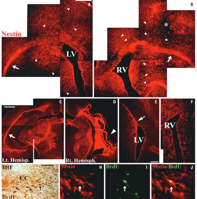
Fig. 3. Unilateral HI promoted the maintenance of cerebral nestin expression. Compared with restricted nestin (red) expression bordering the lateral ventricle in the intact brain of control mice on P8 (F), rapid and robust nestin expression was evident in the periventricular regions, septum, striatum, corpus callosum, external capsules (arrows in A and B), and cerebral cortices of both ipsilateral right (B) and contralateral left hemispheres (A), partic-ularly at the right cortical infarction site (* in B), 1 day after HI. Density of nestin+ radial glia fibers with long processes appeared to be highly increased
in both hemispheres (arrowheads in A and B). While nestin expression in the contralateral hemisphere decreased, except the external capsule (arrow in C), 1 week after HI and almost disappeared, except the dorsolateral SVZ adjacent the LV (the arrow in E), 3 weeks after HI, the robust nestin expres-sion was maintained in HI site (arrowhead in D) and cortical penumbral area of the right hemispheres 1 week and even 3 weeks (data not shown) after HI. Many of the BrdU+ cells (arrows in G, dark brown) in the CC close to IHF showed migration towards HI-injured hemisphere analyzed 1 week after HI.
Immunofluorescent labeling showed that migrating BrdU+ cells (arrow in I, green) in the CC bearing elongated leading processes towards affected
hemisphere were dual-labeled with nestin (arrows in H and J, red). Scale bars: 500 μm in (A, C, E); 100 μm in (G). HI, hypoxic-ischemic brain injury; LV, left lateral ventricle; RV, right lateral ventricle; CC, corpus callosum; IHF, interhemisphere fissue; SVZ, subventricular zone.
many BrdU+/nestin+ cells in the corpus callosum bearing
elon-gated leading processes oriented towards the affected hemi-sphere suggested the migration of NSPCs in response to HI (Fig. 3G-J). These findings suggest that nestin+ newly-born V-SVZ
cells migrate towards adjacent parenchyma and HI site, and that retained nestin+ radial glia fibers in response to HI may provide
a scaffold for the migration of V-SVZ-derived neurons and glia toward non-neurogenic brain regions in the postnatal brain.35 Differentiation pattern of BrdU-labeled NSPCs in response to HI
To help determine the differentiation fate in vivo of HI-gener-ated BrdU-labeled NSPCs, particularly in non-neurogenic re-gions, they were analyzed for their co-expression of neural cell type-specific antigens (Figs. 4 and 5). Over the 3-week period
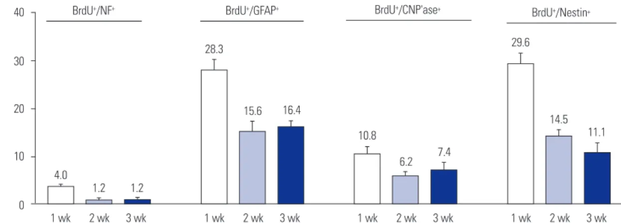
following the final BrdU pulse, many of the cells were induced to proliferate yielded new oligodendrocytes [10.8% (mean), 6.2%, and 7.4% at 1, 2, and 3 wk, respectively] (Fig. 4P-R and Fig. 5), astrocytes (28.3%, 15.6%, and 16.4%, respectively) (Fig. 4S-U and Fig. 5), and neurons (4.0%, 1.2%, and 1.2%, respec-tively; Fig. 4G-O and Fig. 5). These new neurons (likely an un-der-representation given the time course of BrdU pulses) were evident not only in the compromised hemisphere, but in the contralateral hemisphere as well, suggesting the widespread “ripple effect” of signals emanating from even a unilateral le-sion. In some experiments designed to assess even longer term survival of BrdU+ cells, daily ip BrdU pulses for a full week
of post-HI were performed. At 2 months after injury, about 1% of BrdU+ cells were NF+—not significantly diminished from
this value at 3 weeks—suggesting permanence of the new
neu-A B C A‘ H‘ D E F B‘ I‘ G H I C‘ J‘ J K L D‘ K‘ M N O E‘ L‘ P Q R F‘ M‘ S T U G‘ N‘
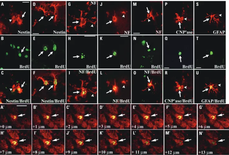
Fig. 4. Differentiation pattern of BrdU-labeled NSPCs in response to HI. A day after unilateral HI and the final BrdU pulse, many cells around the V-SVZ (A-C) and in the cortex (D-F) of both hemispheres showed co-localization of both anti-nestin (A, D) (red) and anti-BrdU (B, E) (green) immunoreac-tivity (arrows), appreciated in merged images (C, F, respectively). One-to-three weeks after HI, many of the BrdU+ cells [arrows in (H, K, N, Q, T)] had
dif-ferentiated into new neurons (G-O), oligodendrocytes (P-R), and astrocytes (S-U) predominantly in the injured hemisphere, supplementing non-BrdU+
[arrowheads in (G, I, J, L, M, O, P, R, S, U)] of the respective cell type with which they seamlessly intermixed, and were often directly juxtaposed. The new neurons, recognized here as NF+ (red) cells [arrows in (G, J, M) and under the dual-wavelength filter in (I, L, O)], were present in the neocortex—
predominantly ipsilateral [arrows in (G-I)] but even contralateral to the lesion [arrows in (J-L)]—and in the ipsilateral hippocampal CA1 area [arrows in (M-O)]—all classically regarded as non-neurogenic regions. The oligodendrocyte marker CNP’ase and the astrocyte marker GFAP were employed in (P) [and under the dual filter in (R)] and (S) [and under the dual filter in (U)], respectively. Arrowheads in (A, C) indicate cellular processes of Nestin+/
BrdU- immature cells. Confocal images showed that a BrdU+ cell (green) (arrows) colocalized with the mature neuronal marker NeuN (red) under the
dual-wavelength filter in (A’-N’). Scale bars: 10 μm in (A, D, H, K, M, A’); 5 μm in (Q, T). NSPCs, neural stem/progenitor cells; HI, hypoxic-ischemic brain injury; V-SVZ, ventricular-subventricular zone.
40 30 20 10 0 1 wk 2 wk 3 wk
BrdU+/NF+ BrdU+/GFAP+ BrdU+/CNP'ase+ BrdU+/Nestin+
4.0 1.2 1.2 28.3 15.6 16.4 10.8 6.2 7.4 29.6 14.5 11.1 1 wk 2 wk 3 wk 1 wk 2 wk 3 wk 1 wk 2 wk 3 wk % of BrdU + cells
Fig. 5. Cellular differentiation patterns of HI-generated BrdU+ newly-born cells in non-neurogenic regions. Counts represent the means and SEM of
the sums of every 6th 15-μm coronal section extending caudally from the rostral tip of the lateral ventricle for the entire expanse of both cerebral corti-ces (ipsilateral and contralateral to HI). HI, hypoxic-ischemic brain injury.
rons. Notably, these analyses were performed caudal to the OBs and rostral to the dentate gyrus of hippocampus to prevent confusion with neurons routinely generated locally in these neu-rogenic regions.5-14,16 Following HI, neurons are the neural cell
type most susceptible to elimination, yet they are the very cells deemed as incapable of being renewed in non-neurogen-ic regions. Given such expectations, the actual numbers were somewhat less consequential than the fact of observing any new neurons in areas that were heretofore regarded as harbor-ing no new neurons, and were considered representative of most of the postnatal brain. The proportion of endogenous NSPCs differentiating into various neural cell types—including the proportion remaining immature and undifferentiated—was consistent with prior studies quantifying the phenotype of pro-liferative cells following exogenous mitogen/growth factor infu-sions.36-38
Tracking of LacZ-expressing V-SVZ NSPCs in response to HI
As a complement to BrdU labeling and to more rigorously track the fate of these newly proliferative HI-responsive V-SVZ NSPCs (as well as to selectively label periventricular cells inde-pendent of local cortical parenchymal progenitors), mitotic cells were identified by their incorporation of a retroviral provi-rus encoding lacZ following infection by a retroviprovi-rus vector21
injected into the ventricles. A cell that was in S-phase at the time of exposure to the retrovirus indicated LacZ expression; therefore, LacZ expression, revealed by an antibody to βgal, provided a direct unambiguous tag for detecting, tracking, and determining the subsequent fate of that cell. The mice were sacrificed at least 1 wk after retrovirus injection into both later-al ventricles of mice being subjected to unilaterlater-al HI (n=5) (Fig. 6). In response to HI, βgal+ V-SVZ cells migrated into the
adjacent striatum (Fig. 6A) and hippocampus, into the cortex ipsilateral to the lesion (Fig. 6D), and into the cortical penum-bra (Fig. 6J). In addition, some LacZ-labeled V-SVZ (often in groups29,30) also migrated into the cortex (Fig. 6G) and overlying
hippocampal CA1 area (Fig. 6M). Providing a broader over-view under low magnification, LacZ-labeled V-SVZ NSPCs fol-lowing severe HI showed a robust radiating migratory pattern towards the overlying hippocampus and neocortex (Fig. 6P and Q). Confirming the observation noted in Fig. 6G-O, a sub-population of the newly proliferative and migratory βgal+ cells
now expressed mature neuronal marker (e.g., NeuN) in all of these non-neurogenic regions (Fig. 6C, F, I, L, O, and Q), sug-gesting de novo neurogenesis. The NeuN+ neurons were
sug-gested to be active, integrated, and activatable in concert with other regional neurons, based on their immunohistochemically detectable upregulation and expression of c-fos and synapsin in the living, behaving animal (Fig. 6R-Z), e.g., in cortex includ-ing the infarcted area (Fig. 6R-T, X-Z) and hippocampus (CA3) where 59.6±3.2% and 25.2±4.2% of LacZ+ cells were dual
im-munoreactive with NeuN and nestin, respectively (Fig. 6U-W). Tracking of NeuroD1/GFP-expressing V-SVZ NSPCs in response to HI
NeuroD1 is a basic helix-loop-helix (bHLH) transcriptional factor that is highly expressed in the developing neurons of pe-ripheral and CNS, supporting its role as a neuronal differenti-ation factor. NeuroD1 may contribute to multiple levels of neural development; nevertheless, it is believed that NeuroD1 mainly plays important roles in the terminal differentiation of postmitotic neuronal cells.39 Therefore, we asked if
NeuroD1-transduced V-SVZ NSPCs could enhance neuronal differentia-tion of internal pool of NSPCs and provide greater numbers of new neurons in non-neurogenic brain regions in response to HI. Retroviral vectors encoding NeuroD1/GFP and GFP were injected into both lateral ventricles of mice being subjected to unilateral HI (n=5 for each group) and analyzed at least 1–2 wk later (Fig. 7). In response to HI, NeuroD1+/GFP+ mitotic
V-SVZ NSPCs migrated into the adjacent striatum, subcortex, and cortical penumbra ipsilateral to the lesion (Fig. 7A, A’, B, B’), and along the external capsule and corpus callosum (Fig. 7A, A”, and B). The majority of newly-born and migratory GFP+
A D G J M B E H K N C F I L O R P Q S T U V W X Y Z
Fig. 6. Immunostaining of retrovirally-infected endogenous mitotic V-SVZ NSPCs with LacZ following HI. In response to HI, LacZ-expressing βgal+
cells (red) migrated widely, including into the striatum (A), the cortex ipsilateral to the lesion (D), and the cortical penumbra of the infarct (J). Some of these βgal+ cells (A, D, J) (red, arrows) expressed the mature neuronal marker NeuN (B, E, K, respectively) (green, arrows) with co-localization of
both antibodies visualized under a dual filter (C, F, L, respectively) (arrows), suggesting de novo neurogenesis. Arrowheads in (B, C) indicate neurons that were not the progeny of retrovirus-infected NSPCs. In the grossly intact contralateral hemisphere, there was also evidence for the migration and differentiation into NeuN+ neurons of some βgal+ cells in the neocortex (G-I) (arrows) and hippocampal CA1 region (M-O) (arrows) in response to
se-vere HI. These phenomena were further illustrated by a low magnification view of the cerebral cortex and hippocampus, in which a significant num-ber of βgal+ V-SVZ NSPCs that migrated to these regions (P) (red) differentiated into NeuN+ neurons (Q) (yellow/orange under a dual filter). Insets in (P)
and (Q) showed the representative βgal+ cell indicated by the arrow in (P) and (Q), respectively, further enlarged to illustrate the typical morphology of a
newly-born neuron derived from a labeled V-SVZ cell that had migrated to the neocortex following injury. (R-Z) The representative βgal+ cells in those
regions [red cells with arrows in (R) and (U, X)] were immunoreactive to an anti-synapsin 1 [green cell with the arrow in (S)] and c-fos nuclear [green cells with arrows in (V, Y)] antibodies, respectively, and visualized under a dual filter [cells with arrows in (T, W, Z)]. Scale bars: 50 μm in A; 5 μm in D, G, J; 10 μm in M, R, U, X; 100 μm in P. V-SVZ, ventricular-subventricular zone; NSPCs, neural stem/progenitor cells; HI, hypoxic-ischemic brain injury.
cells transduced with a retrovirus encoding GFP expressed as-trocyte marker GFAP (65.1±5.6%) (Fig. 7C-E”) and the oligo-dendroglial progenitor marker Olig2 (22.4±3.8%) (Fig. 7F-H’), whereas relatively few expressed early neuronal marker DCX (11.3±2.5%) in the striatum and cortical penumbral area (Fig. 7I-K’). In contrast, most of the newly-born and migratory GFP+
cells transduced with a retrovirus encoding NeuroD1/GFP did not express GFAP or Olig2, but expressed early neuronal mark-er DCX or mature neuronal markmark-er NeuN (95.1±7.4%) in all of these non-neurogenic brain regions (Fig. 7L-X), suggesting enhanced neurogenesis. In addition, very few of GFP+ cells
(1.3±0.7%) expressed nestin (data not shown).
DISCUSSION
This study sought to elucidate the brain’s intrinsic response to HI, particularly as it involves an endogenous V-SVZ NSPC pool and specifically in areas that are designated "non-neurogenic" and hence non-regenerative. Our findings suggest that
neo-natal HI alters developmental programs and does so towards the goal of repopulation and self-repair, allocating new cells to the regions of greatest need; during acute phases of neurode-generation, many factors, such as morphogens, growth factors, neurotrophic factors, erythropoietin, and microRNAs, are elab-orated to which endogenous NSPCs may respond to promote the establishment of new neurons, even within non-neuro-genic regions of the postnatal brain.17,40 Some of these
newly-born neurons, while generated from V-SVZ NSPCs, appear to be targeted specifically to damaged, neuron-deficient regions by redirecting otherwise stereotypical migratory patterns. Such observations may attest to an under-appreciated degree of plasticity and resilience in the mammalian CNS, even be-yond the classical periods and regions of developmental malle-ability. The CNS may, indeed, spontaneously attempt to repair itself with its own endogenous pool of NSPCs, but that supply may simply be insufficient either in number or in factors regu-lating proliferation, migration, and differentiation in the con-text of the most destructive injuries. Therefore, the net impact of the production of new nerve cells may be limited. It is
plau-sible that this type of homeostatic response occurs routinely at a marginally detectable level, and is effective under most circumstances (where CNS insults are modest—e.g., from in-fections, toxins, minor trauma) in preventing such events from coming to clinical attention. However, when the damage is mas-sive, the system—invoked but inadequate—is “overwhelmed.”
Consequently, there is no apparent recovery, although endog-enous NSPCs have responded in a “reparative” fashion to sig-nals directing them towards repopulation of the damaged area. Nevertheless, this does not negate the inherent plasticity of CNS nor the potential for augmenting this response. Therefore, the relationship between the severity of HI and NSPC response
re-A‘ A‘ B‘ B‘ A“ A“ A B C C‘ F I D D‘ G J E E‘ H K E” H‘ K‘ L O R U M P S V N Q T W T‘ X
Fig. 7. Immunostaining of retrovirally-infected endogenous mitotic V-SVZ NSPCs with NeuroD1/GFP and GFP following HI. In response to HI, Neu-roD1/GFP-expressing GFP+ cells (green) migrated into the adjacent striatum, subcortex, and cortical penumbra ipsilateral to the lesion (A, A’, B, B’),
and along the EC and CC (A, A”, B). The boxed areas in A and B are shown at high magnification in A’, A” and B’. The asterisk in A’ and arrowheads in A” indicates the infarction cavity and LV, respectively (C-K’). Many of the newly-born and migratory GFP+ cells (C, C’) transduced with a retrovirus
en-coding GFP expressed astrocyte marker GFAP (D, D’, respectively) (red) with co-localization of both antibodies visualized under a dual filter (E, E’, re-spectively) (yellow or orange) in the cortical penumbral area. Boxed regions in (C-E) are magnified in (C’-E’), respectively. Arrows in (C’-E”) indicate dual-labeled cells (F-H). Some of newly-born GFP+ cells (F) expressed the oligodendrocyte progenitor marker Olig2 (G) (red) with co-localization of both
antibodies visualized under a dual filter (H). Arrows in (F-H) indicate dual-labeled cells. (I-K) A few of newly-born GFP+ cells (I) expressed early neuronal
marker DCX (J) (red) with co-localization of both antibodies visualized under a dual filter (K). Confocal images showed that GFP+ cells (arrows in E”, H’ and
K’) colocalized with differentiation markers under a dual filter (yellow or orange) (L-X). In contrast, most of the newly-born and migratory GFP+ cells
[ar-rows and arrowheads in (R) and (U), respectively] transduced with a retrovirus encoding NeuroD1/GFP expressed early neuronal marker DCX [ar[ar-rows in (S)] (red) or mature neuronal marker NeuN [arrowheads in (V)] (red) with co-localization of both antibodies visualized under a dual filter [arrows in (T)] (yellow or orange) in the striatum and cortical penumbral area. DAPI+ cells [arrowheads in (W)] (blue) indicate dual-labeled cells. Confocal images
showed that GFP+ cells [arrows and arrowheads in (T’) and (X), respectively] colocalized with neuronal markers under a dual filter (yellow or orange).
However, GFP+ cells (L, O) did not express GFAP or Olig2 (M, N, P, Q). Scale bars: 500 μm in (A); 20 μm in (H’, N) 10 μm in (A”, E, E”, H). V-SVZ,
ventricular-subventricular zone; NSPCs, neural stem/progenitor cells; HI, hypoxic-ischemic brain injury; EC, external capsule; CC, corpus callosum; LV, left lateral ventricle; RV, right lateral ventricle.
mains to be explored. The prediction of NSPC response based on the level of severity of HI is an important issue that should be further investigated.
While it is clear that newly-born brain cells are being pro-duced in response to HI, data compiled to date suggest that the long-term survival of migrated neuroblasts or newly-born neu-rons in the injured area is not supported.41-43 The reasons for the
death of newly-born neurons remain elusive and may be due to the inhospitable post-HI environment. HI initiates an inflam-matory cascade, such as microglial activation, immune cells in-filtration, and the release of toxic proinflammatory molecules.44
Microglial activation, proinflammatory factors, and circulating monocytes associated with inflammation not only lead to neu-ron death but also affect the neurogenesis and survival of new-ly-born neurons.40 In addition, some studies have indicated that
SVZ-derived migratory NSPCs differentiate into reactive astro-cytes and contribute to astrocyte scar formation after stroke.45,46
While the role of astrogliosis after stroke or HI is still elusive and requires further investigation, the modulation of pathologic en-vironment after HI by anti-inflammatory or neurogenic factors results in substantial repair of neural tissue.47,48
Radial glial (RG) cells are embryonic neural stem cells express-ing nestin, GFAP, and astrocyte-specific glutamate transporter that extend apical long processes to the pial surface from their soma, which is located in the ventricular zone.33 RG cells are
not only stem cells and progenitors, but during brain develop-ment they also provide a scaffold along which newly generated immature neurons migrate from the periventricular zone into the neocortex and to subcortical structures. In the late fetal pe-riod in the human brain, and in the first postnatal week in the rodent brain, the RG fibers collapse, transform into astrocytes or ependymal cells, and normally disappear soon after birth.49
However, this study showed that signals generated in HI elic-ited robust cerebral nestin expression, and the density of nes-tin+ radial glia fibers with long processes appeared to be highly
increased in both hemispheres. Moreover, the robust nestin-IR was maintained in the lesioned and cortical penumbral ar-eas of ipsilateral hemispheres, even more than 3 weeks after HI. These findings suggest that retained nestin+ radial glia
fi-bers in response to HI may provide a scaffold for the wide-spread migration of V-SVZ-derived neurons toward non-neu-rogenic brain regions in the postnatal brain. Similarly, earlier studies reported an increase in cells expressing RG markers after chronic hypoxia in immature rats, and demonstrated the role of RG cells in both neurogenesis and migration.50,51 A recent
study also showed that RG fiber persisted in neonatal mouse brain after cryogenic injury and acted as a scaffold for the mi-gration of V-SVZ-derived neuroblasts to the lesion site, thereby enhancing neuronal regeneration and functional recovery from neonatal brain injuries.35
Whereas there has been a great increase of studies observ-ing neuronal replacement after neonatal HI, there have been fewer studies assessing the extent of oligodendroglial
replace-ment. This study, along with other reports,52-54 documented an
increase in the number of newly generated oligodendrocytes in the damaged hemispheres and suggested that these newly-born oligodendrocytes were produced by V-SVZ NSPCs. How-ever, none of these studies clearly established whether these newly-born oligodendrocytes proceeded to differentiate into mature myelin-producing cells. In this study, significant astro-gliosis was observed after HI, along with an increase in BrdU+/
GFAP+ cells. It is well-known that reactive astrocytes produce
extracellular matrix components, such as hyaluronic acid and chondroitin sulfate proteoglycan, that inhibit oligodendrocyte differentiation.55,56 In addition, astrocytes produce numerous
cytokines, including fibroblast growth factors, WNT ligands, endothelin-1, and Notch ligands, that inhibit oligodendrocyte progenitor cell maturation.17 There is also an alternative
expla-nation for the lack of myeliexpla-nation depending on SVZ cell speci-fication.57,58 However, further research is critically needed to
fa-cilitate the differentiation into mature, myelin-producing cells from V-SVZ NSPC-derived oligodendrocytes in HI.
Identifying the cues that promote the movement and differ-entiation of this endogenous pool of NSPCs during their nor-mal, developmentally-appropriate migratory course28-30,59,60
and, as we observed here, during their confrontation with neu-rodegeneration, may allow those factors to be provided thera-peutically at even more optimal, sustained levels, over broader periods and terrain. In fact, multiple cytokines, such as Delta/ Notch-1, erythropoietin, interleukin-6, leukemia inhibitory factors (LIF), transforming growth factor, and vascular endo-thelial growth factor (VEGF), were reported to be elaborated to which endogenous NSPCs respond to change in their pro-liferation, migration, and differentiation patterns after neona-tal HI. In addition, the treatment of erythropoietin, brain-de-rived neurotrophic factor, epidermal growth factor, VEGF, LIF, hyperbaric oxygen, or hypothermia has been shown to atten-uate HI by modulating the behavior of endogenous NSPCs.17,40
Lastly, we have demonstrated here that neurogenic transcrip-tional factor NeuroD1 can efficiently convert V-SNZ NSPCs to neurons, providing a new target for cellular conversion strate-gies aimed at enhancing neurogenesis. Therefore, augment-ing the endogenous NSPC population with exogenous neural stem cells,40 factors, and/or providing adequate synaptic
neigh-bors and guidance cues for connectivity and trophic support may enable more significant recovery.
BrdU labeling for mitotic cells is still the most commonly used of the thymidine analogues, and much work on this sub-ject has been done in mice. However, BrdU inhibited the ex-pansion of NSPCs, increased cell death, and repressed neuro-nal and oligodendroglial differentiation.61 Although BrdU
does obviously label dividing cells, BrdU positivity may be due to attempts of cells to repair themselves or a sign of cell death (apoptosis). In addition, BrdU has been well-known to have detrimental effects on chromosomes and DNA stability, as well as the cell cycle, cell differentiation, and survival.62 Besides the
toxicity of BrdU and caveats concerning its utility, BrdU label-ing, particularly at the epicenter of HI, can be hard to interpret due to the reliance on immunodetection in the injury setting, where significant labeling issues can arise. In addition, BrdU is subject to dilution effects, where the absence of labeling or decreased labeling over time can be misinterpreted. There-fore, we also utilized a retroviral vector to label mitotic NSPCs and their progeny, a technique that is not affected by the num-ber of cell divisions. Another advantage of using a retroviral approach is the ability to direct the location of injection into V-SVZ NSPCs. Gene transfer mediated by replication-incompe-tent retroviral vectors has played an important role in develop-mental studies, particularly in revealing lineage relationships among different cell types. Replication-incompetent retroviral vectors have also been used for gain-of-function studies to ex-plore the molecular mechanisms underlying cell differentia-tion in neural tissues of mice. The greatest advantage of this approach is that it can reveal cell-autonomous effects of a transgene, such as NeuroD1, on cell differentiation without dis-rupting the overall context of development.63 However,
gam-ma retroviral vectors have a propensity to integrate near regula-tory regions of active genes. The activity of promoter/enhancer elements within the vector long terminal repeats (LTRs) or ab-errant splicing events can lead to the activation of genes flank-ing the integration site. This is commonly referred to as inser-tional mutagenesis. If the genes near the integration sites are proto-oncogenes, it may lead to increased proliferation of clonal populations of cells. In addition, a number of studies have shown that the promoter regions of gamma retroviral vectors may be subject to epigenetic silencing, which has been associated with DNA methylation of CpG sequences. Such silencing events gradually reduce the expression of transgenes, such as LacZ, GFP, or NeuroD1, under the control of both viral LTR and in-ternal promoters.64 Taken together, it is difficult to find a single
method to accurately and dynamically track the behaviors of endogenous NSPCs in response to HI. Therefore, more sophis-ticated measures including the use of genetic mouse models or live molecular imaging would be required to avoid the com-mon mistake of decom-monstrating the behaviors of endogenous NSPCs in HI.
ACKNOWLEDGEMENTS
This study was supported by a faculty research grant of Yonsei University College of Medicine (6-2019-0096) and Bio & Medi-cal Technology Development Program of the National Research Foundation (NRF) funded by the Korean government (MSIT) (No. NRF-2019M3A9H1032791).
AUTHOR CONTRIBUTIONS
Conceptualization: Kook In Park. Data curation: Jeong Eun Shin,
Haejin Lee, and Kook In Park. Formal analysis: Jeong Eun Shin.
Funding acquisition: Jeong Eun Shin and Kook In Park. Investigation:
Jeong Eun Shin, Haejin Lee, Kwangsoo Jung, Miri Kim, Kyujin Hwang, and Il-Sun Kim. Methodology: Haejin Lee, Kwangsoo Jung, Miri Kim, Kyujin Hwang, and Kwang-Il Lim. Project administration: Kook In Park. Resources: Kook In Park. Supervision: Kook In Park. Validation: Joohee Lim, Jungho Han,Kook In Park. Visualization: Jeong Eun Shin.
Writing—original draft: Jeong Eun Shin and Kook In Park. Writing—
review & editing: Kook In Park. Approval of final manuscript: all
au-thors.
ORCID iDs
Jeong Eun Shin https://orcid.org/0000-0002-4376-8541 Haejin Lee https://orcid.org/0000-0003-1455-4189 Kwangsoo Jung https://orcid.org/0000-0001-7365-7247 Miri Kim https://orcid.org/0000-0002-0380-1677 Kyujin Hwang https://orcid.org/0000-0001-5193-5154 Jungho Han https://orcid.org/0000-0001-6661-8127 Joohee Lim https://orcid.org/0000-0003-4376-6607 Il-Sun Kim https://orcid.org/0000-0003-4033-4323 Kwang-Il Lim https://orcid.org/0000-0002-1895-2080 Kook In Park https://orcid.org/0000-0001-8499-9293
REFERENCES
1. Ferriero DM. Neonatal brain injury. N Engl J Med 2004;351:1985-95.
2. Volpe JJ. Neonatal encephalopathy: an inadequate term for hy-poxic-ischemic encephalopathy. Ann Neurol 2012;72:156-66. 3. Azzopardi DV, Strohm B, Edwards AD, Dyet L, Halliday HL,
Juszc-zak E, et al. Moderate hypothermia to treat perinatal asphyxial encephalopathy. N Engl J Med 2009;361:1349-58.
4. Shankaran S, Pappas A, McDonald SA, Vohr BR, Hintz SR, Yolton K, et al. Childhood outcomes after hypothermia for neonatal en-cephalopathy. N Engl J Med 2012;366:2085-92.
5. Ramon y Cajal S. Degeneration and regeneration of the nervous system. Oxford: Clarendon Press; 1928.
6. Altman J. Autoradiographic and histological studies of postnatal neurogenesis. IV. Cell proliferation and migration in the anterior forebrain, with special reference to persisting neurogenesis in the olfactory bulb. J Comp Neurol 1969;137:433-57.
7. Altman J, Das GD. Autoradiographic and histological evidence of postnatal hippocampal neurogenesis in rats. J Comp Neurol 1965; 124:319-35.
8. Goldman SA, Luskin MB. Strategies utilized by migrating neurons of the postnatal vertebrate forebrain. Trends Neurosci 1998;21: 107-13.
9. Lois C, Alvarez-Buylla A. Proliferating subventricular zone cells in the adult mammalian forebrain can differentiate into neurons and glia. Proc Natl Acad Sci U S A 1993;90:2074-7.
10. Kornack DR, Rakic P. Continuation of neurogenesis in the hippo-campus of the adult macaque monkey. Proc Natl Acad Sci U S A 1999;96:5768-73.
11. Gould E, Reeves AJ, Fallah M, Tanapat P, Gross CG, Fuchs E. Hip-pocampal neurogenesis in adult Old World primates. Proc Natl Acad Sci U S A 1999;96:5263-7.
12. Cameron HA, McKay RD. Restoring production of hippocampal neurons in old age. Nat Neurosci 1999;2:894-7.
13. Eriksson PS, Perfilieva E, Björk-Eriksson T, Alborn AM, Nordborg C, Peterson DA, et al. Neurogenesis in the adult human hippo-campus. Nat Med 1998;4:1313-7.
en-hances adult neurogenesis in the hippocampal formation. Nat Neurosci 1999;2:260-5.
15. Lim DA, Huang Y, Alvarez-Buylla A. Adult subventricular zone and olfactory bulb neurogenesis. Cold Spring Harb Perspect Biol 2008;52:175.
16. Kempermann G, Gage FH, Aigner L, Song H, Curtis MA, Thuret S, et al. Human adult neurogenesis: evidence and remaining ques-tions. Cell Stem Cell 2018;23:25-30.
17. Niimi Y, Levison SW. Pediatric brain repair from endogenous neural stem cells of the subventricular zone. Pediatr Res 2018;83: 385-96.
18. Paredes MF, James D, Gil-Perotin S, Kim H, Cotter JA, Ng C, et al. Extensive migration of young neurons into the infant human fron-tal lobe. Science 2016;354:aaf7073.
19. Sanai N, Nguyen T, Ihrie RA, Mirzadeh Z, Tsai HH, Wong M, et al. Corridors of migrating neurons in the human brain and their de-cline during infancy. Nature 2011;478:382-6.
20. Rice JE, Vannucci RC, Brierley JB. The influence of immaturity on hypoxic-ischemic brain damage in the rat. Ann Neurol 1981;9: 131-41.
21. Price J, Turner D, Cepko C. Lineage analysis in the vertebrate ner-vous system by retrovirus-mediated gene transfer. Proc Natl Acad Sci U S A 1987;84:156-60.
22. Flax JD, Aurora S, Yang C, Simonin C, Wills AM, Billinghurst LL, et al. Engraftable human neural stem cells respond to developmen-tal cues, replace neurons, and express foreign genes. Nat Biotech-nol 1998;16:1033-9.
23. Lim KI, Klimczak R, Yu JH, Schaffer DV. Specific insertions of zinc finger domains into Gag-Pol yield engineered retroviral vectors with selective integration properties. Proc Natl Acad Sci U S A 2010; 107:12475-80.
24. Nam JS, Lee JE, Lee KH, Yang Y, Kim SH, Bae GU, et al. Shifting retroviral vector integrations away from transcriptional start sites via DNA-binding protein domain insertion into integrase. Mol Ther Methods Clin Dev 2018;12:58-70.
25. Shin JE, Jung K, Kim M, Hwang K, Lee H, Kim IS, et al. Brain and spinal cord injury repair by implantation of human neural progeni-tor cells seeded onto polymer scaffolds. Exp Mol Med 2018;50:39. 26. Kuhn HG, Dickinson-Anson H, Gage FH. Neurogenesis in the
dentate gyrus of the adult rat: age-related decrease of neuronal progenitor proliferation. J Neurosci 1996;16:2027-33.
27. Coggeshall RE, Lekan HA. Methods for determining numbers of cells and synapses: a case for more uniform standards of review. J Comp Neurol 1996;364:6-15.
28. Wu W, Wong K, Chen J, Jiang Z, Dupuis S, Wu JY, et al. Directional guidance of neuronal migration in the olfactory system by the protein Slit. Nature 1999;400:331-6.
29. Lois C, García-Verdugo JM, Alvarez-Buylla A. Chain migration of neuronal precursors. Science 1996;271:978-81.
30. Kakita A, Goldman JE. Patterns and dynamics of SVZ cell migra-tion in the postnatal forebrain: monitoring living progenitors in slice preparations. Neuron 1999;23:461-72.
31. McKay R. Stem cells in the central nervous system. Science 1997; 276:66-71.
32. Lendahl U, Zimmerman LB, McKay RD. CNS stem cells express a new class of intermediate filament protein. Cell 1990;60:585-95. 33. Rakic P. Mode of cell migration to the superficial layers of fetal
monkey neocortex. J Comp Neurol 1972;145:61-83.
34. Soriano E, Alvarado-Mallart RM, Dumesnil N, Del Río JA, Sotelo C. Cajal-Retzius cells regulate the radial glia phenotype in the adult and developing cerebellum and alter granule cell migration. Neuron 1997;18:563-77.
35. Jinnou H, Sawada M, Kawase K, Kaneko N, Herranz-Pérez V,
Mi-yamoto T, et al. Radial glial fibers promote neuronal migration and functional recovery after neonatal brain injury. Cell Stem Cell 2018;22:128-37.
36. Craig CG, Tropepe V, Morshead CM, Reynolds BA, Weiss S, van der Kooy D. In vivo growth factor expansion of endogenous sub-ependymal neural precursor cell populations in the adult mouse brain. J Neurosci 1996;16:2649-58.
37. Kuhn HG, Winkler J, Kempermann G, Thal LJ, Gage FH. Epider-mal growth factor and fibroblast growth factor-2 have different ef-fects on neural progenitors in the adult rat brain. J Neurosci 1997; 17:5820-9.
38. Im SH, Yu JH, Park ES, Lee JE, Kim HO, Park KI, et al. Induction of striatal neurogenesis enhances functional recovery in an adult animal model of neonatal hypoxic-ischemic brain injury. Neuro-science 2010;169:259-68.
39. Cho JH, Tsai MJ. The role of BETA2/NeuroD1 in the development of the nervous system. Mol Neurobiol 2004;30:35-47.
40. Huang L, Zhang L. Neural stem cell therapies and hypoxic-isch-emic brain injury. Prog Neurobiol 2019;173:1-17.
41. Plane JM, Liu R, Wang TW, Silverstein FS, Parent JM. Neonatal hypoxic-ischemic injury increases forebrain subventricular zone neurogenesis in the mouse. Neurobiol Dis 2004;16:585-95. 42. Felling RJ, Snyder MJ, Romanko MJ, Rothstein RP, Ziegler AN,
Yang Z, et al. Neural stem/progenitor cells participate in the re-generative response to perinatal hypoxia/ischemia. J Neurosci 2006;26:4359-69.
43. Yang Z, Covey MV, Bitel CL, Ni L, Jonakait GM, Levison SW. Sus-tained neocortical neurogenesis after neonatal hypoxic/ischemic injury. Ann Neurol 2007;61:199-208.
44. Tobin MK, Bonds JA, Minshall RD, Pelligrino DA, Testai FD, Laz-arov O. Neurogenesis and inflammation after ischemic stroke: what is known and where we go from here. J Cereb Blood Flow Metab 2014;34:1573-84.
45. Li L, Harms KM, Ventura PB, Lagace DC, Eisch AJ, Cunningham LA. Focal cerebral ischemia induces a multilineage cytogenic re-sponse from adult subventricular zone that is predominantly glio-genic. Glia 2010;58:1610-9.
46. Faiz M, Sachewsky N, Gascón S, Bang KW, Morshead CM, Nagy A. Adult neural stem cells from the subventricular zone give rise to reactive astrocytes in the cortex after stroke. Cell Stem Cell 2015; 17:624-34.
47. Lee IS, Koo KY, Jung K, Kim M, Kim IS, Hwang K, et al. Neuro-genin-2-transduced human neural progenitor cells attenuate neo-natal hypoxic-ischemic brain injury. Transl Res 2017;183:121-36. 48. Cho M, Jung K, Kim SH, Kim IS, Kim M, Shin M, et al. Safety and
efficacy evaluations of an adeno-associated virus variant for pre-paring IL10-secreting human neural stem cell-based therapeu-tics. Gene Ther 2019;26:135-50.
49. Kriegstein A, Alvarez-Buylla A. The glial nature of embryonic and adult neural stem cells. Annu Rev Neurosci 2009;32:149-84. 50. Fagel DM, Ganat Y, Silbereis J, Ebbitt T, Stewart W, Zhang H, et al.
Cortical neurogenesis enhanced by chronic perinatal hypoxia. Exp Neurol 2006;199:77-91.
51. Ganat Y, Soni S, Chacon M, Schwartz ML, Vaccarino FM. Chronic hypoxia up-regulates fibroblast growth factor ligands in the perina-tal brain and induces fibroblast growth factor-responsive radial gli-al cells in the sub-ependymgli-al zone. Neuroscience 2002;112:977-91. 52. Ong J, Plane JM, Parent JM, Silverstein FS. Hypoxic-ischemic
in-jury stimulates subventricular zone proliferation and neurogene-sis in the neonatal rat. Pediatr Res 2005;58:600-6.
53. Yang Z, Levison SW. Perinatal hypoxic/ischemic brain injury in-duces persistent production of striatal neurons from subventricu-lar zone progenitors. Dev Neurosci 2007;29:331-40.
54. Zaidi AU, Bessert DA, Ong JE, Xu H, Barks JD, Silverstein FS, et al. New oligodendrocytes are generated after neonatal hypoxic-isch-emic brain injury in rodents. Glia 2004;46:380-90.
55. Back SA, Tuohy TM, Chen H, Wallingford N, Craig A, Struve J, et al. Hyaluronan accumulates in demyelinated lesions and inhibits oligodendrocyte progenitor maturation. Nat Med 2005;11:966-72. 56. Pendleton JC, Shamblott MJ, Gary DS, Belegu V, Hurtado A,
Malone ML, et al. Chondroitin sulfate proteoglycans inhibit oli-godendrocyte myelination through PTPσ. Exp Neurol 2013;247: 113-21.
57. Bain JM, Ziegler A, Yang Z, Levison SW, Sen E. TGFβ1 stimulates the over-production of white matter astrocytes from precursors of the "brain marrow" in a rodent model of neonatal encephalopa-thy. PLoS One 2010;5:e9567.
58. Sabo JK, Heine V, Silbereis JC, Schirmer L, Levison SW, Rowitch DH. Olig1 is required for noggin-induced neonatal myelin repair. Ann Neurol 2017;81:560-71.
59. Faigle R, Song H. Signaling mechanisms regulating adult neural
stem cells and neurogenesis. Biochim Biophys Acta 2013;1830: 2435-48.
60. Semple BD, Blomgren K, Gimlin K, Ferriero DM, Noble-Hae-usslein LJ. Brain development in rodents and humans: identifying benchmarks of maturation and vulnerability to injury across spe-cies. Prog Neurobiol 2013;106-107:1-16.
61. Lehner B, Sandner B, Marschallinger J, Lehner C, Furtner T, Couil-lard-Despres S, et al. The dark side of BrdU in neural stem cell bi-ology: detrimental effects on cell cycle, differentiation and surviv-al. Cell Tissue Res 2011;345:313-28.
62. Duque A, Spector R. A balanced evaluation of the evidence for adult neurogenesis in humans: implication for neuropsychiatric disorders. Brain Struct Funct 2019;224:2281-95.
63. Satoh T, Fekete DM. Retroviral vectors to study cell differentia-tion. Front Biosci 2003;8:d183-92.
64. Cooray S, Howe SJ, Thrasher AJ. Retrovirus and lentivirus vector design and methods of cell conditioning. Methods Enzymol 2012; 507:29-57.