1)
서 론
가역성 후두부 뇌병증 증후군 (posterior reversible
en-이하 은 가역적인 뇌부종
cephalopathy syndrome: PRES)
에 따른 오심 및 구토 두통 시각장애 경련 및 의식 저하와 같, , , 은 신경학적 변화와 뇌 영상 검사상 후두 엽과 두정 엽의 피질 하 백질 및 피질의 대칭적인 부종을 특징으로 한다1, 2). PRES 는 주로 갑작스런 혈압 상승, cyclosporin A등과 같은 면역억 제제의 사용 임신 중독증 요독증 체액저류와 연관되어 발생하, , , 접수: 2011년 1월14 ,일 수정: 2011년5월2일 승인: 2011년 6월3일 책임저자 박형천 서울시 강남구 언주로: 712 연세의대 강남세브란스병원 신장내과 Tel: 02)2019-3310, Fax: 02)3463-3882 E-mail: [email protected] 는 것으로 알려져 있으며 드물게 급성 간헐성 포르피린증이나 만성간경화, linezolid 사용과 연관된 PRES 증례가 보고되어 있다5-7). 면역억제제 사용과 연관된 경우를 제외하고는 대부분 의 환자에서 혈압 상승을 동반하며 혈압 조절 후 호전되는 가역 성 변화를 보였다1, 4). 혈압 상승과 연관된 국내 PRES 증례는 혈액투석과 복막투 석 환자 루프스신염 및 소아 연쇄구균 감염 후 사구체 신염 환, 자에서 보고되었고 면역억제제나 항암제 사용과 연관된, PRES 로서 스테로이드 저항성 신증후군 환아에서 cyclosporine 투 여 후 발생한 증례와gemcitabine치료 중인 담낭암 환자 증 례가 각각 보고되었다8-13).횡문근융해증은 임상에서 드물지 않 게 접하는 질병으로 대부분 투석치료 없이 호전되는 양호한 임 상경과를 보이나 일부에서는 급성 신손상이 발생하여 투석치료 가 필요한 경우가 있다 저자들은 횡문근융해증으로 인한 급성. 신손상 환자에서 급격한 혈중 칼슘 농도 증가에 따른 혈압상승
횡문근융해증에 의한 급성 신손상 환자에서 발생한
고혈압성가역적후두부뇌병증 증후군 예
1
연세대학교 의과대학 내과학교실 강남세브란스병원 신장내과,김우정 박승교 최훈영 하성규 박형천
ㆍ
ㆍ
ㆍ
ㆍ
Rhabdomyolysis-Induced Acute Kidney Injury Associated
with Posterior Reversible Encephalopathy Syndrome
Woo Jeung Kim, M.D., Seong Kyo Park, M.D., Hoon Young Choi, M.D., Sung Kyu Ha, M.D. and Hyeong Cheon Park, M.D.
Department of Internal Medicine, Yonsei University, Gangnam Severance Hospital, College of Medicine, Seoul, Korea
Posterior reversible encephalopathy syndrome (PRES) is characterized by altered mental status, head-ache, vomiting, visual loss, seizure and reversible posterior subcortical white matter and cortex edema in brain image studies. It is often associated with malignant hypertension and immunosuppression. We present a 30-year-old male with PRES. He was admitted to our hospital with rhabdomyolysis and acute kidney injury (AKI). During the hospital course, he developed acute malignant hypertension accom-panied by visual loss and generalized seizure. Brain MRI demonstrated increased signal intensity with gyral swelling in cerebellar hemisphere, parieto-occipital cortex, and subcortical white matter area. Fol-lowing aggressive blood pressure control and hemodialysis the patient recovered fully without any neurologic or visual complications. We report a case of PRES associated with AKI due to rhabdomyolysis. Key Words: Posterior reversible encephalopathy syndrome, Acute kidney injury, Rhabdomyolysis
후 나타난PRES 1예를 경험하였기에 보고하고자 한다. 증 례 환 자: 남자, 30세 주 소: 양 하지 부종과 핍뇨 현병력: 환자는 내원 일전 지리산 종주 후 내원 일 전부터3 1 발생한 양 하지 부종과 압통 및 핍뇨를 주소로 입원하였다 과거력: 환자는10년 전 망상형 정신 분열증 진단받았고 년2 간olanzapine 2.5 mg 투약 하였다. 가족력: 특이 사항 없음 진찰 소견: 내원 당시 혈압130/94 mmHg,맥박수88회/ 분 호흡수, 18 회 분 체온 섭씨/ , 37도 였다 급성 병색을 보였. 고 의식은 명료하였으며 양측 하지에 부종이 관찰되었다. 검사 소견: 입원 당시 시행한 말초혈액 검사에서 백혈구 20,870/mm3 (중성구 91.9%), 혈색소 15 g/dL, 혈소판 150,000/mm3 였다 생화학 검사에서 혈중 요소 질소. 164.8 크레아티닌 mg/dL, 13.9 mg/dL, calcium 4.9 mg/dL, inor-ganic phosphate 11.6 mg/dL, aspartate aminotrans-ferase 4,304 IU/L, alanine aminotransaminotrans-ferase 1,255 IU/ L, total bilirubin 0.5 mg/dL, creatine kinase 880,200 U/L, creatine kinase myocardial isoenzyme 206.5 g/μ L, troponin T 2.31 g/L, lactate dehydrogenase 22,741μ
혈청
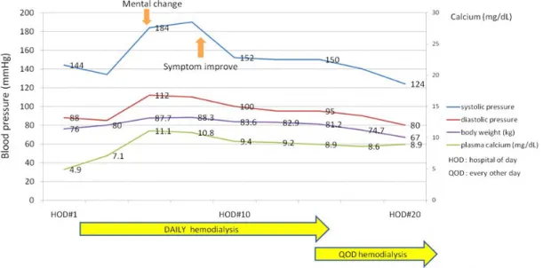
IU/L, sodium 118 mmol/L, potassium 8.2 mmol/L, 였다 동맥혈 가스 분석에서 chloride 84 mmol/L . pH 7.35, pCO2 19.5 mmHg, pO2 102.8 mmHg, bicarbonate 10.9 였으며 요검사상 이상 측 mmol/L myoglobin 30,000 g/Lμ 정되었다. 치료 및 경과: 내원 당시 시행한 혈액검사상 횡문근융해증에 의한 급성 신손상이 의심되었다 핍뇨 및 폐부종과 추적 혈액검. 사상 악화되는 고칼륨혈증 소견 관찰되어 응급 혈액투석 시행하 였으며 혈액투석 후 대사성 산혈증과 전해질 장애가 호전되었다. 입원 일째 혈청 칼슘8 11.1 mg/dL,수축기 혈압180 mmHg 이상으로 고칼슘혈증 및 혈압의 급격한 상승 소견이 관찰되었다 이후 경구 항고혈압제 투여에도 불구하고 혈압 상승 (Fig. 1). 이 지속되었으며 입원 일째 양측 시야 장애를 동반한9 1-2분 간의 의식 저하와 강직성 경련이 관찰되었다 컴퓨터 전산화 뇌. 혈관 단층촬영상 후교통동맥 경색증 의심되었으나(Fig. 2) 뇌 자기공명영상 촬영상 확산 강조영상 에서는 양측 두정 엽과 후( ) 두엽 그리고 소뇌의 대칭적인 부종이 피질하백질에 관찰되어 뇌, 경색증이 아닌 뇌혈관 부종으로 판단하였다(Fig. 3A). 정주 과 투여 후 nicardipine (0.5 mg/kg) labetalol (1 mg/kg) 혈압 조절 시작되었고 추가적인 경련은 관찰되지 않았다 시력. 장애 발생 후 시행한 세극등 안저 검사상 망막의 부분적인 함몰 소견 관찰되었으나 출혈반과 유두부종은 없었고 시야 검사에서 우안의 시야 결손 관찰되었다 정주 항고혈압 제제 치료 시작 후. 일째 입원 일째 부터 양측 시각장애 호전되었고 입원 2 ( 10 ) 15 일째에는 시력은 정상으로 회복 되었다. 14일째부터 수축기 혈 압130 mmHg,이완기 혈압80 mmHg으로 조절되었으며 입 원 19일째 부종 소실 및 소변양 증가와 혈중요소질소 40.2
Fig. 1. Blood pressure, body weight and corrected serum calcium level changes during hospitalization (HOD: hospital of day, QOD: every other day).
크레아티닌 혈청 mg/dL, 4.91 mg/dL, potassium 4.3 로 호전되어 혈액투석 치료를 중단하였다 입원 일 mmol/L . 26 째 시행한 추적 뇌 자기공명 영상 검사상 뇌부종 소견 소실되었 으며 (Fig. 3B) 전신상태 호전되어 입원30일째 퇴원하였다. 퇴원 개월 후 외래 추적 관찰 중 시행한 혈청 크레아티닌 수치1 는1.01 mg/dL로 정상 소견 보였다. 고 찰 는 악성 고혈압 임신중독증 요독증 면역억제제나 PRES , , , in-을 투여하는 간 신장 및 골수이식 환자들에서 주로 보 terferon , 고되고 있다1). 본 증례는 횡문근융해증에 의해 유발된 급성 신 손상 환자에서 급격한 혈압 상승과 동반된PRES발생 증례이다. 의 임상 증상은 주로 두통 오심 구토 의식변화 발작 PRES , , , , , 시각장애 등이며 급성으로 나타나는 특성을 보인다 발작의 경우. 주로 대발작양상이며 시각장애는 반맹 시각 무시 피질 맹 등이, , 나타난다1).본 증례의 경우 내원 당시 혈압은130/94 mmHg 였으나 내원 일째부터 평소 혈압보다7 30% 이상 증가한180/ 로 혈압 상승이 지속되었고 항고혈압 치료 중 오 100 mmHg , 심 의식변화 발작 양측 시각장애가 발생하였다 컴퓨터 전산화, , , . 뇌혈관 단층촬영상 및 뇌 자기공명 영상 검사에서 후두부에 뇌 부종이 관찰되었고 혈압 조절 후 증상과 후두부 부종이 호전된, 것으로 미루어 볼 때 환자는 횡문근융해증에 의한 급성 신손상 및 혈압 상승과 연관된PRES가 발병한 것으로 판단된다. 급격한 혈압상승에 의한 PRES 의 병태생리학적 발병 기전 은 아직까지 명확하게 밝혀져 있지 않으나 현재 두 가지의 상반 된 가설이 제시되고 있다 첫째 기전은 뇌혈관의 자동조절. (au-기능 소실로 인한 혈액 뇌 장벽
toregulation) - (blood brain
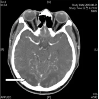
장애 및 뇌부종의 발생이다 뇌혈관의 자동조절은 혈 barrier) . 압이 급격하게 상승하거나 감소할 경우 뇌혈관의 수축과 이완 작용을 통해 뇌혈류를 일정하게 유지하는 것이다 뇌혈관 저항의. 약80%는 소동맥과 모세혈관이 담당하며 나머지20%는 모세 관후세정맥과 정맥에 의해 조절된다 즉 자동조절 작용은 주로. , 직경 30-300 mμ 의 소동맥에서 나타나며 하한 및 상한 평균 혈압의 범위가 각각40-60 mmHg, 150-160 mmHg 사이 에서 그 작용이 유지된다 그러나 혈압이 뇌혈관 자동조절 능력. 의 한계점 이상으로 급격히 상승하면 수축된 뇌혈관이 수동적으 로 이완되고 소동맥의 확장으로 인한 혈관 벽의 압력 증가가 모, 세혈관의 혈액 뇌 장벽 손상을 일으킨다 혈액 뇌 장벽의 손상- . -은 차적으로 뇌실 질 내부로의 단백질 적혈구 및 수액 유출시2 , 켜 뇌부종을 발생한다14, 15). 둘째 기전은 급작스러운 혈압 상승 Fig. 2. Brain angiography CT performed after seizure
shows low density lesion on bilateral posterior cerebral artery territory.
Fig. 3. (A) T2 weighted MR image performed 1 day after seizure shows bilateral patchy increased signal intensity with gyral swelling in both cerebellar hemisphere, parieto-occipital cortex and subcor-tical white matter area. (B) T2 weighted MR image performed 26 days after seizure shows markedly decreased T2 hyperintense lesion and gyral swel-ling in cerebellum and parietooccipital lobe.
에 의한 가역적인 뇌혈관 수축과 뇌혈류 감소 그리고 이에 따른, 허혈과 혈관성 뇌부종으로 PRES가 발생하는 것이다 자간증. 환자와 중증 고혈압 환자에서 컴퓨터 전산화 뇌혈관 단층촬영상 큰 뇌혈관들의 수축과 두정 후두부 저 음영 소견이 보고되어 -이와 같은 기전이 제안되었다 즉 급격한 혈압 상승에 대한 보. , 상과정으로 뇌혈관 수축이 유발되고 이에 따른 뇌허혈 및 뇌혈 관 부종이 발생 한다는 것을 유추해 볼 수 있다15-17). 국내 증례로는 혈액투석과 복막투석 환자에서 급격한 PRES 혈압 상승과 의식변화 및 두통 등의 신경학적 증상을 호소한 증 례가 있었으며 두 증례 모두 혈압 조절 후 증상이 호전되었다. 본 증례는 고혈압 당뇨 신부전 등의 기저 질환이 없는 남자에, , 서 심한 운동 후 발생한 횡문근융해증 및 급성 신 손상 치료 중 가 나타났으며 당시 혈청 칼슘 농도의 급격한 변동과 혈 PRES 압 상승이 동반되었다 (Fig. 1). 횡문근융해증 발병 초기에는 손상된 근육 조직에 칼슘이 침착되고 골조직의 부갑상선 호르몬 에 대한 반응 저하로 저칼슘혈증이 나타날 수 있다 한편 회복. , 기에는 근육에 침착되었던 칼슘이 혈중으로 유리되면서 고칼슘 혈증이 발생할 수 있다 실제 급성 신손상이 동반된 횡문근융해. 증 환자에서 고칼슘혈증에 의한 급격한 혈압상승이 보고되어 있 다18). 본 증례에서도 급성 신손상에 의한 체내 수분 저류 증가 와 고칼슘혈증 발생으로 인한 급격한 혈압 상승이 초래되었을 것으로 추정되었다. 는 혈관성 부종으로 인한 오심 구토 시각장애 등의 PRES , , 증상이 발생하기 때문에 뇌경색과 비교하여 방사선학적 소견과 임상 양상의 중증도와 연관성은 거의 없다 뇌 자기공명 영상. 검사상(T2강조영상 확산 강조영상 후 뇌동맥 경색의 전형, ) 적인 소견은 후 교통혈관이 관여하는 부위의 고신호강도가 나타 날 수 있으나PRES 에서는 주로 양측 두정 후두부 백질에 부 -종 소견을 보이면서 새발톱고랑과 측정 중 후두 엽은 보존되어 후뇌동맥 경색과 구별된다 할 수 있다 후두부 영역의 혈관성 부. 종 호발 원인은 뇌의 전방순환에 비해 후방순환인 추골기저동맥 계에 교감신경 분포가 적어 혈관 수출 유도가 잘 되지 않아 후대 뇌동맥영역인 후두 엽에 뇌부종이 호발 하는 것으로 설명되고 있다19). 또한 피질은 피질하백질에 비해 조직이 좀 더 치밀하, 여 부종은 피질하백질에 더 자주 발생한다. PRES진단에 컴퓨 터 전산화 뇌혈관 단층촬영만으로는 특이성이 떨어지며 자기공 명영상 특히, FLAIR (fluid attenuated inversion recovery)
영상은T2강조영상에 비하여 뇌척수액 신호를 저 강도로 낮추 어 뇌척수액 주변부의 경미한 뇌부종 소견을 감별진단 할 수 있 어 PRES 진단에 도움이 된다2, 20). 본 증례도 컴퓨터 전산화 뇌혈관 단층촬영만으로는 뚜렷한 감별 진단 어려웠고 추가적으 로 시행한 뇌 자기공명영상 관찰된 두정 후두부 피하백질 및 -피질 부종이 진단에 도움을 주었다. 와 감별 진단해야 할 질환들로 혈압상승이 동반된 경 PRES 련성 질환 두개강내 종양 중추 신경계 감염성 질환 다발성 경, , , 화증 뇌경색 등이 있으며 상기 질환들은 뇌척수액 검사 영상학, , 적 검사 및 혈압 조절 후 증상의 호전 여부로PRES와 감별할 수 있다 대부분의. PRES 환자는 수일 또는 수 주 내에 가역적 인 경과를 보이나 조기에 적절한 처치 및 진단을 하지 못하여 뇌 출혈이나 뇌경색이 발병할 경우 심각한 신경학적 후유증이 발생 하거나 사망에 이르기도 한다21). 한편 병력상 극심한 운동 후 발병한 급성 신손상 환자에서, 혈압 상승과 무관하게 발병한PRES 증례 보고가 있었다 이와. 같은 운동유발 급성 신손상은 혈관내피세포 기능 부전에 의한 신장 혈관 수축이 그 원인으로 알려져 있으며 creatine kinase 와myoglobin 등의 검사실 검사가 정상범위이고 극심한 허리 통증이 동반되어 횡문근융해증과 감별할 수 있다22). 최근PRES환자의 단기 및 장기 예후에 대한 연구가 진행되 었으며 PRES환자의84%에서 경련이 발생하였고 진단 당시, 영상학적 검사에서 이상소견을 동반한 환자가72%였다 경련. 및 시각장애는 증상 발생 후7.5 ,일 영상학적 이상소견은41일 내에 호전되었으며 평균 년간의 추적관찰 기간 중7 PRES재발 은 두 명의 환자(8%)에서만 관찰되었던 것으로 미루어 볼 때 조기 진단과 치료된PRES의 단기 및 장기 예후는 비교적 양호 하다고 할 수 있다23). 저자들은 횡문근융해증에 의한 급성 신손상 환자에서 급격한 혈압 상승과 동반된 두통 경련 및 시각장애 등의 신경학적 증상, 을 보인PRES 환자를 치험 하였기에 보고하는 바이다. 참 고 문 헌
1) Hinchey J, Chaves C, Appignani B, Breen J, Pao L, Wang A, Pessin MS, Lamy C, Mas JL, Caplan LR: A reversible posterior encephalopathy syndrome.N Engl J Med 334:494-500, 1996
2) Casey SO, Sampaio RC, Michel E, Truwit CL: Posterior reversible encephalopathy syndrome: utility of fluid-attenuated inversion recovery MR imaging in the detec-tion of cortical and subcortical lesions. AJNR Am J Neuroradiol 21:1199-1206, 2000
3) Celik M, Forta H, Dalkili T, Babacan G: MRI revealsç reversible lesions resembling posterior reversible ence-phalopathy in porphyria. Neuroradiology 44:839-841, 2002
4) Park SK, Rha JH, Han SR, Kim BW, Kim CC: Immu-nosuppressants induced reversible posterior dominant encepalopathy. J Korean Neurol Assoc 15:1125-1135, 1997
5) ML Ishimori, BD pressman, DJ wallace, Posterior rever-sible encephalopathy syndrome: another manifestation of CNS SLE. LUPUS 16:436-443, 2007
6) Rashimi Chawla, Daniel Smith, Paul E Marik, Near fatal posterior reversible encephalopathy syndrome complicating chronic liver failure and treated by induced hypothermia and dialysis. Jounal of Medical Case Reports 3:6623, 2009
7) Simon Nagel, Martin Kohrmann, Hagen B: Linezolid-induced posterior reversible leukoencephalopathy synd-rome. Arch Neurol64:746-748, 2007
8) Koo YS, Kim DH, Chang YK, Yang JO, Kang MG, Hwang PJ, Song CJ, Lee KW, Shin YT: A Case of Reversible Posterior Leukoencephalopathy Syndrome in Patient with Chronic Renal Failure. Korean J Nephrol 20:127-131, 2001
9) Choi SY, Park JY, Kwak SD, Yim JK, Chun JH, Kim TW, Seo BJ: A Case of Posterior Reversible Encephalo-pathy Syndrome in a Patient having Continuous Am-bulatory Peritoneal Dialysis. Korean J Nephrol 28:531-535, 2009
10) Yun BS, Lee SJ, Kim Y, Kim KH, Jung HJ: A Case of Posterior Reversible Encephalopathy Syndrome with Post Streptoccocal Glomerulonephritis. J Korean Child Neurol Soc 16:229-234, 2008
11) Jeong MH, Lee JH, Yum MS, Ko TS, Park YS: A Case of Posterior Reversible Encephalopathy Syndrome during Cyclosporine Therapy in a Child with Steroid Resistant Nephrotic Syndrome.J Korean Soc Pediatr Nephrol11: 92-99, 2007
12) Yun BS, Lee SJ, Kim Y, Kim KH, Jung HJ: A Case of Posterior Reversible Encephalopathy Syndrome with Post Streptoccocal Glomerulonephritis. J Korean Child Neurol Soc 16:229-234, 2008
13) Kwon EJ, Kim SW, Kim KK, Seo HS, Kim DY: A Case
of gemcitabine associated posterior reversible encepha-lopathy syndrome.Korean J Med(Suppl)75:S375, 2008 14) Hauser RA, Lacey DM, Knight MR: Hypertensive en-cephalopathy: magnetic resonance imaging demonstration of reversible cortical and white matter lesions. Arch Neurol 45:1078-1083, 1988
15) Bartynski WS: Posterior reversible encephalopathy syndrome, Part 2: controversies surrounding pathophy-siology of vasogenic edema. AJNR Am J Neuroradiol 29:1043 1049, 2008–
16) MacKenzie ET, Strandgaard S, Graham DI. Jones JV, Harper AM, Farrar JK: Effects of acutely induced hy-pertension in cats on pial arteriolar caliber, local cere-bral blood flow, and the blood-brain barrier. CircRes 39:33-41, 1976
17) Ijima T, Kubota Y, Kuroiwa T, Sankawa H: Blood-brain barrier opening following transient reflex sympa-thetic hypertension. Acta Neurochir Suppl(Wien) 60: 142-144, 1994
18) Bedani PL,Gilli P: Hypertensive emergency due to hy-percalcemia after acute renal failure secondary to rhab-domyolysis. Nephron 69:120-121, 1995
19) Sheth RD, Riggs JE, Bodenstenier JB, Gutierrez AR, Ketonen LM, Ortiz OA: Parietal occipital edema in hypertensive encephalopathy: a pathogenic mechanism. Eur Neurol 36:25-28, 1996
20) Chambers KA, Cain TW: Postpartum blindenss: two cases. Ann Emerg Med 43:243-246, 2004
21) Covarrubias DJ, Luetmer PH, Campeau NG: Posterior reversible encephalopathy syndrome: Prognostic utility of quantitative diffusion-weighted MR images. Am J Neuroradiol 23:1038-1048, 2002
22) Kimura T, Iio K, Imai E, Rakugi H, Isaka Y, Hayashi T: Exercise-induced acute kidney injury with reversible posterior leukoencephalopathy syndrome. Clin Exp Nephrol 14:173-5, 2010
23) Roth C, Ferbert A: Posterior reversible encephalopathy syndrome long-term follow-up. J Neurol Neurosurg Psychiatry 81:773-777, 2010