Case Report
Correspondence to: Yong Chan Lee
Department of Internal Medicine, Yonsei University College of Medicine, 134, Shinchon-dong, Seodaemun-gu, Seoul 120-752, Korea
Tel: +82-2-2228-1960, Fax: +82-2-393-6884, E-mail: [email protected] Received on May 18, 2009. Accepted on August 3, 2009.
DOI: 10.5009/gnl.2009.3.4.329
Achalasia Combined with Esophageal Cancer Treated by
Concurrent Chemoradiation Therapy
Jun Chul Park*, Yong Chan Lee*, Sang Kyum Kim†
, Yu Jin Kim*, Sung Kwan Shin*, Sang Kil Lee*, Hoguen Kim†, and Choong Bai Kim‡
Departments of *Internal Medicine, Institute of Gastroenterology, †Pathology and ‡Surgery, Yonsei University College of Medicine, Seoul, Korea
Achalasia is a rare neurological deficit of the esoph-agus that produces an impaired relaxation of the low-er esophageal sphinctlow-er and decreased motility of the esophageal body. Achalasia is generally accepted to be a pre-malignant disorder, since, particularly in the mega-esophagus, chronic irritation by foods and bac-terial overgrowth may contribute to the development of dysplasia and carcinoma. We present a case of a 51-year-old man with achalasia combined with esoph-ageal cancer who has had dysphagia symptoms for more than 20 years. Since there was a clinically high possibility of supraclavicular lymph node metastasis, concurrent chemoradiation therapy was scheduled. After the third cycle of chemoradiation therapy, trans- thoracic esophageolymphadenectomy was performed. Histopathological examination of the main esophagus specimen revealed no residual carcinoma. And the entire regional lymph node areas were free of carci-noma except for one azygos metastatic lymph node. In summary, achalasia is a predisposing factor for esophageal squamous cell carcinoma. Although sur-veillance endoscopy in achalasia patients is still con-troversial, periodic screening for cancer development in long-standing achalasia patients might be advisable.
(Gut and Liver 2009;3:329-333)
Key Words: Esophageal achalasia; Esophageal
neo-plasms
INTRODUCTION
Achalasia is a disease of unknown cause in which
aper-istalsis in the distal esophagus and a failure of relaxation of the lower esophageal sphincter (LES) occurs. Since the etiology is unclear, treatment focuses on reliving symp-toms. Nonetheless, despite treatment, food stasis often persists, causing development of chronic inflammation, dysplasia and possibly esophageal cancer.
The correlation between achalasia and esophageal can-cer was first noted by Fagge in 1872.1 Since this initial
report, achalasia has frequently been described as a pre-disposing factor for esophageal cancer.2,3 When
esoph-ageal cancer develops in patients with underlying acha-lasia, diagnosis tends to be in the more advanced stages of cancer, compared to cases with no achalasia, because both physicians and patients often regard symptoms such as dysphagia and chest discomfort as attributable to the achalasia, rather than to other causes. Therefore, addi-tional approaches that would lead to earlier diagnosis might be pursued less aggressively.
Here we report a case of achalasia combined with esophageal cancer treated by concurrent chemoradiation therapy.
CASE REPORT
A 51-year-old man was admitted to our hospital be-cause of mild dysphagia. He was diagnosed as esophageal motor disorder at a local clinic when he was teenager. The symptoms, which occurred for for both solids and liquids simultaneously, had begun more than 20 years ago. And whenever swallowing solid or liquid diet, he
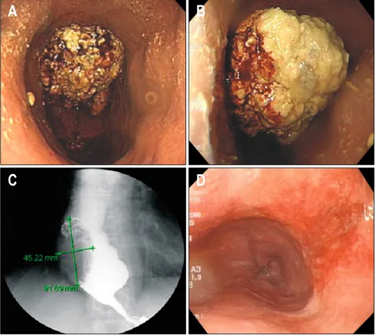
al-Fig. 1. (A, B) Endoscopic view
of fungating mass lesion with di-lated lower esophagus. (C) Fun-gating mass at distal esophagus and dilated distal esophagus with bird-beak sign. (D) Follow-up endoscopy showing disappearance of the previous cancer mass after the third cycle of concu-rrent chemoradiation therapy. ways felt the food was retained in the esophagus in a
mo-ment and passing through after a few seconds. However, there was no epigastric pain or chest pain. Sometimes vomiting and regurgitation developed after meal. Above symptoms had continued for several decades without sig-nificant change of their patterns and severities. Since his initial disagnosis at a local clinic, he did not go to hospi-tal thereafter till this visit to our institute.
Physical examination revealed a small, round, palpable mass at the right supraclavicular fossa and routine blood tests were unremarkable. The patient underwent upper gastroendoscopy, showing a fungating, exophytic mass le-sion with an irregular and friable surface involving 90% of the luminal circumference at 42 cm from the upper in-cisors, just above the gastroesophageal (GE) junction (Fig. 1A, B). The scope could be passed through the GE junction into the stomach body with mild resistance. Multiple biopsies were taken from the fungating mass, which allowed identification of squamous cell carcinoma upon histopathological examination. An esophagogram al-so showed a fungating mass of approximately 9 cm at the distal esophagus, and a dilated thoracic esophagus with bird-beak sign (Fig. 1C). Based on these results, esoph-ageal squamous cell carcinoma with achalasia was consid-ered and esophageal manometric study and positron
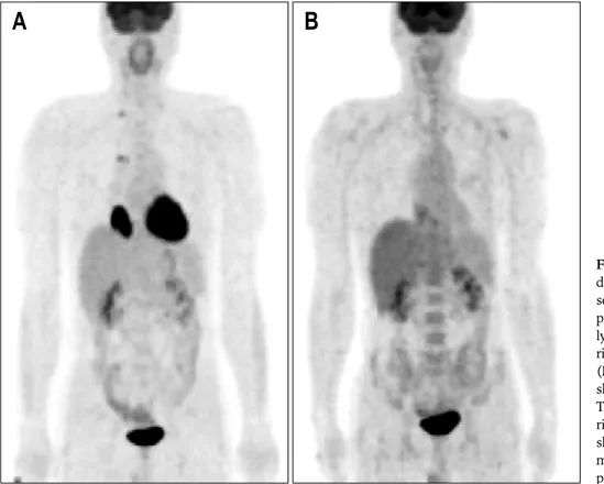
emis-sion tomography- computed tomography (PET-CT) were undertaken. The result of manometry was compatible with achalasia, specifically simultaneous contraction (mean pressure 14.48 mm Hg) and incomplete lower esophageal sphincter relaxation. PET-CT showed a large, hypermetabolic mass lesion at distal esophagus which was compatible with esophageal cancer (Fig. 2A). Hypermetabolism at the right hilar lymph node area and the right supraclavicular lymph node were observed. Although the aspiration biopsy at the right supraclavicular lymph node was negative for malignancy, there was a clinically high possibility of lymph node metastasis. For this reason, concurrent chemoradiation therapy was sug-gested instead of esophagectomy. By this time, the pa-tient’s dysphagia symptoms had worsened, so dilation of the lower gastroesophageal junction was performed using a 30 mm achalasia balloon dilator (6 psi for 5 seconds, Boston Scientific MicrovasiveⓇ, Natick, MA, USA).
Dysphagia symptoms greatly improved after dilation. Thereafter, concurrent chemoradiation therapy of 800 mg/m2/d fluorouracil was administered by continuous
in-fusion on day 1 to 3, and cisplatin 80 mg/m2
intrave-nously on day 2. Radiotherapy was delivered in five daily fractions per week of 1.8 Gy over 5.5 weeks, for a total of 50.4 Gy. Radiation fields also included the right
supra-Fig. 2. (A) Intense 18 F-fluoro-deoxyglucose (FDG) uptake is seen in the primary distal eso-phagus lesion with right hilar lymph node area and in the right supraclavicular lymph node. (B) The right hilar lymph node shows no definite FDG uptake. The main esophageal mass with right supraclavicular lymph node shows a decrease in size and mild FDG uptake compared to previous imaging.
clavicular lymph node area. After the third cycle of che-motherapy with radiation, followup PET-CT was under-taken. The right hilar lymph node showed no definite
18F-fluorodeoxyglucose (FDG) uptake. The main
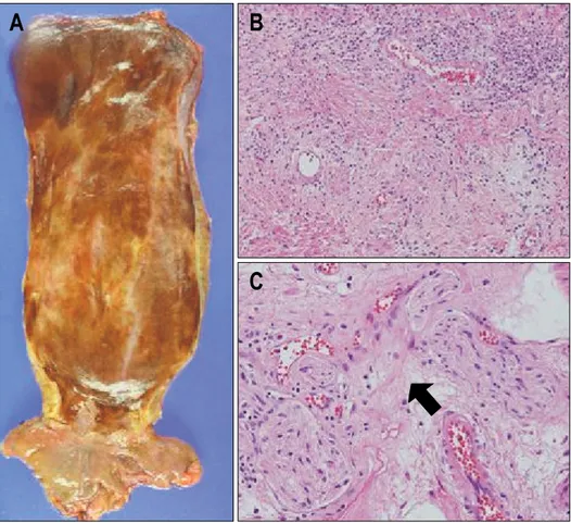
esoph-ageal mass with the right supraclavicular lymph node demonstrated decreased size and mild FDG uptake, com-pared to the previous analysis (Fig. 2B). The follow-up gastroendoscopy also showed disappearance of the distal esophageal mass, with mild erythematous mucosal change at the previous cancer site (Fig. 1D). The patient had no serious complications during the third cycle of concurrent chemoradiation therapy. He was informed about his dis-ease status and trans-thoracic esophageolymphadenectomy was performed (Fig. 3A). Histopathological examination of the main esophagus specimen revealed no residual car-cinoma and negative resection margin. Chronic inflamma-tory cell infiltration with stromal edema, telangiectasia and fibroblast proliferation was observed from secondary changes of radiation therapy (Fig. 3B). The entire para-esophageal, subcarnial and regional lymph node areas were free of carcinoma except for one azygos metastatic lymph node. Myenteric inflammation with lymphocytic in-filtration within the myenteric plexus was observed, and in particular, ganglion cells were absent (Fig. 3C). The patient was discharged on the 29th postoperative day with no serious complications. Further postoperative
chemotherapy was scheduled.
DISCUSSION
Although achalasia is a common functional disorder of the esophageal body and lower esophageal sphincter (LES), it is still a relatively rare disease. Epidemiological data have demonstrated an annual incidence in the range of 0.5 cases per 100,000 people, with a prevalence of about 8 cases per 100,000 people per year.4
The risk of esophageal malignancy for patients with long-standing achalasia is between 14- and 140-fold over the rest of the population.3,5 A recent large cohort study
showed male achalasia patients have substantially greater risk for both squamous cell carcinoma and adenocarci-noma of the esophagus.6 Several autopsy studies have
re-ported an esophageal carcinoma prevalence of 20-29% in achalasia patients,7 and evaluation of esophagectomy
specimens from patients with end-stage achalasia has demonstrated squamous hyperplasia.8 Most likely, in the
late phase of achalasia, chronic irritation by food and bac-terial overgrowth causes epithelial proliferation of the mucosa that may progress from squamous hyperplasia to dysplasia and carcinoma, similar to that seen in sporadic esophageal squamous cell carcinoma.9
ia-Fig. 3. (A) Gross specimen of
the surgically removed esopha-gus after concurrent chemoradia-tion therapy. (B) Microscopic findings of achalasia-associated squamous cell carcinoma after concurrent chemoradiation the-rapy. Chronic inflammatory cell infiltration with stromal edema, telangiectasia and fibroblast pro-liferation is shown. (C) Myen-teric inflammation with lympho-cytes infiltration observed in the myenteric plexus (arrow). Gang-lion cells are absent.
trogenic reflux after myotomy or pneumatic dilatation has been also been suggested as cause of adenocarcinoma.10
In a Dutch study of 331 achalasia patients treated with pneumatic dilation, 28 (8.5%) developed endoscopical evi-dence of Barrett’s metaplasia with intestinal metaplasia, seen in histological samples.11 Other study also reported
adenocarcinoma in Barrett’s metaplasia in achalasia.2
Barrett’s esophagitis is probably the result of LES tone-lowering therapy, which may induce significant sphincter insufficiency, which, in theory, may lead to worsened gastroesophageal reflux.
In patients with underlying achalasia, esophageal can-cers are generally diagnosed in advanced stages because the two diseases have similar symptoms, so rigorous di-agnostic evaluation of symptoms might be undertaken less frequently. Patients also have a large amount of re-tained food in the mega-esophagus, making visualization difficult. Therefore, reports of operability have been in-frequent and 80% of patients are inoperable at initial diagnosis.3,5 Nonetheless, whether surveillance endoscopy
should be generally recommended for all patients with esophageal achalasia is still controversial because of the long interval between initial symptoms of achalasia and the development of carcinoma. A previous study on 1,062
achalasia patients reported that the mean age at entry was 57.2 years and the mean age at cancer diagnosis was 71 years.12 Other studies also showed an interval between
the first symptoms of achalasia and the diagnosis of esophageal cancer of at least 15 years.3,5 Based on these
observations, a recommendation of regular surveillance endoscopy might be beneficial in long-standing achalasia patients. The benefits of surveillance endoscopy in these patients are seen in a higher prevalence of early esoph-ageal cancer stages.3
The treatment of choice for early esophageal cancer is curative resection. Surgery for advanced esophageal carci-noma has been disappointing, however, because of low resectability and a high risk of distant metastasis, which is seen in about half of the patients at the time of initial diagnosis.13 Surgery has limited therapeutic effect, and
therefore effective multimodality treatment is required to obtain better survival in advanced stage esophageal carci-noma cases.
Radiation and chemotherapy may synergistically en-hance anti-tumor effects against esophageal cancer, so con-current chemoradiotherapy (CRT) is an attractive strategy for radiosensitization and control of micrometastatic disease.14 Recent studies have shown that radiotherapy
with chemotherapy is superior to radiotherapy alone, based on the results of a number of randomized trials.15-17 A
landmark study by the Radiation Therapy Oncology Group (RTOG) compared radiation alone versus chemoradiation treatment (RTOG 85-01).16 At 5 years of follow-up, the
overall survival rates were 26% for patients who received combined modality treatment and 0% for those who re-ceived radiotherapy alone.
Cytotoxic agents, for example 5-Fluorouracil (5-FU) and cisplatin, have been used as radiosensitizers in many tumors, and a synergistic effect has been shown between the two drugs.15,17 Based on these results, continuous
5-FU and cisplatin can be used in concurrent chemo-radiotherapy. In phase II studies on advanced or recurrent esophageal squamous cell carcinoma, this combination in-duced a response rate of about 33-35%, leading to a me-dian overall survival of 6-8 months, although complete re-sponse was rare.18,19 This case presented here also
repre-sents an excellent response to preoperative, concurrent chemoradiation therapy. A large main esophageal mass disappeared with no remaining malignant cells. We also observed loss of ganglion cells in the myenteric plexuses, surrounded by lymphocytes representing achalasia (Fig. 3C). This inflammatory degeneration preferentially in-volves the nitric oxide-producing, inhibitory neurons that induce the relaxation of esophageal smooth muscle. Thus, the cholinergic neurons that contribute to LES tone by causing smooth muscle contraction are spared.20
Our case represents achalasia with squamous esoph-ageal carcinoma and mild dysphagia, which the patient experienced for more than 20 years. The long duration of dysphagia symptoms interfered with early diagnosis of esophageal cancer, and long-term food stasis might have been a factor in the precancerous state. Although surveil-lance endoscopy in achalasia patients is still controversial, periodic screening for cancer development might be advis-able in long-standing achalasia patients.
REFERENCES
1. Fagge CH. A case of simple stenosis of the oesophagus, followed by epithelioma. Guy's Hosp Rep 1872;17:413. 2. Ellis FH Jr, Gibb SP, Balogh K, Schwaber JR. Esophageal
achalasia and adenocarcinoma in Barrett's esophagus: a re-port of two cases and a review of the literature. Dis Esophagus 1997;10:55-60.
3. Brücher BL, Stein HJ, Bartels H, Feussner H, Siewert JR. Achalasia and esophageal cancer: incidence, prevalence, and prognosis. World J Surg 2001;25:745-749.
4. Mayberry JF. Epidemiology and demographics of achalasia. Gastrointest Endosc Clin N Am 2001;11:235-248.
5. Meijssen MA, Tilanus HW, van Blankenstein M, Hop WC, Ong GL. Achalasia complicated by oesophageal squamous
cell carcinoma: a prospective study in 195 patients. Gut 1992;33:155-158.
6. Zendehdel K, Nyrén O, Edberg A, Ye W. Risk of esoph-ageal adenocarcinoma in achalasia patients, a retrospective cohort study in Sweden. Am J Gastroenterol. Forthcoming 2007.
7. Carter R, Brewer LA. Achalasia and esophageal carcinoma. Studies in early diagnosis for improved surgical manage-ment. Am J Surg 1975;130:114-120.
8. Goldblum JR, Whyte RI, Orringer MB, Appelman HD. Achalasia. A morphologic study of 42 resected specimens. Am J Surg Pathol 1994;18:327-337.
9. Pajecki D, Zilberstein B, dos Santos MA, et al. Megaeso-phagus microbiota: a qualitative and quantitative analysis. J Gastrointest Surg 2002;6:723-729.
10. Guo JP, Gilman PB, Thomas RM, Fisher RS, Parkman HP. Barrett's esophagus and achalasia. J Clin Gastroenterol 2002;34:439-443.
11. Scholten P, Leeuwenburgh I, Vaessen R, et al. Barrett’s esophagus after pneumo-dilatation for achalasia. Gastroen-terology 2004;126 Suppl 2:A635.
12. Sandler RS, Nyrén O, Ekbom A, Eisen GM, Yuen J, Josefsson S. The risk of esophageal cancer in patients with achalasia. A population-based study. JAMA 1995;274:1359- 1362.
13. Kelsen DP, Ginsberg R, Pajak TF, et al. Chemotherapy fol-lowed by surgery compared with surgery alone for lo-calized esophageal cancer. N Engl J Med 1998;339:1979- 1984.
14. Leichman L, Steiger Z, Seydel HG, Vaitkevicius VK. Combined preoperative chemotherapy and radiation ther-apy for cancer of the esophagus: the Wayne State Univer-sity, Southwest Oncology group and Radiation Therapy Oncology Group experience. Semin Oncol 1984;11:178- 185.
15. al-Sarraf M, Martz K, Herskovic A, et al. Progress report of combined chemoradiotherapy versus radiotherapy alone in patients with esophageal cancer: an intergroup study. J Clin Oncol 1997;15:277-284.
16. Cooper JS, Guo MD, Herskovic A, et al. Chemoradiother-apy of locally advanced esophageal cancer: long-term fol-low-up of a prospective randomized trial (RTOG 85-01). Radiation Therapy Oncology Group. JAMA 1999;281:1623- 1627.
17. Herskovic A, Martz K, al-Sarraf M, et al. Combined che-motherapy and radiotherapy compared with radiotherapy alone in patients with cancer of the esophagus. N Engl J Med 1992;326:1593-1598.
18. Hayashi K, Ando N, Watanabe H, et al. Phase II evalua-tion of protracted infusion of cisplatin and 5-fluorouracil in advanced squamous cell carcinoma of the esophagus: a Japan Esophageal Oncology Group (JEOG) Trial (JCOG9407). Jpn J Clin Oncol 2001;31:419-423.
19. Bleiberg H, Conroy T, Paillot B, et al. Randomised phase II study of cisplatin and 5-fluorouracil (5-FU) versus cis-platin alone in advanced squamous cell oesophageal cancer. Eur J Cancer 1997;33:1216-1220.
20. Holloway RH, Dodds WJ, Helm JF, Hogan WJ, Dent J, Arndorfer RC. Integrity of cholinergic innervation to the lower esophageal sphincter in achalasia. Gastroenterology 1986;90:924-929.