Copyright © 2016 The Korean Society of Environmental Health and Toxicology
Introduction
Herbal medicines are commonly used to treat various diseases worldwide and are often co-administered with therapeutic drugs. However, the co-administration of herbs and drugs raises the potential of herb–drug interactions, which may affect the clinical safety of therapeutic drugs. Herb–drug interactions can be predicted by evaluating pharmacokinetic parameters, includ-ing absorption, metabolism, distribution, and excretion of drugs. Several drugs or herbs may affect the activity of drug-me-tabolizing enzymes, especially cytochrome P450 (CYP), result-ing in herb–drug or drug–drug interactions [1]. Among the
CYP subfamily, CYP3A is one of the most important drug-me-tabolizing enzymes in humans. It metabolizes more than 50% of clinically administered drugs [2]. Midazolam (MDZ), a short-acting, water-soluble benzodiazepine, is widely used as a probe substrate for CYP3A in humans and mice [3,4]. MDZ is mainly metabolized to 1′-hydroxymidazolam (1′-OH-MDZ) by the CYP3A subfamily [5]. Therefore, MDZ and 1′-OH-MDZ are typical probes used to measure the activity of CYP3A both in vivo and in vitro [6,7].
Commonly known as Banha-sasim-tang (BST) in Korea, or Ban-xia-xie-xin tang in China, the herb under study is one of the most popular herbal formulae mentioned in old herbal
prescrip-Decreased absorption of midazolam in the
stomach due to low pH induced by
co-administration of Banha-sasim-tang
Jun Hyeon Jo, Sun Joo Kim, Woong Shik Nam, Eun Ji Seung, Sangkyu Lee
BK21 Plus KNU Multi-Omics Based Creative Drug Research Team, College of Pharmacy and Research Institute of Pharmaceutical Sciences, Kyungpook National University, Daegu, Korea
• Original Article
http://dx.doi.org/10.5620/eht.e2016016eISSN: 2233-6567
Objectives Banha-sasim-tang (BST), which consists of seven different herbs, is one of the most popular herbal formulae for treating gastrointestinal disorders in Eastern Asia. The commonly used herbal medicine is often co-administered with other therapeutic drugs, which raises the possibility of herb–drug interactions and may modify the clinical safety profile of therapeutic drugs.
Methods We investigated the potential herb–drug interactions between BST extract and midazolam (MDZ) in mice. The area under the plasma concentration-time curve (AUC) of MDZ and 1ʹ-hydroxymidazolam (1ʹ-OH-MDZ) was evaluated for both oral and intra-peritoneal administration of MDZ, following oral administration of BST (0.5 and 1 g/kg).
Results It was found that the AUC of MDZ and 1ʹ-OH-MDZ was lower in case of oral administration of MDZ. Administration of BST extract was not associated with hepatic cytochrome P450 activity. BST extract induced a strong reduction in pH and it has been reported that oral mucosal absorption of MDZ is lower at low pH. The decreased ab-sorption rate of MDZ might be caused by the ingredients of BST and may not be related to other factors such as increased excretion of MDZ by P-glycoprotein.
Conclusions The altered pharmacokinetics of midazolam caused by co-administration with BST in vivo could be attributed to a decrease in pH and subsequent reduction of MDZ absorption rate.
Keywords Herb-drug interaction, Pharmacokinetics, Banha-sasim-tang, Midazolam
Correspondence: Sangkyu Lee 80 Daehak-ro, Buk-gu, Daegu 41566, Korea Tel: +82-53-950-8571 Fax: +82-53-950-8557 E-mail: [email protected] Received: May 24, 2016 Accepted: August 4, 2016 Published: August 9, 2016
tion literature [8]. BST consists of seven different herbs, includ-ing Pinellia ternata, Zingiber officinale, Glycyrrhizae uralensis, Cop-tis japonica, Scutellaria baicalensis, Panax ginseng, and Zizyphus jujuba. In East Asia, BST has been used to treat gastrointestinal
disorders [8]. In addition, BST has displayed preventive action against diarrhea induced by irinotecan [9]. BST has been used to treat chronic hypofunction of the gastrointestinal tract (GI) and functional abnormalities of the upper and lower GI system by positively improving GI hormone levels [10]. Recently in China, modified BST was shown to cause symptomatic im-provement in patients with functional dyspepsia [11].
To prevent or minimize adverse herb–drug interactions, herb-al medicine that interacts with drugs should be investigated in both in vivo and in vitro systems. Despite extensive clinical
re-search conducted on BST, its potential drug interactions remain to be elucidated [8,12]. BST has been more frequently pre-scribed in combination therapy with other herbs or drugs than on its own. Moreover, BST can be obtained easily without a prescription in Korea and patients sometimes self-prescribe medicines, which can lead to the co-administration of several different kinds of drugs. Here, a potential herb–drug interaction in BST therapy was investigated by evaluating the pharmacoki-netics of model drug MDZ when simultaneously administered with BST on mouse plasma parameters, using an liquid chroma-tography-tandem mass spectrometry (LC-MS/MS) system.
Materials and Methods
Chemicals and Reagents
BST solid extract was obtained from Hanzung Pharmaceutical (Daejeon, Korea) and prepared in accordance with the Korean Pharmacopoeia (KP), 10th edition. The preparation contained the following ingredients: water extract of P. ternate (1.67 g,
KP), Zingiber officinale (0.83 g, KP), Glycyrrhizae uralensis (1.0 g,
KP), Coptis japonica (0.33 g, KP), Scutellaria baicalensis (1.0 g,
KP), Panax ginseng (1.0 g, KP), and Zizyphus jujuba (1.0 g, KP).
MDZ and 1′-OH-MDZ were obtained from Bukwang Pharma-ceutical (Seoul, Korea) and Cayman Chemical (Ann Arbor, MI, USA), respectively.
Animals
Specific pathogen-free, 5-week-old male ICR mice (24 to 26 g) were obtained from Orient Bio (Seongnam, Korea) and accli-mated for at least seven days before the experiment. Upon arriv-al, the animals were randomly housed in cages, with four or five per cage. The animal quarters were strictly maintained at 23±3°C and 50±10% relative humidity with a 12 hours light/ dark cycle (intensity: 150–300 Lux). All animal procedures were
performed in accordance with the Society of Toxicology guide-lines of 1989. The study was approved by the institutional re-view board of the Kyungpook National University (2015-0099).
Pharmacokinetics Study
A total of 18 male ICR mice (30±2 g) were randomly divided into a vehicle-treated, and two MDZ-treated groups, which also received BST extract dissolved in saline. In the first group, MDZ (2.0 mg/kg) was orally administered to nine mice after 5 min-utes of oral administration of BST extract (0, 0.5 or 1.0 g/kg, n=3). In the second group, nine mice were intraperitoneally treated with MDZ (2.0 mg/kg) after 5 minutes of oral adminis-tration of BST extract (0, 0.5 or 1.0 g/kg, n=3). After the mice received MDZ, blood samples were collected from the tail vein into heparinized capillary tubes at 0.08-, 0.167-, 0.25-, 0.5-, 1-, and 2 hour time-points. The collected blood was immediately centrifuged at 4000 g for 10 minutes and 10 μL of plasma was obtained for each sample. The samples were stored at −20°C until analysis.
Plasma samples (10 μL) were added to 90 μL of acetonitrile (ACN) with 0.1% formic acid and 5 μM reserpine solution (in-ternal standard [IS], 97.5:2.5, v/v) was added. The solution was mixed and centrifuged at 13000 rpm for 10 minutes at 4°C, and 90 μL of the supernatant was obtained. The supernatants were transferred to autosampler vials, and 5 μL aliquots were injected into the LC system.
Liquid Chromatography-tandem Mass Spectrometry
Analysis System
All measurements were performed using an LC-MS/MS sys-tem in the selective reaction monitoring (SRM) mode. A Triple Stage Quadrupole Vantage mass spectrometer an HESI-II spray source coupled to an Accela™ LC system (Thermo Fisher Scien-tific, Waltham, MA, USA) was employed. The vaporizer and cap-illary temperatures were set to 150°C and 300°C, respectively. Electrospray ionization was performed in the positive mode at a spray voltage of 3500 V. Nitrogen was used as the sheath and aux-iliary gas, and was set to 45 and 20 (arbitrary units), respectively. Data were analyzed using the Xcalibur software (Thermo Fisher Scientific). An ACE® 5C18 column (5 μm, 50 mm×2.1 mm, Ad-vanced Chromatography Technologies, Scotland, UK) and a guard C18 column (2 μm, 2.1 mm ID, Phenomenex, Torrance, CA, USA) were employed for LC separation. The mobile phase consisted of ACN with 0.1% FA (mobile phase A) and water (mobile phase B) at a flow rate of 220 μL/min. The gradient was as follows: 0 minute 5% A, 0.5 minutes 5% A, 1.5 minutes 95% A, 3.0 minutes 95% A, 3.5 minutes 5% A, 5.0 minutes 5% A. Ions monitored in the SRM mode were m/z 326.0→ 291.0 for MDZ,
342.0→ 203.0 for 1’-OH-MDZ m/z, and 609.4→ 174.1 for the IS, respectively, at an SRM collision energy of 29 eV for MDZ, 15 eV for 1ʹ-OH-MDZ and 42 eV for IS. Data procurement was con-trolled using the Xcalibur software (Thermo Fisher Scientific).
Method Validation
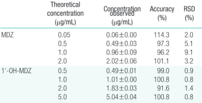
The calibration curves of MDZ and 1′-OH-MDZ ranged from 0.05 to 2.0 μg/mL, respectively. Calibration curves were con-structed by plotting the peak-area ratios of analyte or IS vs. the concentrations of MDZ and 1’-OH-MDZ in mouse plasma. The equations for the calibration curves of MDZ and 1ʹ-OH-MDZ were y =25114 x–13.85 (r2=0.999) and y =19380x –
9.12 (r2=0.998), respectively. The lower limits of quantification
values of MDZ and 1ʹ-OH-MDZ were 50 and 50 ng/mL, re-spectively. The quality control samples of the methodology were evaluated by analyzing five replicates of mouse plasma spiked with known concentrations of MDZ (0.05, 0.5, 1.0, and 2.0 μg/mL) or 1’-OH-MDZ (0.05, 0.5, 1.0, and 2.0 μg/mL) (Table S1).
PH Determination of the Mixture Containing Banha-sasim-tang Extract
The pH of BST extract was measured using a Metter Toledo S220 SevenCompact™ pH/Ion pH meter (Metter-Toledo Inter-national Inc., Columbus, OH, USA). The pH of each concen-tration of BST extract in the mixture was measured immediately after it dissolved.
Pharmacokinetic Parameters
A non-compartmental model was used to calculate the
phar-macokinetic parameters, using WinNonlin version 2.1 (Scientif-ic Consulting Inc., Cary, NC, USA), and includes maximum plasma concentration (Cmax), time to reach maximum plasma
concentration (Tmax), area under the plasma concentration-time
curve (AUC), and half-life. Statistics
The mean value ±standard error was determined for each treatment group of a given experiment. Dunnett’s t-test was
per-formed to compare the statistical significance of the data. The
p-values <0.05 were considered statistically significant. These are represented by asterisks .
Results
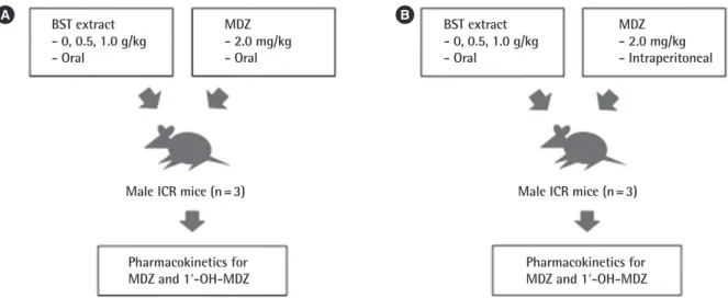
To investigate the potential herb–drug interactions observed during co-administration of BST with MDZ, two different ad-ministration methods were evaluated (Figure 1). Firstly, MDZ (2.0 mg/kg) was orally administered after treatment with BST extract (0, 0.5, or 1.0 g/kg) by oral gavage (Figure 1A). By mon-itoring the plasma concentration of MDZ and 1ʹ-OH-MDZ, first-pass metabolism of BST can be determined. In the other group, MDZ was intraperitoneally administered following oral administration of BST, to focus on the effects of BST in the met-abolic system and eliminate the absorption step (Figure 1B).
Mean plasma concentration–time profiles of MDZ and 1′-OH-MDZ were obtained (Figure 2) after simultaneous ad-ministration of a single-dose of BST (0, 0.5, or 1.0 g/kg, peroral [PO]) or vehicle control and a single dose of MDZ (2 mg/kg, PO) in mice. The pharmacokinetic parameters of MDZ and
MDZ - 2.0 mg/kg - Intraperitoneal
Pharmacokinetics for
MDZ and 1ʹ-OH-MDZ Pharmacokinetics forMDZ and 1ʹ-OH-MDZ
Figure 1. Experimental design. (A) Male ICR mice were orally administered MDZ (2.0 mg/kg) following administration of BST extract (0, 0.5, or 1.0 g/kg) or vehicle (saline) by oral gavage. (B) Male ICR mice were intraperitoneally treated with MDZ (2.0 mg/kg) following administration of BST extract (0, 0.5, or 1.0 g/kg) or vehicle (saline) by oral gavage. The blood samples were obtained and the concentrations of MDZ and 1ʹ-OH-MDZ (metabolite) were determined. MDZ, midazolam; BST, Banha-sasim-tang; 1ʹ-OH-MDZ, 1ʹ-hydroxymidazolam.
BST extract - 0, 0.5, 1.0 g/kg - Oral BST extract - 0, 0.5, 1.0 g/kg - Oral MDZ - 2.0 mg/kg - Oral
Male ICR mice (n=3) Male ICR mice (n=3)
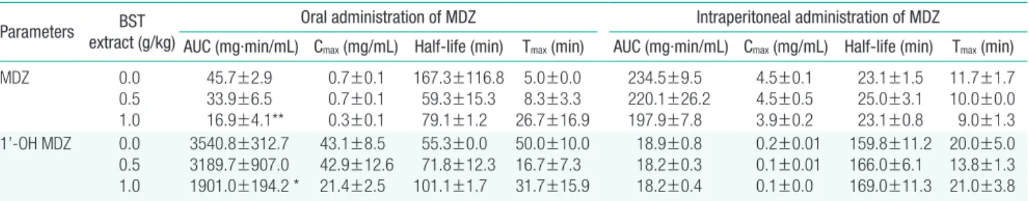
1′-OH-MDZ were compared between the vehicle- and BST-treated groups (Table 1). Following oral co-administration of BST and MDZ, the mean Cmax and AUC values were
signifi-cantly lower than those observed in the vehicle-treated group. The mean AUC decreased by 76% in the BST co-treated group (1.0 g/kg, PO). This corresponds to the plasma concentration-time profile of 1′-OH-MDZ, in which Cmax and AUC were
sig-nificantly decreased by 51% and 53%, respectively in the BST
co-treated group (1.0 g/kg, PO). Here, both the AUC and Cmax
of MDZ and its metabolite, 1′-OH-MDZ, decreased in the BST co-administered group, indicating decreased absorption of the parent drug MDZ.
ICR male mice were simultaneously treated with MDZ (2.0 mg/kg, intraperitoneal [IP]) following oral administration of BST (0.5 or 1.0 g/kg), to investigate the inhibitory effect of BST
1 0.1 0.01 MDZ ( μg/mL) 0 30 60 90 120 Time (min) 0 g/kg 0.5 g/kg 1.0 g/kg A 100 10 1 0.1 1ʹ -OH-MDZ ( μg/mL) 0 30 60 90 120 Time (min) 0 g/kg 0.5 g/kg 1.0 g/kg B
Figure 2. Mean plasma concentration-time profiles of MDZ and 1 ʹ-OH-MDZ in mice simultaneously treated with BST (0, 0.5, or 1.0 g/kg, PO) and MDZ (2.0 mg/kg, PO). Data are presented as mean±standard error (n=3). MDZ, midazolam; 1ʹ-OH-MDZ, 1ʹ-hydroxymidazolam; BST, Banha-sasim-tang; PO, peroral.
Figure 3. Mean plasma concentration-time profiles of MDZ and 1 ʹ-OH-MDZ in mice simultaneously treated with BST (0, 0.5, or 1.0 g/kg, PO) and MDZ (2.0 mg/kg, IP). Data are presented as mean ± standard error (n=3). MDZ, midazolam; 1ʹ-OH-MDZ, 1ʹ-hydroxymidazolam; BST, Banha-sasim-tang; PO, peroral; IP, intraperitoneal.
10 1 0.1 0.01 MDZ ( μg/mL) 0 30 60 90 120 Time (min) 0 g/kg 0.5 g/kg 1.0 g/kg A 1 0.1 0.01 1ʹ -OH-MDZ ( μg/mL) 0 30 60 90 120 Time (min) 0 g/kg 0.5 g/kg 1.0 g/kg B
Table 1. Effects of co-administration of BST extract with MDZ on the pharmacokinetic parameters of MDZ and 1’-OH-MDZ (n=3)
Parameters extract (g/kg)BST Oral administration of MDZ Intraperitoneal administration of MDZ AUC (mg·min/mL) Cmax (mg/mL) Half-life (min) Tmax (min) AUC (mg·min/mL) Cmax (mg/mL) Half-life (min) Tmax (min)
MDZ 0.0 0.5 1.0 45.7±2.9 33.9±6.5 16.9±4.1** 0.7±0.1 0.7±0.1 0.3±0.1 167.3±116.8 59.3±15.3 79.1±1.2 5.0±0.0 8.3±3.3 26.7±16.9 234.5±9.5 220.1±26.2 197.9±7.8 4.5±0.1 4.5±0.5 3.9±0.2 23.1±1.5 25.0±3.1 23.1±0.8 11.7±1.7 10.0±0.0 9.0±1.3 1’-OH MDZ 0.0 0.5 1.0 3540.8±312.7 3189.7±907.0 1901.0±194.2 * 43.1±8.5 42.9±12.6 21.4±2.5 55.3±0.0 71.8±12.3 101.1±1.7 50.0±10.0 16.7±7.3 31.7±15.9 18.9±0.8 18.2±0.3 18.2±0.4 0.2±0.01 0.1±0.01 0.1±0.0 159.8±11.2 166.0±6.1 169.0±11.3 20.0±5.0 13.8±1.3 21.0±3.8
Data are presents as mean±standard errors.
BST, Banha-sasim-tang; MDZ, midazolam; 1ʹ-OH-MDZ, 1ʹ-hydroxymidazolam; AUC, area under the plasma concentration-time curve; Cmax, maximum plasma concentration;
Tmax, maximum plasma concentration.
on CYP3A4 activity. The concentration-time profiles of MDZ and 1′-OH-MDZ in plasma are shown in Figure 3. The pharma-cokinetics parameters of MDZ and 1′-OH-MDZ in the BST-treated group did not differ significantly from those seen in the vehicle-treated group (Table 1). In addition, BST did not exert an inhibitory effect on CYP1A, 2B, 2C, 2D, or 3A4 at concen-trations of 0 to 0.5 g/mL in human liver microsomes, based on the use of a cocktail of probe substrate and LC-MS/MS analysis (data not shown) [7].
Discussion
Our study showed that the pharmacokinetic parameters of MDZ and 1′-OH-MDZ decreased following oral co-administra-tion with BST, but not in case of IP administraco-administra-tion. When MDZ was orally administrated, the plasma concentration of MDZ was regulated by two mechanisms: absorption rate and metabolic stability in the liver. The plasma concentration of MDZ was found to be mainly affected by a mechanism involving metabol-ic conversion of MDZ in the liver. The plasma concentration of MDZ and its metabolites was not changed by BST IP co-ad-ministration, which indicates that BST does not have a strong inhibitory action on CYP activities in the liver. Moreover, a de-crease in plasma concentration of MDZ and 1’-OH-MDZ is ob-served following oral co-administration with BST. The findings therefore indicate that BST affects the absorption of MDZ, but not its metabolism.
The reduction in plasma concentration of MDZ and 1′-OH-MDZ in animals co-administered with 1′-OH-MDZ and BST (1.0 g/ kg, PO) indicates that the absorption of MDZ might be inhibit-ed by BST, and that this inhibition might not be associatinhibit-ed with metabolic activity or CYP3A4 enzyme. The reduced absorption of MDZ may be attributed to several phenomena. One is the formation of insoluble particles from the components of BST extract interacting with MDZ at physiological pH conditions (pH 1-2 in the stomach, or pH 5-6 in the intestines). However, the concentrations of MDZ in the supernatants of the BST-MDZ mixture in artificial gastric juice or intestinal juice was not significantly altered (Figure S1). In this way, the ingredients in the extract were found not to form insoluble particles that could reduce the absorption of MDZ in the stomach and intestines.
Another hypothesis is that the reduced pH induced by BST extract in the stomach or intestines affects the oral mucosal ab-sorption of MDZ, since it has been reported that MDZ absorp-tion is strongly inhibited under low pH condiabsorp-tions [13]. Fur-thermore, changes in local pH affect intestinal absorption of wa-ter-soluble, weakly acidic compounds. The bioavailability of MDZ at pH 2.8, 3.2, and 3.9 solutions was 6.2, 18.7, and 22.6%
respectively [14]. As shown in Figure 4, pH significantly re-duced with increasing concentrations of BST extract (1-100 mg/mL). The pH value of normal saline was 5.9, whereas that of 1 and 10 mg/mL BST extract was 5.0 and 4.7, respectively. Since BST consists of seven herbs, it exhibits varying concentra-tion ranges for each of its diverse ingredients, and thus might contain acidic compounds that decrease pH.
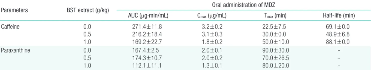
The other possible mechanism involved in the phenomena under investigation is that the ingredients in BST inhibits the absorption of MDZ in the stomach or intestine. The complex ingredients in BST might compete with the absorption of MDZ. Since MDZ is not a substrate of P-glycoprotein (P-gp), the reduced plasma concentration cannot be related to in-creased excretion by P-gp [13]. Further, following co-adminis-tration with BST (1.0 g/kg, PO), the absorption of caffeine was found to be inhibited, since the bioavailability of caffeine has been shown to be almost 100% in vivo [14]. Moreover, we
eval-uated the change in the pharmacokinetic parameters of caffeine and paraxanthine after BST administration Table S2. The plas-ma concentration of caffeine and its metabolite, paraxanthine, decreased following the administration of BST (0.5 and 1.0 g/ kg) by oral gavage, as was seen in case of MDZ-BST co-admin-istration. In a previous study, Kampo extract used in traditional Japanese medicines, originated from Glycyrrhiza uralensis,
re-duced the Cmax and AUC of talinolol, a P-gp substrate drug [15].
Decreased intestinal talinolol absorption induced by Kampo ex-tract might be caused by the inhibition of organic anion trans-porting peptides, and not by inhibition of P-gp. The low plasma concentration of MDZ or caffeine following co-administration with BST indicated that the absorption rate was inhibited by components of the BST extract.
Here, we investigated the herb–drug interactions between BST extract and MDZ in vivo, following oral administration of
BST extract (1.0 g/kg). BST extract did not exert effects on the Figure 4. PH values of the Banha-sasim-tang (BST) extract mixtures (0– 100 mg/mL). 6 5 4 pH 0 5 10 15 20 25 BST extract (mg/mL)
activity of an enzyme crucial to drug interactions, CYP, in the liver of the mouse model studied. Nevertheless, the pharmaco-kinetic parameters of MDZ were altered when it was orally ad-ministered following BST extract treatment (1.0 g/kg), as evi-denced by the observed reduction in the total absorption of MDZ and 1′-OH-MDZ. The reduction of pH induced by the administration of BST extract indicates mechanisms behind the decreased rate of mucosal absorption of MDZ. In conclusion, the oral administration of BST extract together with MDZ might cause potential herb–drug interactions in vivo.
Acknowledgements
This work was supported by the Korea Institute of Planning and Evaluation for Technology in Food, Agriculture, Forestry and Fisheries (IPET) through Export Promotion Technology Development Program, funded by the Ministry of Agriculture, Food and Rural Affairs (MAFRA) (grant no. 316017-3).
Conflict of Interest
The authors have no conflicts of interest associated with mate-rial presented in this paper.
ORCID
Jun Hyeon Jo https://orcid.org/0000-0003-2007-0076
Sun Joo Kim https://orcid.org/0000-0001-7679-0270
Woongshik Nam https://orcid.org/0000-0002-6475-7139
Eun Ji Seung https://orcid.org/0000-0003-1233-9530
Sangkyu Lee https://orcid.org/0000-0001-5343-701X
References
1. Chen XW, Sneed KB, Pan SY, Cao C, Kanwar JR, Chew H, et al. Herb-drug interactions and mechanistic and clinical consider-ations. Curr Drug Metab 2012;13(5):640-651.
2. Wacher VJ, Wu CY, Benet LZ. Overlapping substrate specificities and tissue distribution of cytochrome P450 3A and P-glycopro-tein: implications for drug delivery and activity in cancer chemo-therapy. Mol Carcinog 1995;13(3):129-134.
3. Nordt SP, Clark RF. Midazolam: a review of therapeutic uses and
toxicity. J Emerg Med 1997;15(3):357-365.
4. Perloff MD, von Moltke LL, Court MH, Kotegawa T, Shader RI, Greenblatt DJ. Midazolam and triazolam biotransformation in mouse and human liver microsomes: relative contribution of CY-P3A and CYP2C isoforms. J Pharmacol Exp Ther 2000;292(2): 618-628.
5. Kronbach T, Mathys D, Umeno M, Gonzalez FJ, Meyer UA. Oxi-dation of midazolam and triazolam by human liver cytochrome P450IIIA4. Mol Pharmacol 1989;36(1):89-96.
6. Kato R, Yamashita S, Moriguchi J, Nakagawa M, Tsukura Y, Uchida K, et al. Changes of midazolam pharmacokinetics in Wistar rats treated with lipopolysaccharide: relationship between total CYP and CYP3A2. Innate Immun 2008;14(5):291-297.
7. Song M, Hong M, Lee MY, Jee JG, Lee YM, Bae JS, et al. Selective inhibition of the cytochrome P450 isoform by hyperoside and its potent inhibition of CYP2D6. Food Chem Toxicol 2013;59:549-553.
8. Park JW, Ko SJ, Han G, Yeo I, Ryu B, Kim J. The effects of Banha-sasim-tang on dyspeptic symptoms and gastric motility in cases of functional dyspepsia: a randomized, double-blind, placebo-con-trolled, and two-center trial. Evid Based Complement Alternat Med 2013;2013:265035.
9. Mori K, Kondo T, Kamiyama Y, Kano Y, Tominaga K. Preventive effect of Kampo medicine (Hangeshashin-to) against irinotecan-induced diarrhea in advanced non-small-cell lung cancer. Cancer Chemother Pharmacol 2003;51(5):403-406.
10. Naito T, Itoh H, Yasunaga F, Takeyama M. Hange-shashin-to raises levels of somatostatin, motilin, and gastrin in the plasma of healthy subjects. Biol Pharm Bull 2002;25(3):327-331.
11. Zhao L, Zhang S, Wang Z, Wang C, Huang S, Shen H, et al. Effica-cy of modified ban xia xie xin decoction on functional dyspepsia of cold and heat in complexity syndrome: a randomized controlled trial. Evid Based Complement Alternat Med 2013;2013:812143. 12. Park JW, Ryu B, Yeo I, Jerng UM, Han G, Oh S, et al.
Banha-sasim-tang as an herbal formula for the treatment of functional dyspepsia: a randomized, double-blind, placebo-controlled, two-center trial. Trials 2010;11:83.
13. Kirby B, Kharasch ED, Thummel KT, Narang VS, Hoffer CJ, Un-adkat JD. Simultaneous measurement of in vivo P-glycoprotein and
cytochrome P450 3A activities. J Clin Pharmacol 2006;46(11): 1313-1319.
14. Blanchard J, Sawers SJ. The absolute bioavailability of caffeine in man. Eur J Clin Pharmacol 1983;24(1):93-98.
15. Iwanaga K, Arimune K, Miyazaki M, Shibano M, Taniguchi M, Baba K, et al. Effects of furanocoumarins in Kampo extract-based medicines on rat intestinal absorption of CYP3A and P-glycopro-tein substrate drugs in vivo. Arch Pharm Res 2012;35(6):1055-1064.
Table S1. The accuracy and RSD of quality control samples of MDZ and 1’-OH MDZ Theoretical concentration (μg/mL) Concentration observed (μg/mL) Accuracy (%) RSD (%) MDZ 0.05 0.5 1.0 2.0 0.06±0.00 0.49±0.03 0.96±0.09 2.02±0.06 114.3 97.3 96.2 101.1 2.0 5.1 9.1 3.2 1’-OH-MDZ 0.5 1.0 2.0 5.0 0.49±0.01 1.01±0.00 1.83±0.03 5.04±0.04 99.0 100.8 91.6 100.8 0.9 0.8 1.4 0.8
RSD, relative standard deviation; MDZ, midazolam; 1ʹ-OH-MDZ, 1ʹ-hydroxym- idazolam.
Table S2. Effects of co-administration of BST extract on the pharmacokinetic parameters of caffeine and paraxanthine
Parameters BST extract (g/kg) Oral administration of MDZ
AUC (μg·min/mL) Cmax (μg/mL) Tmax (min) Half-life (min)
Caffeine 0.0 0.5 1.0 271.4±11.8 216.2±18.4 169.2±22.7 3.2±0.2 3.1±0.3 1.8±0.2 22.5±7.5 30.0±0.0 50.0±10.0 69.1±0.0 48.9±6.8 88.1±0.0 Paraxanthine 0.0 0.5 1.0 167.4±2.5 174.3±10.7 112.1±11.1 2.0±0.1 2.0±0.2 1.3±0.1 90.0±30.0 70.0±26.5 80.0±20.0
-Nine male ICR mice were orally administered with caffeine (2.0 mg/kg) after 5 minutes of oral administration of BST extract (0, 0.5 or 1.0 g/kg, n=3); After the mice received caffeine, blood samples were collected from the tail vein into heparinized capillary tubes at 0.08- 0.25- 0.5- 1.0- and 2 hours; The collected bloods were immediately centrifuged at 4000 g for 10 minutes and 10 μL of plasma was obtained for each sample and stored at -20°C until analysis.
BST, Banha-sasim-tang; MDZ, midazolam; AUC, area under the plasma concentration-time curve; Cmax, maximum plasma concentration; Tmax, maximum plasma
25 20 15 10 5 0 MDZ ( μg/mL) 0 0.025 0.05 0.1 BST extract (mg/mL) A 50 40 30 20 10 0 MDZ ( μg/mL) 0 0.025 0.05 0.1 BST extract (mg/mL) B
Figure S1. The concentration of MDZ in the supernatant of artificial gas-tric juice (A) and intestinal juice (B) with MDZ and BST. The data shown are the means of duplicates. MDZ, midazolam; BST, Banha-sasim-tang.