Pancreatic Paraganglioma: a Case
Report and Literature Review
INTRODUCTION
Paraganglioma is a rare tumor derived from neural crest cells in sympathetic or parasympathetic ganglions (1). During the development of paraganglioma, neural crest cells get dispersed throughout the body and aggregate to form paraganglia (2). Therefore, paragangliomas can be found in every part of the body where paraganglia are known to arise, frequently in the posterior mediastinum and thoracolumbar paravertebral region, including Zuckerkandl’s body (1).
The incidence of paraganglioma is about one in 2,000,000 adults, and that of paraganglioma arising within the pancreas is even more rarely reported (3). To the best of our knowledge, about 31 cases of primary pancreatic paragangliomas have been described in the English literature over the past decades and even lesser cases were reported with gadoxetic-acid-enhanced magnetic resonance imaging (MRI) and diffusion-weighted images (DWI) sequences (1, 3-11).
Given the rarity and the overlapping appearance of pancreatic paraganglioma with other pancreatic neoplasms, the preoperative diagnosis can be often challenging, especially in nonfunctional cases. Pancreatic paragangliomas have been reported as well-defined hypervascular soft tissue masses, frequently with cystic areas (2). These hyper-enhancing tumors are often misdiagnosed as either pancreatic neuroendocrine tumor (pNET) or vascular lesions such as aneurysm (2).
We herein report about computed tomography (CT), gadoxetic-acid-enhanced MRI and DWI sequence, as well as endoscopic ultrasound (EUS) findings of pancreatic paraganglioma, with clearly defined draining veins, washout pattern enhancement, and
This is an Open Access article distributed under the terms of the Creative Commons Attribution Non-Commercial License (http://creativecommons.org/licenses/ by-nc/4.0/) which permits unrestricted non-commercial use, distribution, and reproduction in any medium, provided the original work is properly cited.
Received: January 2, 2021 Revised: January 25, 2021 Accepted: February 5, 2021
Correspondence to:
Joon Suk Park, M.D.
Department of Radiology, Hallym University Dongtan Sacred Heart Hospital, 7, Keunjaebong-gil, Hwaseong-si, Gyeiniggi-do 18450, Korea.
Tel. +82-10-3993-8959 E-mail: parkjoonsuk8856@
gmail.com
Copyright © 2021 Korean Society of Magnetic Resonance in Medicine (KSMRM)
Case Report
Paraganglioma is a rare tumor of paraganglia, derived from neural crest cells in sympathetic or parasympathetic ganglions. It can be widely distributed from the skull base to the bottom of the pelvis. The pancreas, however, is a rare location of this neoplasm, and only a limited number of cases have been reported in the English literature, especially with gadoxetic-acid-enhanced magnetic resonance imaging (MRI) and diffusion-weighted images (DWI). We herein report a case of pathologically proven paraganglioma in the pancreas head with a literature review on endoscopic ultrasonography (EUS), computed tomography (CT), gadoxetic-acid-enhanced MRI, and DWI sequence.
Keywords: Pancreas; Paraganglioma; Computed tomography; Magnetic resonance imaging
Joon Suk Park1, Seon Jeong Min1, Soo Kee Min2, Jung-Ah Choi1
1Department of Radiology, Hallym University Dongtan Sacred Heart Hospital, Hwaseong, Korea 2
multiple areas of signal void interspersed with hyperintense foci within the mass as ‘salt-and-pepper’ appearance on MRI.
CASE REPORT
A 46-year-old man presented to our hospital with abdominal discomfort for over 2 months. The patient’s past medical history and physical examination were unremarkable. Initial laboratory studies including levels of tumor marker as well as pancreatic and other endocrine hormones were within their normal range.
The patient underwent contrast-enhanced dynamic
abdominal CT; the imaging revealed the presence of a 2.8 × 2.6 × 3.2 cm sized irregular solid lesion within the head of the pancreas (Fig. 1). On the pre-contrast image, the lesion appeared as a low attenuated area compared with the surrounding pancreas parenchyma, with an average of 27 Hounsfield units (HU). On the arterial phase, the lesion showed intense enhancement compared with the adjacent normal pancreas, and non-enhancing irregular shaped inner portions. Prominent peritumoral vessels including early enhancing draining veins were also observed; there was relatively early contrast filling of the main portal vein and draining vein around the lesion. On the portal phase, it showed decreased but still higher enhancement than the adjacent normal pancreas. Also, the extent of the
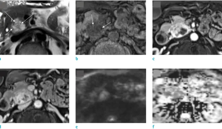
non-Fig. 1. Pancreatic paraganglioma in a 46-year-old man. The axial contrast-enhanced CT image during the arterial phase reveals the presence of (a), about 2.8 × 2.6 × 3.2 cm sized well defined irregular shape mass in the head of the pancreas with marked enhancement and non-enhancing inner portions. Prominent peritumoral vessels including early enhancing draining veins are also noted (arrows). In addition, on the arterial phase (b), there was relatively early contrast filling of the main portal vein (arrowhead) and draining vein (arrow) around the mass. No dilatation of the main pancreatic duct or biliary duct is observed. Moreover, note that the normal parenchyma completely surrounds the tumor (arrowheads), indicating that the mass (arrows) is located at the intra-pancreatic area (c), which is further proved based on the surgical findings. On the portal phase (d), the lesion shows decreased enhancement and the extent of the non-enhancing portions shrank (arrow) compared with those on the arterial phase. Also, note the prominent peritumoral draining vessels (arrows) around the lesion on the coronal image (e).
a b c
enhancing inner portion was noted to shrink on the portal phase. The lesion was completely surrounded by the normal pancreas parenchyma, indicating the location within the pancreas, which was further proved based on surgical findings.
On MRI, after intravenous administration of contrast agent, gadobutrol (Gadovist®; Bayer-Healthcare, Berlin, Germany), dynamic images were obtained (Fig. 2). In comparison with the remainder of the pancreas, the lesion showed marked heterogeneous enhancement on the arterial phase and gradual faint enhancement on the portal phase and transitional phase with a slow washout pattern. The lesion showed relatively higher enhancement than the normal pancreas on the portal phase and transitional phase. Compared with the contiguous normal pancreas, the solid portion showed mild high signal intensity (SI) on T2-weighted image (T2WI) and mild low SI on T1-T2-weighted
image (T1WI). We assumed that the lesion demonstrated interspersed inner high- and low SI foci, the so-called ‘salt and pepper appearance’ on both T1WI and T2WI images. The ‘pepper’ component denotes the spots of vascular flow signal void and the ‘salt’ denotes high SI foci due to slow flow or hemorrhage on both T1WI and T2WI images. The lesion showed high SI on DWI at a high b-value (1000 s/mm2) and low value on the ADC map, reflecting the
cellularity.
No abnormalities were found in the adjacent bile duct or pancreatic duct and the liver. There were no enlarged lymph nodes around the mass or any evidence of local or distant metastasis. Based on CT and MRI, we made a preoperative diagnosis of benign paraganglioma, but also considered nonfunctioning pNET as a differential diagnosis.
EUS showed a well-demarcated hyperechoic mass of the pancreatic head, where a fine needle biopsy (FNB) was
Fig. 2. MRI of the patient. On MRI, the main solid portion of the lesion shows mild high SI on T2WI image and mild low SI on T1WI image. The lesion suspiciously shows inner high- and low SI foci (arrows), the so-called ‘salt and pepper appearance’, on both T2WI image (a) and T1WI image (b); the ‘pepper’ represents the interspersed areas of signal void and the ‘salt’ represents the hyperintense foci due to slow flow or hemorrhage. On gadoxetic-acid-enhanced fat-suppressed T1WI image, the lesion shows marked heterogeneous enhancement on the arterial phase (c) and gradual faint enhancement on the portal phase (d) and transitional phase (not shown), but still relatively well enhanced compared with the remainder of the pancreas. The lesion does not show a clear washout pattern that is typically shown in pNET. The lesion shows high SI on DWI (e) at a high b-value (1000 s/mm2) and low value on ADC map (f), reflecting the cellularity.
a b c
done (Fig. 3). The pathology report of FNB at the pancreas head suggested some nests of pleomorphic cells with neuroendocrine morphology and differentiation, suggesting both NET and paraganglioma. Although pancreatic paraganglioma is rare, the possibility could not be excluded completely, as both paraganglioma and NET share a similar neuroendocrine morphology based on pathology.
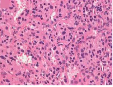
The patient underwent pylorus-preserving pancreati-coduodenectomy. Gross pathological examination revealed a well-circumscribed pink tanned rubbery mass surrounded by normal pancreatic parenchyma (Fig. 4). The final pathology report described an intra-pancreatic mass, composed of clustered multinucleated cells in the nesting pattern with a typical zellballen pattern. The cells contained finely granular cytoplasm and formed clusters with peripheral flattened sustentacular cells. This lesion was considered to be benign based on the histopathologic analysis: all the resection margins were negative, both lymphovascular and perineural invasions were absent, the cellularity was moderate, and comedo necrosis was absent. It was not possible to evaluate the Ki-value67 labeling index at the time of preparation of the pathologic report.
According to the Grading of Adrenal Pheochromocytoma and Paraganglioma (GAPP) scoring system by Tischler and deKrijger (12), which predicts the prognosis and metastatic potential, our patient scored 1 (1 for moderate cellularity; Ki-value67 labeling index was ignored) indicating non-metastatic tumor or low non-metastatic potential at the highest including K-value67 labeling index.
Since the lesion was completely surrounded by and in continuity with normal pancreatic parenchyma, the origin was reported as pancreatic paraganglioma. The
lesion contained inner hemorrhagic foci; the findings were consistent with our MRI findings of ‘salt and pepper appearance’. We suspected that the ‘salt’ portion could correlate with pathologic findings of intratumoral hemorrhage, but the ‘pepper’ portion such as the intratumoral vessel was not fully evaluated pathologically. Immunohistochemistry showed positive for synaptophysin, chromogranin, and S-100 protein. The final histopathologic diagnosis was benign pancreatic paraganglioma without metastasis.
The patient’s post-operative recovery was good and he was not re-admitted for similar abdominal discomfort. Repeated follow-up CT scans were performed that showed no evidence of recurrence or metastasis. The patient has been disease-free for more than 5 years.
DISCUSSION
Pancreatic paraganglioma occurs between the ages of 19 and 85 years, and the male to female incidence ratio is approximately 1:2 (8). The majority of them are nonfunctional and are discovered either incidentally or in a patient with abdominal pain (2). They are frequently found in the pancreatic head, as in our case, and rarely occur in the pancreatic body or tail; the size ranges from 1.5cm to 13cm in diameter (6, 8).
Ultrasound findings of pancreatic paraganglioma show a hypoechoic mass with abundant blood supply which can contain inner anechoic areas (13). CT findings demonstrate a clear or an ill-defined isodense or hypodense mass with strong enhancement. Pancreatic paragangliomas commonly exhibit cystic changes and necrosis, but rarely contain calcification (1).
On CT and MRI, pancreatic paraganglioma may have prominent intratumoral vessels and peritumoral draining vessels (10). Although details on the mechanism of the vessel induction remain to be elucidated, the feature of peritumoral draining veins has been reported to be useful in distinguishing pancreatic paraganglioma from pNET (10). To the best of our knowledge, only three cases have been reported to have ‘early contrast filling of peritumoral draining veins’ (1, 3, 10). In accordance with these previous reports, our case also confirmed early contrast filling of draining veins in primary pancreatic paraganglioma. As Kim et al suggested, we agree that the feature, ‘early contrast filling of peritumoral draining vein’, can be a clue in the diagnosis of pancreatic paraganglioma and a distinguishing
Fig. 3. EUS of the patient. EUS shows well marginated hyperechoic mass (arrows) in the pancreas head, where the biopsy was done.
feature from pNET on dynamic image studies (3, 10).
Paragangliomas occur in a variety of locations, and they may share similar imaging features at any location, including a hypervascular mass with multiple areas of signal void interspersed within the tumor mass with a ‘salt and pepper’ appearance (13). To the best of our knowledge, our case is unique in that it is the first case report to suspect ‘salt and pepper appearance’ of pancreatic paraganglioma
on MRI. This feature was further proved with pathologic examination, where spots of intratumoral hemorrhage could be correlated with the ‘salt’ portion; however, we could not evaluate intratumoral vessel to represent the ‘pepper’ portion in pathologic specimen which remains to be further elucidated.
The available radiologic images were limited in previous reports, especially on dynamic contrast-enhanced MRI
Fig. 4. Gross pathological examination (a) revealed a well-circumscribed pink tanned rubbery soft tissue mass (arrows) surrounded by the normal pancreatic parenchyma with foci of inner hemorrhage, which was consistent with the image findings. Microscopic examination (b) demonstrated the presence of a cluster of cells in a nesting or trabecular pattern, with a typical zellballen pattern composed of bizarre or multinucleated cells (Hematoxylin and Eosin stain (H&E), × 400). The cells contained finely granular cytoplasm and formed clusters with peripheral flattened sustentacular cells. Immunohistochemistry showed positive for chromogranin (c) and S-100 protein (d) (immunohistochemical stain, × 200). The pathological finding was consistent with the presence of pancreatic paraganglioma.
a b
or DWI of pancreatic paraganglioma. In line with the previous report by Liang et al. (1), our case showed marked enhancement on the arterial phase and reduced enhancement on the portal and transitional phase, showing a washout pattern. The lesion showed high SI on DWI and low SI on ADC, representing high cellularity. This finding of DWI and ADC with further exploration may be of value in the diagnosis or differential diagnosis of pancreatic paraganglioma and the differentiation of benign and malignant paraganglioma in the future.
Furthermore, although the mass was located at the head of the pancreas, it was not associated with biliary duct dilatation and the patient was not jaundiced. This feature also helped in differential diagnosis from pancreatic carcinoma (4).
As can be seen in our case, the pathological diagnosis is necessary for confirmation, because FNB is frequently either inaccurate or non-diagnostic (14). The first differential diagnosis of FNB was suspicion of pNET, but since the cells of paraganglioma also share similar neuroendocrine morphology and differentiation, the possibility of paraganglioma could not be excluded. After surgery, the tumor was finally diagnosed as benign pancreatic paraganglioma with a GAPP score of 1 indicating a non-metastatic tumor as described in the case report section.
Surgical excision is the main treatment of choice as there exists a possibility of malignant transformation. Although paragangliomas are typically benign tumors, malignant transformation is not fully predictable and can be definitively diagnosed only when metastases occur (2). Therefore, pancreaticoduodenectomy is recommended for pancreatic head lesions, whereas distal pancreatectomy is recommended for pancreatic body and tail lesions.
In summary, we herein report a case of a patient with primary pancreatic paraganglioma. The tumor was characterized by hypervascularity with a slow washout pattern, early contrast filling of peritumoral draining vessels, a ‘salt, and pepper’ appearance on MRI, and high cellularity on DWI. Preoperative diagnosis of pancreatic paraganglioma is challenging due to its rarity and its similar appearance with another hypervascular tumor such as pNET. However, we conclude that our findings may be useful in making a correct preoperative diagnosis of pancreatic paraganglioma.
REFERENCES
1. Liang W, Xu S. CT and MR imaging findings of pancreatic paragangliomas: a case report. Medicine (Baltimore) 2016;95:e2959
2. Manning MA, Paal EE, Srivastava A, Mortele KJ. Nonepithelial neoplasms of the pancreas, part 2: malignant tumors and tumors of uncertain malignant potential from the radiologic pathology archives. Radiographics 2018;38:1047-1072
3. Kim SY, Byun JH, Choi G, et al. A case of primary paraganglioma that arose in the pancreas: the color Doppler ultrasonography and dynamic CT features. Korean J Radiol 2008;9 Suppl:S18-21
4. Al-Jiffry BO, Alnemary Y, Khayat SH, Haiba M, Hatem M. Malignant extra-adrenal pancreatic paraganglioma: case report and literature review. BMC Cancer 2013;13:486 5. Ginesu GC, Barmina M, Paliogiannis P, et al. Nonfunctional
paraganglioma of the head of the pancreas: a rare case report. Int J Surg Case Rep 2016;28:81-84
6. He J, Zhao F, Li H, Zhou K, Zhu B. Pancreatic paraganglioma: a case report of CT manifestations and literature review. Quant Imaging Med Surg 2011;1:41-43
7. Lightfoot N, Santos P, Nikfarjam M. Paraganglioma mimicking a pancreatic neoplasm. JOP 2011;12:259-261 8. Lin S, Peng L, Huang S, Li Y, Xiao W. Primary pancreatic
paraganglioma: a case report and literature review. World J Surg Oncol 2016;14:19
9. Meng L, Wang J, Fang SH. Primary pancreatic paraganglioma: a report of two cases and literature review. World J Gastroenterol 2015;21:1036-1039
10. Misumi Y, Fujisawa T, Hashimoto H, et al. Pancreatic paraganglioma with draining vessels. World J Gastroenterol 2015;21:9442-9447
11. Sangster G, Do D, Previgliano C, Li B, LaFrance D, Heldmann M. Primary retroperitoneal paraganglioma simulating a pancreatic mass: a case report and review of the literature. HPB Surg 2010;2010:645728
12. Tischler AS, deKrijger RR. 15 years of paraganglioma: pathology of pheochromocytoma and paraganglioma. Endocr Relat Cancer 2015;22:T123-133
13. Lee KY, Oh YW, Noh HJ, et al. Extraadrenal paragangliomas of the body: imaging features. AJR Am J Roentgenol 2006;187:492-504
14. Tumuluru S, Mellnick V, Doyle M, Goyal B. Pancreatic paraganglioma: a case report. Case Rep Pancreat Cancer 2016;2:79-83