Inflammatory cytokine and osmolarity
changes in the tears of dry eye patients
treated with topical 1%
methylprednisolone
Ji Hwan Lee
Department of Medicine
Inflammatory cytokine and osmolarity
changes in the tears of dry eye patients
treated with topical 1%
methylprednisolone
Directed by Professor Tae-im Kim
The Master's Thesis
submitted to the Department of Medicine
the Graduate School of Yonsei University
in partial fulfillment of the requirements for the degree
of Master of Medicine
Ji Hwan Lee
This certifies that the Master's Thesis of
Ji Hwan Lee is approved.
---
Thesis Supervisor : Tae-im Kim
---
Eung Kweon Kim
---
Myeong Heon Shin
The Graduate School
Yonsei University
ACKNOWLEDGEMENTS
This dissertation could not be fructified without the supports,
advices, and encouragements of many people and colleagues.
I would like to express my gratitude to those who have
contributed to this work in various ways.
First of all, I would like to express sincere gratitude to my
academic advisor, Prof. Tae-im Kim, who has provided
advices, supports, and encouragements over years. Next, I
would like to thank the other members of my dissertation
committee, Prof. Eung Kweon Kim and Prof. Myeong Heon
Shin. Especially, Prof. Eung Kweon Kim gave me accurate
advices on the weak points of study design.
Likewise, I would like to thank my colleagues at clinical
medicine research center in Yonsei university. Kyung Min
gave me a lot of help and advices in experimental techniques.
Sung Hyun Hong at Zeiss also helped me with performing
cytokine analysis. Min Ji Song supported me on arranging
PACS machine utilization.
I would like to extend my thanks to my residency colleagues,
Dr. Se kyung Kim, Dr. Jong Seo Park, Dr. Young Jun Choi,
Dr. Suk Jin Kim, and assistant nurse Eun Hee Lee, who
contributed on obtaining tear sample and measuring
osmolarity at outpatient clinic.
My family always gave me love, support and encouragement,
and I would like to thank to my parents, my brother Tae
Hwan, and my grandmother.
<TABLE OF CONTENTS>
ABSTRACT ··· 1
I. INTRODUCTION ··· 4
II. MATERIALS AND METHODS ··· 6
1. Patient Selection ··· 6
2. Study Design ··· 7
3. Study Materials ··· 8
4. Clinical Assessment ··· 8
A. Corneal Staining ··· 8
B. Conjunctival Staining ··· 9
C. TFBUT and Schirmer Test ··· 9
D. Tear Osmolarity ··· 9
5. Tear Collection and Multiplex Immunobead Assay ··· 10
6. Safety Monitoring ··· 10
7. Statistical Analysis ··· 11
III. RESULTS ··· 12
1. Patients ··· 12
2. Changes of Dry Eye Signs and Tear Osmolarity ··· 14
3. Changes in Inflammatory Cytokine Concentrations ··· 17
4. Safety Results ··· 20
IV. DISCUSSION ··· 20
V. CONCLUSION ··· 24
REFERENCES ··· 25
LIST OF FIGURES
Figure 1. Changes in corneal staining score (A), conjunctival
staining score (B), TFBUT (C), and tear osmolarity (D) ··· 16
Figure 2. Changes in inflammatory cytokine concentrations.
IL-1β, IL-6, IL-17, IFN-γ, and TNF-α (A), IL-8 and MCP-1 (B)
··· 19
LIST OF TABLES
Table 1. Descriptive statistics for signs, tear osmolarity, and
inflammatory cytokines in dry eye patients··· 13
Table 2. Changes in dry eye signs and tear osmolarity ··· 15
Table 3. Changes in inflammatory cytokine concentrations ··· 18
ABSTRACT
Inflammatory cytokine and osmolarity changes in the tears of dry eye
patients treated with topical 1% methylprednisolone
Ji Hwan Lee
Department of Medicine
The Graduate School, Yonsei University
(Directed by Professor Tae-im Kim)
Dry eye syndrome is a common condition, affecting approximately
10-20% of the adult population, and its pathogenesis has not been clearly
established. However, there is increasing evidences that ocular
inflammation plays a key role in the pathogenesis of dry eye syndrome.
This dissertation deals with changes in clinical outcome, inflammatory
cytokine levels, and tear osmolarity in the tears of patients with dry eye
syndrome both before and after the application of topical 1%
methylprednisolone.
Thirty-two patients (64 eyes) with moderate to severe dry eye were
enrolled. Topical 1% methylprednisolone was applied four times per
day for 1 month and tapered for the next month. Preservative-free 0.1%
sodium hyaluronate was also applied 4-6 times per day. Corneal and
conjunctival staining scores, tear film breakup time (TFBUT), and tear
osmolarity were assessed at baseline, week 4, and week 8. Tear samples
were collected at every visit for cytokine analysis.
Corneal and conjunctival staining scores and TFBUT showed statistically
significant improvement at weeks 4 and 8. Schirmer test scores were
improved at weeks 4 and 8, but there were no statistically significant
differences. Tear osmolarity decreased significantly at week 8. All
cytokine levels decreased at weeks 4 and 8, and interleukin (IL)-1β,
IL-8, IL-17, and monocyte chemoattractant protein-1 were significantly
decreased at week 8 compared with baseline. No adverse events were
observed during the study period.
Short-term treatment with topical 1% methylprednisolone not only
improved clinical outcome but also decreased tear osmolarity and
inflammatory cytokine levels without significant adverse events.
Measurement of inflammatory cytokine levels and tear osmolarity, in
addition to widely used clinical parameters, might be used as an
objective method to evaluate the efficacy of treatment in dry eye
patients.
---
Key words: dry eye, inflammatory cytokine, osmolarity, 1%
methylprednisolone
Inflammatory cytokine and osmolarity changes in the tears of dry eye
patients treated with topical 1% methylprednisolone
Ji Hwan Lee
Department of Medicine
The Graduate School, Yonsei University
(Directed by Professor Tae-im Kim)
I. INTRODUCTION
Dry eye syndrome is a common condition, affecting approximately 10-20% of the adult population,1 and is associated with subjective symptoms including ocular
discomfort, visual disturbance, dryness, and soreness.2,3 Objective signs
including tear film instability and ocular surface inflammation often accompany these symptoms.3
The pathogenesis of dry eye syndrome has not been clearly established. However, there is increasing evidence that ocular surface inflammation plays a key role in the pathogenesis of dry eye. Increased levels of inflammatory cytokines, including interleukin (IL)-1, IL-6, and tumor necrosis factor alpha (TNF-α), and chemokines such as IL-8, CXC chemokine ligand 9 (CXCL9), CXCL10, and CXCL11, have been detected in the tears of dry eye patients.4-6
Immunopathological changes have also been detected in the conjunctiva of dry eye syndrome patients, including increased expression of major
histocompatibility class II human leukocyte antigen-DR and intercellular adhesion molecule 1, and increased infiltration of T lymphocytes.7
Clinical evidence has shown that anti-inflammatory treatments such as topical corticosteroids and cyclosporine are effective in the treatment of dry eye syndrome.8-10 Topical methylprednisolone has been reported to reduce the
expression of matrix metalloproteinase as well as levels of inflammatory cytokines in experimental murine dry eye.11
In previous studies, tear osmolarity correlated significantly with dry eye severity grade12 and was found to be the best marker of disease severity.13 With recent
technological advances, minimally invasive measurements of tear osmolarity have become available.
dry eye syndrome patients were confirmed by previous studies,4-6,12,13 there have
not been studies that investigate changes in inflammatory cytokine levels and tear osmolarity both before and after treatment. The purpose of this study is to determine whether there are differences in tear cytokine levels and osmolarity both before and after treatment with topical 1% methylprednisolone and preservative-free artificial tears.
II. MATERIALS AND METHODS
1. Patient Selection
Thirty-two patients (64 eyes) with moderate to severe dry eye whose symptoms and signs were unresponsive to aqueous enhancement therapy were enrolled in this study. Inclusion criteria were as follows: tear film breakup time (TFBUT) less than 5 seconds and positive corneal and conjunctival staining. Exclusion criteria included the presence of any ocular disease other than dry eye syndrome, history of ocular surgery, use of contact lenses, use of other topical ocular medications, and history of hypersensitivity or adverse events to the study medication. The study was approved by the Institutional Review Board of
Severance Hospital and conducted according to the Declaration of Helsinki and Good Clinical Practices. Informed consent was obtained from all patients.
2. Study Design
At the initial visit, patients were assessed for whether they met the previously outlined inclusion and exclusion criteria, and the following parameters were also assessed: corneal and conjunctival fluorescein staining, TFBUT, tear osmolarity, and tear collection for cytokine analysis. All of these parameters were evaluated at baseline, week 4, and week 8.
Patients were instructed to apply topical 1% methylprednisolone four times per day for the first 4 weeks and were re-evaluated at week 4. Based on their symptoms and signs, the methylprednisolone eyedrops were continued or gradually applied at a lower frequency. Preservative-free 0.1% sodium hyaluronate was also applied 4-6 times per day during the study period. All examinations were performed and recorded by the same examiner, and safety
was assessed by monitoring any adverse events throughout the duration of the study.
3. Study Materials
Topical methylprednisolone was prepared by diluting intravenous
methylprednisolone sodium succinate in non-preserved sterile normal saline at a final concentration of 1%.8 Patients were intructed to keep the steroid solution
refrigerated and to discard it after 4 weeks. Preservative-free 0.1% sodium hyaluronate (Kynex, Alcon Laboratory, Seoul, Korea) was used in this study.
4. Clinical Assessment
A. Corneal Staining
The degree of staining was measured for each of the five regions of the cornea: central, superior, temporal, nasal, and inferior. The degree of staining was based on the following: grade 0 (normal): no staining; grade 1 (mild): superficial stippling and micropunctate staining; grade 2 (moderate): macropunctate staining with some coalescent areas; and grade 3 (severe): numerous coalescent macropunctate areas and/or patches. Each of the five regions was graded on a scale from 0 to 3. The maximum score for each area was 3. The scores of the five areas were added to obtain a total score for each eye (the maximum score
for each eye was 15).
B. Conjunctival Staining
The degree of staining was separately assessed for the three portions of the temporal conjunctiva and the three portions of the nasal conjunctiva on a scale from 0 to 3 (the maximum score for each area is 3). The scores for each of the six areas were added to obtain a total score for each eye (the maximum score for each eye was 18).
C. TFBUT and Schirmer test
The investigator monitored the integrity of the tear film and measured the time from the last blink to the point where one or more dry spots appeared in the precorneal tear film. Schirmer Ⅱ test with topical anesthesia was performed to evaluate the basal tear secretion.
D. Tear Osmolarity
San Diego, CA, USA). 50 nL tear sample was obtained from the inferior tear lake near the lateral canthus and was collected at the bottom tip of the test card. An analysis was performed using a disposable lab-on-a-chip, and a read-out was quickly generated by a separate desktop machine.
5. Tear Collection and Multiplex Immunobead Assay
30 μL of phosphate-buffered saline was instilled into the conjunctival sac. A 20 μL volume of tear fluid and buffer were collected with a micropipette at the lateral canthus, avoiding an additional tear reflex. The fluid was placed into a 1.5 mL Eppendorf tube, which was stored at -70°C until further examination. Cytokine concentrations were measured using multiplex immunobead assay
(BDTM Cytometric Bead Array Human Soluble Protein Flex Set, BD Biosciences,
San Jose, CA, USA) and flow cytometry (BDTM FACS LSR II, BD Biosciences,
San Jose, CA, USA). Cytokines and chemokines analyzed included IL-1β, IL-6, IL-17, interferon gamma (IFN-γ), TNF-α, IL-8, and monocyte chemotactic protein-1 (MCP-1).
All adverse events that could arise during the course of the study, including elevation of intraocular pressure, were assessed by the examiner using slit lamp examination and tonometry using a pneumatic tonometer at every visit. Patients were also encouraged to report any unfavorable or unintended symptoms or signs such as a decrease of vision, hyperemia, and ocular pain.
7. Statistical Analysis
Changes in clinical parameters, tear osmolarity, and cytokine levels both before and after treatment were compared using a paired t-test. Dry eye signs, tear osmolarity, and inflammatory cytokine concentrations at week 4 and week 8 were compared with the baseline values. All statistical analyses were performed using SPSS version 12.0 (SPSS Inc., Chicago, IL, USA). A p-value less than 0.05 was considered statistically significant.
III. RESULTS
1. Patients
Thirty-two patients (64 eyes) were enrolled in this study, but five patients were lost to follow up after their baseline visit. Twenty-seven patients (54 eyes) who completed the study were eligible for analysis.
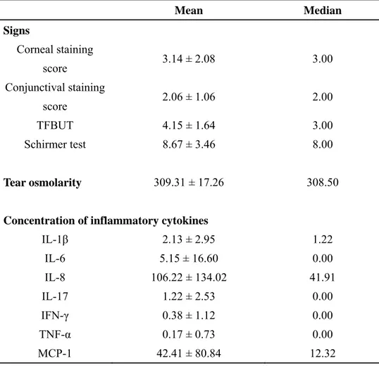
Mean values at baseline are presented in Table 1. As inclusion criteria included TFBUT less than 5 seconds and positive corneal and conjunctival staining, mean corneal staining score and conjunctival staining score were 3.14 ± 2.08 and 2.06 ± 1.06, respectively. Mean TFBUT and Schirmer test score were 4.15 ± 1.64 seconds and 8.67 ± 3.46, respectively. Mean tear osmolarity at baseline (309.31 ± 17.26 mOsm/L) was similar to that found by Suzuki et al. (309.7 ± 22.3 mOsm/L).12 Seven cytokines were detected; IL-8 (106.22 ± 134.02 pg/mL) and
TABLE 1. Descriptive Statistics for Signs, Tear Osmolarity, and Inflammatory
Cytokines in Dry Eye Patients
Mean Median Signs Corneal staining score 3.14 ± 2.08 3.00 Conjunctival staining score 2.06 ± 1.06 2.00 TFBUT 4.15 ± 1.64 3.00 Schirmer test 8.67 ± 3.46 8.00 Tear osmolarity 309.31 ± 17.26 308.50
Concentration of inflammatory cytokines
IL-1β 2.13 ± 2.95 1.22 IL-6 5.15 ± 16.60 0.00 IL-8 106.22 ± 134.02 41.91 IL-17 1.22 ± 2.53 0.00 IFN-γ 0.38 ± 1.12 0.00 TNF-α 0.17 ± 0.73 0.00 MCP-1 42.41 ± 80.84 12.32
2. Changes of Dry Eye Signs and Tear Osmolarity
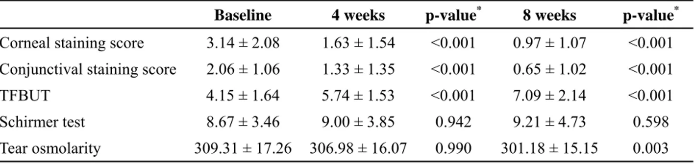
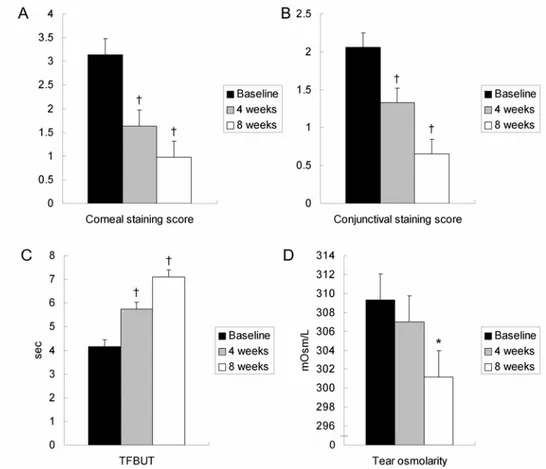
Corneal staining score, conjunctival staining score, and TFBUT showed statistically significant improvement at week 4 and 8 compared with baseline. Schirmer test scores were increased at week 4 and 8, but there were no statistically significant differences. A statistically significant decrease in tear osmolarity was observed at week 8 compared with baseline (p=0.003). Tear osmolarity decreased at week 4 and 8, which was statistically significant at week 8 (Table 2, Figure 1).
TABLE 2. Changes in Dry Eye Signs and Tear Osmolarity
Baseline 4 weeks p-value* 8 weeks p-value*
Corneal staining score 3.14 ± 2.08 1.63 ± 1.54 <0.001 0.97 ± 1.07 <0.001 Conjunctival staining score 2.06 ± 1.06 1.33 ± 1.35 <0.001 0.65 ± 1.02 <0.001
TFBUT 4.15 ± 1.64 5.74 ± 1.53 <0.001 7.09 ± 2.14 <0.001
Schirmer test 8.67 ± 3.46 9.00 ± 3.85 0.942 9.21 ± 4.73 0.598
Tear osmolarity 309.31 ± 17.26 306.98 ± 16.07 0.990 301.18 ± 15.15 0.003 * paired t-test was used.
FIGURE 1.Changes in corneal staining score (A), conjunctival staining score
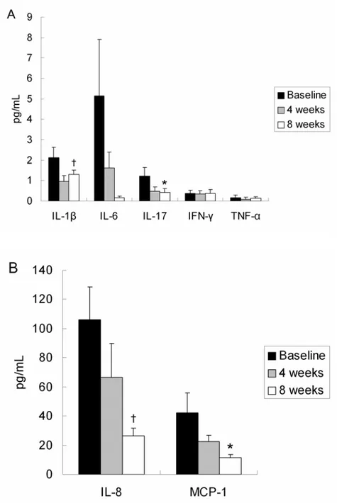
3. Changes in Inflammatory Cytokine Concentrations
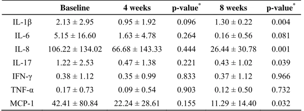
IL-1β, IL-8, IL-17, and MCP-1 were significantly decreased at week 8 compared with baseline (p=0.004, 0.001, 0.039, 0.032 respectively). The concentrations of these cytokines also decreased at week 4, but the differences were not
statistically significant. Other cytokines (IL-6, IFN-γ, and TNF-α) also decreased at week 4 and 8 but was not statistically significant (Table 3, Figure 2).
TABLE 3. Changes in Inflammatory Cytokine Concentrations
Baseline 4 weeks p-value* 8 weeks p-value*
IL-1β 2.13 ± 2.95 0.95 ± 1.92 0.096 1.30 ± 0.22 0.004 IL-6 5.15 ± 16.60 1.63 ± 4.78 0.264 0.16 ± 0.56 0.081 IL-8 106.22 ± 134.02 66.68 ± 143.33 0.444 26.44 ± 30.78 0.001 IL-17 1.22 ± 2.53 0.47 ± 1.38 0.221 0.43 ± 1.02 0.039 IFN-γ 0.38 ± 1.12 0.35 ± 0.99 0.833 0.37 ± 1.12 0.966 TNF-α 0.17 ± 0.73 0.09 ± 0.54 0.903 0.12 ± 0.50 0.732 MCP-1 42.41 ± 80.84 22.24 ± 28.61 0.155 11.29 ± 14.40 0.032 * paired t-test was used.
FIGURE 2. Changes in inflammatory cytokine concentrations. 1β, 6, IL-17, IFN-γ, and TNF-α (A), IL-8 and MCP-1 (B). *p < 0.05, †p < 0.01.
4. Safety Results
No adverse events were observed by the examiner or reported by the patients during the study period; the study medications were well tolerated.
IV. DISCUSSION
This study showed that treatment with topical 1% methylprednisolone not only improves clinical indices, such as corneal staining score, conjunctival staining score, and TFBUT, but also decreases tear osmolarity and levels of inflammatory cytokines.
Tear hyperosmolarity is regarded as the central causing ocular surface
inflammation, damage, and symptoms in dry eye syndrome.3 Hyperosmolarity
stimulates a cascade of inflammatory events in the epithelial surface cells, involving mitogen-activated protein kinases (MAPKs), nuclear factor-kappa B signaling pathway,14 and the generation of inflammatory cytokines (IL-1α, IL-1β,
and TNF-α) and MMP-9,11 which activate inflammatory cells at the ocular
surface.15 Increased levels of multiple pro-inflammatory cytokines in the tears of
been confirmed by previous studies.4-6
As it is not correlated to subsets of dry eye syndrome, tear osmolarity is believed to be a global indicator of the disease3 and has been proposed as the gold
standard for diagnosis of dry eye.16 Previously, measurement of tear osmolarity
was thought to require at least 5 μL of tear sample, not practical for use in a clinical setting.17 It has also been difficult to determine tear osmolarity
accurately due to reflex tearing.18 With technological advances, it is now
possible to measure osmolarity using smaller amounts of tears (e.g., 50 nL in this study) and more conveniently by collecting tear samples from the inferior tear lake with the tip of the test card. Recent studies showed significant correlations between tear osmolarity and dry eye severity.12,13
It is unique in this study that we proposed a possible objective approach to evaluate the efficacy of topical methylprednisolone in the treatment of patients with dry eye syndrome. Recent studies have shown that inflammatory cytokines and tear osmolarity are increased in dry eye syndrome and that tear osmolarity has a significant correlation with dry eye severity.4-6,12,13 It is anticipated that
improvements in dry eye syndrome will result in a decrease in inflammatory cytokine levels and tear osmolarity. By measuring the changes in inflammatory cytokine levels and tear osmolarity, we could objectively confirm a decrease in tear osmolarity resulting from the anti-inflammatory effect of
methylprednisolone.
Methylprednisolone has been shown to have great efficacy in preserving corneal epithelial barrier function. De Paiva et al.11 reported that methylprednisolone
decreased levels of MMP-9, IL-1α, IL-1β, and TNF-α transcripts as well as activation of MAPKs. MMP-9 plays a key role in corneal barrier disruption in dry eye via the lysis of tight junction proteins in the apical epithelium, and the therapeutic effect of methylprednisolone may be primarily the result of MMP-9 inhibition.19 Moreover, methylprednisolone might secondarily inhibit MAPKs by
decreasing the production of IL-1 and TNF-α that activate MAPKs, as
corticosteroids have not been recognized as direct MAPKs inhibitors.11,20 In this
study, we confirmed a significant decrease in IL-1β levels after treatment with topical methylprednisolone.
IL-17 is a potent pro-inflammatory cytokine, and interaction of IL-17 with its receptor evokes activation of IL-8, resulting in recruitment of neutrophils to the injury site.21 MCP-1 has been identified as a key molecule for the chemotaxis of
monocytes to the site of inflammation.22 The results of this study showed that
IL-17, IL-8, and MCP-1 levels were all significantly decreased by topical methylprednisolone. These findings suggest that topical methylprednisolone improved ocular surface inflammation by decreasing the recruitment of inflammatory cells.
Other pro-inflammatory cytokines including IL-6, IFN-γ, and TNF-α also decreased, but the differences were not statistically significant in this study. Further and additional studies are required to define the changes in inflammatory cytokines after treatment with topical methylprednisolone.
Tear hyperosmolarity arises in situations of low aqueous tear flow, excessive evaporation, or a combination of these events.3 A previous study demonstrated
that patients with faster tear film thinning time may be more susceptible to evaporation of tear film, thus increasing tear film evaporation, which may lead to a more concentrated tear film and increased osmolarity.23 In this study, the
stability of tear film (i.e., TFBUT) was significantly improved at weeks 4 and 8, and tear osmolarity significantly decreased at week 8.
Preservatives in topical agents such as benzalkonium chloride excite inflammatory cell markers at the ocular surface, causing epithelial cell damage and a decrease in mucin expression in addition to any direct effect on goblet cells.3 In this study,
topical methylprednisolone was prepared as a non-preserved aqueous solution to avoid the toxicity associated with benzalkonium chloride. It should be
considered that preservative-free 0.1% sodium hyaluronate was also used in this study. As sodium hyaluronate has a huge capacity to bind water, with an affinity 1000-fold of its own weight,24 it was used to retain the aqueous layer of tear film
to the therapeutic effects of methylprednisolone.
V. CONCLUSION
In this study, we objectively evaluated the anti-inflammatory effects of topical methylprednisolone in the treatment of dry eye patients by measuring the changes in inflammatory cytokine levels and tear osmolarity. With more technological advances, the changes in tear inflammatory cytokine levels and osmolarity, in addition to the widely used clinical parameters such as corneal and conjunctival staining, TFBUT, and Schirmer test, could be used as objective methods to evaluate the efficacy of treatment in dry eye patients.
REFERENCES
1. Johnson ME, Murphy PJ. Changes in the tear film and ocular surface from dry eye syndrome. Prog Retin Eye Res 2004;23:449-74.
2. Lemp MA. Report of the National Eye Institute/Industry Workshop on clinical trials in dry eye. CLAO J 1995;21:221-32.
3. DEWS definition and classification. The definition and classification of dry eye disease: report of the definition and classification subcommittee of the
international dry eye workshop (2007). Ocul Surf 2007;5:179-93.
4. Massingale ML, Li X, Vallabhajosyula M, Chen D, Wei Y, Asbell PA. Analysis of inflammatory cytokines in the tears of dry eye patients. Cornea
2009;28:1023-7.
5. Lam H, Bleiden L, De Paiva CS, Farley W, Stern ME, Pflugfelder SC. Tear Cytokine profiles in dysfunctional tear syndrome. Am J Ophthalmol
2009;147:198-205.
6. Yoon KC, Park CS, You IC, Choi HJ, Lee KH, Im SK, et al. Expression of CXCL9, -10, -11, and CXCR3 in the tear film and ocular surface of patients with dry eye syndrome. Invest Ophthalmol Vis Sci 2010;51:643-50.
7. Stern ME, Gao J, Schwallb TA, Ngo M, Tieu DD, Chan CC, et al. Conjunctival T-cell subpopulations in Sjögren’s and non-Sjögren’s patients with dry eye.
Invest Ophthalmol Vis Sci 2002;43:2609-14.
8. Marsh P, Pflugfelder SC. Topical nonpreserved methylprednisolone therapy for keratoconjunctivitis sicca in Sjögren syndrome. Ophthalmology 1999;106:811-6. 9. Lee HK, Ryu IH, Seo KY, Hong S, Kim HC, Kim EK. Topical 0.1%
prednisolone lowers nerve growth factor expression in keratoconjunctivitis sicca patients. Ophthalmology 2006;113:198-205.
10. Sall K, Stevenson OD, Mundorf TK, Reis BL. Two multicenter, randomized studies of the efficacy and safety of cyclosporine ophthalmic emulsion in moderate to severe dry eye disease. CsA phase 3 study group. Ophthalmology 2000;107:631-9.
11. De Paiva CS, Corrales RM, Villarreal AL, Farley WJ, Li DQ, Stern ME, et al. Corticosteroid and doxycycline suppress MMP-9 and inflammatory cytokine expression, MAPK activation in the corneal epithelium in experimental dry eye.
Exp Eye Res 2006;83:526-35.
12. Suzuki M, Massingale ML, Ye F, Godbold J, Elfassy T, Vallabhajosyula M, et al. Tear osmolarity as a biomarker for dry eye disease severity. Invest
Ophthalmol Vis Sci 2010;51:4557-61.
13. Sullivan BD, Whitmer D, Nichols KK, Tomlinson A, Foulks GN, Geerling G, et al. An objective approach to dry eye disease severity. Invest Ophthalmol Vis
14. Li DQ, Chen Z, Song XJ, Luo L, Pflugfelder SC. Stimulation of matrix metalloproteinases by hyperosmolarity via a JNK pathway in human corneal epithelial cells. Invest Ophthalmol Vis Sci 2004;45:4302-11.
15. Baudouin C. The pathology of dry eye. Surv Ophthalmol 2001;45 Suppl 2:S211-20.
16. Farris RL. Tear osmolarity: a new gold standard? Adv Exp Med Biol 1994;350:495-503.
17. Farris RL, Stuchell RN, Mandel ID. Tear osmolarity variation in the dry eye. Trans Am Ophthalmol Soc 1986;84:250-68.
18. Nelson JD, Wright JC. Tear film osmolarity determination: an evaluation of potential errors in measurement. Curr Eye Res 1986;5:677-81.
19. Pflugfelder SC, Farley W, Luo L, Chen LZ, de Paiva CS, Olmos LC, et al. Matrix metalloproteinase-9 knockout confers resistance to corneal epithelial barrier disruption in experimental dry eye. Am J Pathol 2005;166:61-71. 20. Liacini A, Sylvester J, Li WQ, Zafarullah M. Inhibition of
interleukin-1-stimulated MAP kinases, activating protein-1 (AP-1) and nuclear factor kappa B (NF-kappa B) transcription factors down-regulates matrix metalloproteinase gene expression in articular chondrocytes. Matrix Biol 2002;21:251-62. 21. Nguyen CQ, Yin H, Lee BH, Chiorini JA, Peck AB. IL17: potential
transfer. Lab Invest 2011;91:54-62.
22. Lagu B, Gerchak C, Pan M, Hou C, Singer M, Malaviya R, et al. Potent and selective CC-chemokine receptor-2 (CCR2) antagonists as a potential treatment for asthma. Bioorg Med Chem Lett 2007;17:4382-6.
23. Nichols JJ, Sinnott LT. Tear film, contact lens, and patient-related factors associated with contact lens-related dry eye. Invest Ophthalmol Vis Sci 2006;47:1319-28.
24. Nakamura, M, Hikida M, Nakano T, Ito S, Hamano T, Kinoshita S. Characterization of water retentive properties of hyaluronan. Cornea 1993;12:433-6.