Original P
aper
β-Catenin and Its Alteration in an Experimental Model of
Diabetic Nephropathy
Se Jin Park,1 Eun-Mi Ahn,2 Tae-Sun Ha,2 Jae Il Shin3
Introduction. The aim of our study was to determine whether β-catenin, a subunit of the cadherin protein complex in the podocyte cytoskeleton, would be altered by hyperglycemia and advanced glycation endproducts (AGE) in glomerular epithelial cells and podocytes in vitro.
Materials and Methods. Rat glomerular epithelial cells and mouse podocytes on bovine serum albumin-coated or AGE-coated plates with normal (5 mM) and high (30 mM) glucose doses were cultured and examined for the distribution of β-catenin using confocal microscopy and changes in β-catenin production by western blotting and reverse transcription-polymerase chain reaction, at 48 hours, 4 weeks, and 10 weeks.
Results. Immunofluorescent staining revealed that β-catenin and α-actinin were colocalized around the cell membrane, and that β-catenin staining was most intense along the capillary loops, but moved internally toward the inner actin filaments in the presence of AGE and hyperglycemia. In western blot analysis, AGE and hyperglycemia significantly decreased the amount of β-catenin proteins by 31.5% at 48 hours, compared with normal control conditions (P < .01). The expression for β-catenin mRNA in AGE and hyperglycemia was also decreased by 59.6% at 24 hours, compared with that of normal glucose conditions (P < .01). No significant changes were seen in the osmotic controls.
Conclusions. Our results suggest that AGE and hyperglycemia may induce the cytoplasmic redistribution of β-catenin and inhibit the production of β-catenin at the transcriptional and posttranslational levels, which may result in the development of kidney dysfunction in diabetic conditions.
IJKD 2014;8:299-309 www.ijkd.org 1Department of Pediatrics,
Ajou University Hospital, Ajou University School of Medicine, Suwon, Korea
2Department of Pediatrics, Chungbuk National University College of Medicine, Cheongju, Korea
3Department of Pediatrics, Severance Children’s Hospital, Yonsei University College of Medicine, Seoul, Korea
Keywords. β-catenin,
advanced glycation endproducts, glomerular epithelial cells, podocytes, diabetic nephropathy
INTRODUCTION
β-catenin, a subunit of the cadherin protein complex, is a multifunctional protein is encoded by the CTNNB1 gene that in humans.1 When β-catenin
was sequenced, it was found to have an armadillo repeat domain (residues 141-664) composed of 12 armadillo repeats, and an N-terminal and a C-terminal domain, which are specialized for protein-protein binding.2 This protein plays a
crucial role in adherens junctions of the cell, where it interacts with the cytoplasmic region of cadherin and acts as an intracellular signal transducer in the Wnt signaling pathway.3-5 In the absence of Wnt
signaling, cytosolic β-catenin binds the destruction complex, where it is phosphorylated by casein kinase 1α and glycogen synthase kinase-3β and thus labels it for ubiquitin-dependent degradation by the proteasome.6 When a Wnt signal is present,
however, β-catenin moves into the nucleus, where it binds to a DNA-binding member of the lymphocyte enhancer factor-1/T cell factor transcription factor family and other transcription factors, resulting in the activation of Wnt-target genes.7
The majority of β-catenin in the podocyte binds to P-cadherin and α-catenin at adherens junctions, and α-catenin binds to actin as a homodimer.8
Recent evidence suggests that β-catenin anchors the actin cytoskeleton and may be responsible for transmitting the contact inhibition signal that causes cells to stop dividing once the epithelial sheet is complete.2 Importantly, the
β-catenin-binding site on α-catenin overlaps with the α-catenin homodimerization interface.8 Thus, α-catenin
cannot simultaneously be bound to both actin and β-catenin in the cell, which is required for its interaction only with actin, or bound to P-cadherin through β-catenin.9
The distribution and changes of cytoskeletal and adherens junctions proteins in glomerular podocytes have been proposed to function as the key regulators in the pathophysiology of diabetic nephropathy9,10; however, to date, there have
been few studies on the role of β-catenin in the pathogenesis of diabetic nephropathy. Therefore, we investigated through in vitro culture experiments whether there are any pathological changes in the β-catenin of glomerular podocytes in diabetic conditions.
MATERIALS AND METHODS
Cell Culture of Rat Glomerular Epithelial Cells and Mouse Podocytes
Rat glomerular epithelial cells (GECs), cloned from primary rat glomerular cultures, were characterized and provided by Kreisberg and coworkers.11 The cells were characterized by
sensitivity to puromycin aminonucleoside, positive for Heymann antigen (gp330) and podocalyxin, and negative staining for factor VIII.11,12 The
GECs were maintained in RPMI 1640 (Gibco BRL, Gaithersburg, MD, USA) supplemented with 10% heat-inactivated fetal bovine serum, 1 M Hepes, and antibiotics. Fetal bovine serum was reduced to 0.5% at least 24 hours before each experiment to reduce background. Fresh media was supplied once every 2 days, and for the subculture, 0.05% trypsin was used to detach cells from the culture dishes.
Conditionally immortalized mouse podocytes were provided by Harvard University, Cambridge, MA, USA (Dr Peter Mundel) and were cultured and differentiated as described previously.10
To stimulate podocyte proliferation, cells were cultivated at 33°C (permissive conditions) in a culture medium supplemented with 10 U/ mL mouse recombinant γ-interferon (Roche, Mannheim, Germany) to induce expression of temperature-sensitive large T antigens. To induce differentiation, the podocytes were maintained at 37°C (nonpermissive conditions) for at least 2 weeks.
Culture Additives
Cells were serum-deprived to reduce background 24 hours before each experiment, then exposed to glucose or advanced glycation endproducts (AGE), or both. Rat GECs and mouse podocytes were incubated in culture media containing either 5 mM (normal) or 30 mM (high) of glucose without insulin. The AGE were produced by the addition of glucose-6-phosphate to a final concentration of 0.2 M in a phosphate buffer solution (pH 7.4) containing 50 mg/mL of bovine serum albumin and protease inhibitors (100 mM 6-aminohexanoic acid, 10 mM benzamidine hydrogen chloride, and 1 mM phenylmethylsulfonyl fluoride), and then incubated in the reaction tube at 37°C for 6 weeks under sterile conditions.13 The formation of AGE was
confirmed by measuring the AGE emission peak at 440 nm with a fluorescence spectrophotometer. To imitate long-term diabetic conditions, AGE (5 μg/ mL) was added, and controls were established using unmodified bovine serum albumin (5 μg/mL). To exclude the effect of additionally glycated proteins produced in culture conditions, an incubation period no longer than 48 hours was used. Fetal bovine serum was reduced to 0.5% on the last media change to reduce background before protein and RNA extraction. For the purpose of identification, AGE and bovine serum albumin are denoted as ‘A’ and ‘B’, and glucose at 5 mM and 30 mM as ‘5’ and ‘30’, respectively. In addition, the osmotic control with 5-mM glucose and 25-mM mannitol is expressed as ‘osm.’ With these combinations, the experimental conditions are expressed as A5, A30, Aosm, B5, and B30, defined as follows: (1) A5 (mM) represents the condition that may have a long-term diabetic condition to form AGE but remains in normal range of glucose; (2) A30 (mM) means
long-term diabetes mellitus and hyperglycemia; (3) Aosm represents the osmotic control group; (4) B5 (mM) means no diabetic condition and normal glucose levels; and (5) B30 (mM) represents initial diabetic condition with hyperglycemia.
Confocal Microscopy for β-catenin Distribution
Rat GECs and mouse podocytes were grown on type I collagen-coated glass cover slips (Upstate Biotechnology Incorporation, Lake Placid, NY, USA) incubated for 24 hours and then fixed in 4% paraformaldehyde, permeabilized in phosphate buffered saline, blocked with 10% normal goat serum (GIBCO BRL, Rockville, MD, USA), and labeled with polyclonal rabbit anti-rat β-catenin (Santa Cruz Biotechnology Inc, Santa Cruz, CA, USA) or TRITIC-phalloidin (Sigma Chemical Co, St Louis, MO, USA) for F-actin staining. Primary antibody-bound specimens were incubated with 1:500 (v/v) Alexa 594 for red conjugates (Molecular Probes, Invitrogen, Carlsbad, CA, USA) and Alexa 488 for green, respective of secondary antibodies, at room temperature for 1 hour. Cover slips were mounted in aqueous mountant and viewed with a fluorescence microscope (TCS SP2 AOBS, Leica Microsystems, Wetzlar, Germany) at wavelengths of 496-534 nm (emission) and 488 nm (excitation) for Alexa 488-conjugated β-catenins, or at 555-635 nm (emission) and 594 nm (excitation) for Alexa 594-conjugated β-catenins. Differentiated mouse podocytes were also prepared as rat GECs for confocal images with the same antibody.
Western Blotting
The confluently grown cell layers were incubated with additives for 48 hr for β-catenin, and for various durations for AGE, and were extracted with a protein extraction solution (PRO-PREP, Intron Biotechnology Inc., Seongnam, Kyeonggi, Korea) at 4°C over night and stored at -20°C until further analysis. Protein concentrations were determined with a Bio-Rad kit (Bio-Rad Laboratories, Hercules, CA, USA).13 To perform
western blotting for β-catenin, 30 μg of boiled extracts were applied on 10% SDS-PAGE gels and transferred to polyvinylidene fluoride membranes (Bio-Rad Laboratories, Hercules, CA, USA). The membranes were air-dried and blocked in 5% fat-free milk before incubation with anti-β-catenin antibody. After incubation with goat anti-rabbit
IgG-HRP (Santa Cruz Biotechnology Inc, Santa Cruz, CA, USA), bands were detected using the LAS-3000 imaging system (Fujifilm Life Science Inc, Minato-ku, Tokyo, Japan). The actin bands confirmed the loading of comparable amounts of extracted protein. Three independent experiments were performed.
Reverse Transcriptase-Polymerase Chain Reaction Analysis of β-catenin
Total RNA was extracted from cultured rat GECs and mouse podocytes. After estimating its concentration by ultraviolet spectrophotometry, 5 μg of total RNA was used for first-strand cDNA synthesis. Aliquots of the cDNA were amplified using primers: (1) CTNNB1: sense 5’-TGGACTACCACGCAGCGAAC-3’, antisense 5’-GCTCTGGCTTAGGCAACGC-3’; (2) GAPDH: sense 5’-CTCTACCCACGGCAAGTTCAA-3’, antisense 5’-GGATGACCTTGCCCACAGC-3’ (Bionics Inc, Seoul, Korea). The housekeeping gene GAPDH served as an internal control. Polymerase chain reaction products were visualized on 1.5% agarose gels, and band density was measured using a densitometry program (Labworks 4.0, UVP Inc, Upland, CA, USA). Standard polymerase chain reaction assays were normalized to GAPDH mRNA band densities. Three independent experiments were performed.
Statistical Analysis
The results were presented as mean ± standard deviation, as appropriate under different conditions. Statistical significance was evaluated by the nonparametric Kruskal-Wallis analysis or Mann-Whitney U test. P values less than .05 were considered significant.
RESULTS
β-catenin Distribution on Confocal Microscopy
The podocytes for β-catenin and F-actin were double-stained and the cell nucleus was stained with 4’-6-diamidino-2-phenylindole. β-catenin as well as α-actinin was located along the glomerular capillary walls (Figure 1). Staining for β-catenin was most intense in the capillary walls. From 48 hours in diabetic conditions to 4 weeks and 10 weeks, β-catenin staining became blurry, indicating a reduction in β-catenin. In vitro, β-catenin and F-actin did not overlap and were located at
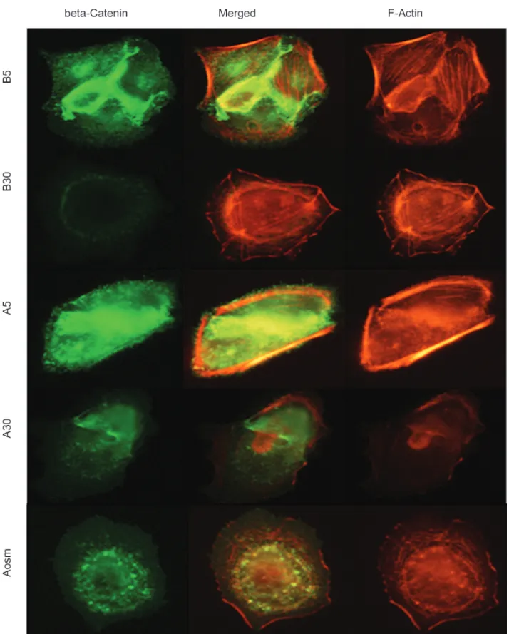
different sites on the confocal image. In the diabetic condition with rat GECs, β-catenin moved toward the cytoplasm from the cell membrane at A5 and A30, compared with B5 (Figure 2). A similar redistribution of β-catenin was also observed in the differentiated mouse podocytes, especially at A30 (Figure 3). In the merged views, β-catenin was distributed at the cell membrane at B5 but redistributed into the cytoplasm at A5, B30, and A30, suggesting that high glucose and AGE may have an impact on the redistribution of β-catenin molecules in GECs and podocytes with F-actin.
β-catenin Protein Assayed by Western Blotting
A major band of β-catenin protein was found at
90 kDa and the results were compared with B5. In both GECs and podocyte, density values for the β-catenin protein of representative immunoblots from each condition tended to decrease in the presence of high glucose and AGE levels in a dose- and time-dependent manner. Densitometric analysis of western blots showed 23.7% and 27.7% decreases in A5 and A30 at 24 hours (P < .05; P < .01; n = 3) and 20.5% and 31.5% decreases in B30 and A30 at 48 hours (P < .05; P < .01; n = 3), respectively, compared with B5 in rat GECs (Figure 4 Top). In mouse podocytes, the density values of β-catenin were also decreased in a dose-dependent manner. High doses (30 mM) of glucose and AGE decreased cellular β-catenin
Figure 1. Immunofluorescent staining of α-actinin and β-catenin. Double-staining of α-actinin and β-catenin show a decrease in the foot
Figure 2. Confocal microscopy with β-catenin and F-actin in rat glomerular epithelial cells. The merged views show that β-catenin is
relocalized inside the cytoplasm in B30, A5, and A30. Advanced glycation endproducts and bovine serum albumin are denoted as ‘A’ and ‘B’, and glucose at 5 and 30 mM by ‘5’ and ‘30’, respectively (× 1000).
Figure 3. Confocal microscopy with β-catenin and F-actin in mouse podocytes. The overlap shows that β-catenin was redistributed from
the sites of cell-cell contact areas between adjacent podocytes into the cytoplasm in B30, A5, and A30. The intensity of β-catenin is decreased in B30 and A30 (× 1000).
protein levels by 23.7% at 24 hours and 13.7% at 48 hours in B30 (both P < .05; n = 3), and by 42.1%
at 24 hours and 27.3% at 48 hours in A30 (both P < .01; n = 3; Figure 4 Bottom). However, these Figure 4. Effects of glucose and advanced glycation endproducts (AGE) on β-catenin protein in cultured rat glomerular epithelial cells
(Top) and mouse podocytes (Bottom) assayed by Western blotting. β-catenin levels were significantly decreased in B30, A5, and A30, compared with that of B5 condition. The density values for β-catenin protein of the representative immunoblots from each group showed a decrease in cellular β-catenin protein levels in B30, A5, and A30, compared with the value for B5. Data on the densitometric analysis of the β-catenin-β-tubulin ratio are expressed as mean ± standard deviation. Control (100%) is shown as “(-).”
*P < .05 **P < .01
changes were not found in the osmotic control (Aosm). The β-catenin reduction in A30 was more than that in B30, suggesting that AGE may have a more substantial impact on the amount of β-catenin protein in GECs and podocytes than high glucose levels.
β-catenin mRNA Assayed by Reverse Transcriptase-polymerase Chain Reaction
In both rat GECs and mouse podocytes, bands of mRNA for β-catenin were observed at 300 bp and the expression levels of β-catenin mRNA in each condition were compared with that of B5 after adjusting for the amount of polymerase chain reaction products relative to GAPDH. Values for mRNA expression of β-catenin and representative gels showed a significant decrease in rat reverse transcriptase-polymerase chain reaction products of β-catenin in B30 and A30 at 48 hours (25.6%, P < .05 and 20.8% P < .05; n = 3; Figure 5 Top). In addition, the expression of mouse β-catenin mRNA was also suppressed by 35.0% and 59.6% at 24 hours in B30 and A30 (P < .05; P < .01, respectively), and by 26.3% and 25.7% at 48 hours in A5 and A30 (both P < .05; Figure 5 Bottom). As in the western blot analysis, the expression of β-catenin mRNA was much less in A30 than in B30, and these changes were not observed in the osmotic condition. These outcomes of reverse transcriptase-polymerase chain reaction analysis indicate that genetic regulation of the β-catenin molecule may be more influenced by AGE and hyperglycemia than hyperglycemia alone at both the transcriptional level and posttranslational phase.
DISCUSSION
The purpose of this study was to investigate whether there are any pathological changes in β-catenin in diabetic conditions. We demonstrated the redistribution, relocation, and reduction of β-catenin in GECs and podocytes. The exposure of β-catenin to AGE and hyperglycemia caused β-catenin to move internally toward the cytoplasmic actin filament, suggesting that the disintegration of the β-catenin complex at the podocyte membrane may be one of the proteinuric causes in the pathogenesis of diabetic nephropathy. These results are in line with a previous study by one of our authors that found that the expression of
α-actinin was also affected and suppressed by high glucose and AGE levels either individually or in combination.14
Proteinuria in diabetic nephropathy has a variety of causes,15,16 including glomerular basement
membrane changes, retraction and effacement of the interdigitating foot processes of podocytes, disruption of charge and size selectivity, and alteration of the slit diaphragm and cytoskeletal molecules.17,18 Recent structural and biochemical
studies have revealed how β-catenin acts as a crucial regulator of cell adhesion19,20 and is the
central effector of the canonical Wnt signaling pathway.21-23 The N terminal domain of β-catenin
harbors glycogen synthase kinase-3 and casein kinase 1α phosphorylation sites for transducin repeats-containing proteins ubiquitin ligase and the binding site for α-catenin.24-26 The central domain
of the armadillo repeat is the binding site for most β-catenin-binding partners, including adenomatous polyposis coli protein, inhibitor of β-catenin and TCF4 (ICAT), axin, and B-cell CLL/lymphoma 9 (BCL9) gene.21 The cytoplasmic domain of cadherin
interacts with the groove of the β-catenin armadillo repeat domain. The 3-dimensional structure of the β-catenin complex in the β-catenin destruction complex and transcriptional complex has also been revealed.21
Upon binding with a ligand, the phosphorylation of β-catenin is inhibited, leading to cytoplasmic accumulation and subsequent nuclear translocation.27
In the nucleus, β-catenin binds to the lymphocyte enhancer factor-1/T cell factor transcription factor family and other associated transcription factors to mediate gene transcription. This results in the activation of Wnt-target genes, causing the development of diabetes, chronic kidney disease, and diabetic kidney disease.28 In the absence of
Wnt signaling, β-catenin binds to the destruction complex, where it is phosphorylated by casein kinase 1α and glycogen synthase kinase-3β, which causes it to be ubiquitylated by the β-transducin repeats-containing proteins ubiquitin ligase and subsequently degraded by the proteasome.29,30
Recently, Kato and colleagues reported that mice with podocyte-specific expression of stabilized β-catenin showed proteinuria and glomerular basement membrane abnormalities, whereas deletion of β-catenin in cultured podocytes increased the expression of podocyte differentiation markers
and enhanced susceptibility to apoptosis, suggesting
that Wnt/β-catenin signaling in podocytes plays a critical role in integrating cell adhesion, motility, differentiation, and cell death.22 Unlike that study, Figure 5. Effects of glucose and advanced glycation endproducts (AGE) levels on the mRNA expression of β-catenin in cultured rat
glomerular epithelial cells (Top) and mouse podocytes (Bottom) assayed by reverse transcription-polymerase chain reaction. The mRNA expression levels of β-catenin and a representative gel show that AGE and hyperglycemia significantly decreased the reverse transcriptase-polymerase chain reaction products of β-catenin in B30, A5, and A30, compared with the B5 condition. Data on the densitometric analysis of the β-catenin-β-tubulin ratio are expressed as mean ± standard deviation. Control (100%) is shown as “(-).” *P < .05
we focused not on the Wnt/β-catenin signaling, but on the distribution, translocation, and quantity of β-catenin in the cadherin-catenin complex in the adherens junctions of podocytes after exposure to diabetic conditions.
As one of the linking molecules in cytoskeletal organization, β-catenin is located between P-cadherin and an actin filament through α-actinin. The extracellular domain of P-cadherin, involved in hemophilic calcium-dependent cell-cell adhesion, acts as a zipper-like structure on both sides of the foot process in the slit diaphragm.31,32 The
intracellular domain of P-cadherin plays the role of a structural and functional molecule of the podocyte by binding to the actin cytoskeleton via catenins.33 The catenin complex is composed
of α-catenin (102 kDa) located on chromosome 5q31, β-catenin (92 kDa) on chromosome 3p21, and γ-catenin/plakoglobin (83 kDa) on chromosome 17q21.34 P-cadherin binds to β-catenin and/or
γ-catenin, whereas α-catenin only binds to β-catenin or γ-catenin and F-actin via α-actinin.34
In a previous study, β-catenin showed distinct down-regulation at 3 days of induction in rat models of puromycin aminonucleoside nephrosis.35
In cultured human podocytes, β-catenin was dislocated from and relocated back to plasma membranes in a similar fashion during cell-cell contact disruption and reformation.36 However,
there was few report on the changes in β-catenin related to the P-cadherin complex, particularly in diabetic conditions. In the present study, β-catenin expression was reduced by diabetic conditions in rat GECs and mouse podocytes. A lack of previous reports limits comparisons of our results with those of other animal models or human models; however, our previous findings from a study of α-actinin and zonula occludens-1 exhibited similar results in vitro: high glucose and AGE may induce pathological changes in multiple molecular proteins composing podocytes.14,37 We speculated that
the irreversible AGE used in this study reacted with receptors on the podocyte surface to cause changes in the structure and quantity of β-catenin and subsequent cellular pathways to glomerular damage. However, as the precise pathomechanism remains elusive, further studies are necessary to improve our knowledge of the pathophysiology of β-catenin changes caused by AGE through in vivo diabetic nephropathy.
CONCLUSIONS
Our study provides one of the bases for understanding β-catenin molecule in the podocyte. Both AGE and hyperglycemia induced the redistribution, translocation, and decrease of β-catenin at transcriptional and translational levels in diabetic conditions, suggesting that these in vitro changes may be one of the major causes in the cytoskeletal and functional alteration of podocytes in long-term diabetic nephropathy. Based on these findings, it should be possible to find additional proteins that cause diabetic nephropathy and to better understand kidney diseases in which a loss of glomerular molecules results in the development of proteinuria.
ACKNOWLEDGEMENTS
This research was supported by the Basic Science Research Program through the National Research Foundation of Korea, funded by the Ministry of Education, Science and Technology (2010-0005009), Korea Science and Engineering Foundation (KOSEF R01-2007-000-20856-0), and a faculty research grant from Yonsei University College of Medicine for 2010 (6-2010-0048).
CONFLICT OF INTEREST
None declared
REFERENCES
1. Kraus C, Liehr T, Hulsken J, et al. Localization of the human beta-catenin gene (CTNNB1) to 3p21: a region implicated in tumor development. Genomics. 1994;23:272-4.
2. Xu W, Kimelman D. Mechanistic insights from structural studies of beta-catenin and its binding partners. J Cell Sci. 2007;120:3337-44.
3. Mundel P, Shankland SJ. Podocyte biology and response to injury. J Am Soc Nephrol. 2002;13:3005-15.
4. Stamos JL, Weis WI. The β-catenin destruction complex. Cold Spring Harb Perspect Biol. 2013;5:a007898. 5. Surendran K, Schiavi S, Hruska KA. Wnt-dependent
beta-catenin signaling is activated after unilateral ureteral obstruction, and recombinant secreted frizzled-related protein 4 alters the progression of renal fibrosis. J Am Soc Nephrol. 2005;16:2373-84.
6. Dai C, Stolz DB, Kiss LP, Monga SP, Holzman LB, Liu Y. Wnt/beta-catenin signaling promotes podocyte dysfunction and albuminuria. J Am Soc Nephrol. 2009;20:1997-2008. 7. MacDonald BT, Tamai K, He X. Wnt/beta-catenin
signaling: components, mechanisms, and diseases. Dev Cell. 2009;17:9-26.
Alpha-catenin is a molecular switch that binds E-cadherin-beta-catenin and regulates actin-filament assembly. Cell. 2005;123:903-15.
9. Gates J, Peifer M. Can 1000 reviews be wrong? Actin, alpha-Catenin, and adherens junctions. Cell. 2005;123:769-72.
10. Mundel P, Reiser J, Zuniga Mejia Borja A, et al. Rearrangements of the cytoskeleton and cell contacts induce process formation during differentiation of conditionally immortalized mouse podocyte cell lines. Exp Cell Res. 1997;236:248-58.
11. Kreisberg JI, Hoover RL, Karnovsky MJ. Isolation and characterization of rat glomerular epithelial cells in vitro. Kidney Int. 1978;14:21-30.
12. Singh AK, Mo W, Dunea G, Arruda JA. Effect of glycated proteins on the matrix of glomerular epithelial cells. J Am Soc Nephrol. 1998;9:802-10.
13. Ha TS, Song CJ, Lee JH. Effects of advanced glycosylation endproducts on perlecan core protein of glomerular epithelium. Pediatr Nephrol. 2004;19:1219-24. 14. Ha TS. High glucose and advanced glycosylated
end-products affect the expression of alpha-actinin-4 in glomerular epithelial cells. Nephrology (Carlton). 2006;11:435-41.
15. Zakkerkish M, Shahbazian HB, Shahbazian H, Latifi SM, Moravej Aleali A. Albuminuria and its correlates in type 2 diabetic patients. Iran J Kidney Dis. 2013;7:268-76. 16. Choudhary N, Ahlawat RS. Interleukin-6 and C-reactive
protein in pathogenesis of diabetic nephropathy: new evidence linking inflammation, glycemic control, and microalbuminuria. Iran J Kidney Dis. 2008;2:72-9. 17. Oh J, Reiser J, Mundel P. Dynamic (re)organization of the
podocyte actin cytoskeleton in the nephrotic syndrome. Pediatr Nephrol. 2004;19:130-7.
18. Asanuma K, Mundel P. The role of podocytes in glomerular pathobiology. Clin Exp Nephrol. 2003;7:255-9. 19. Wang Z, Havasi A, Gall JM, Mao H, Schwartz JH, Borkan SC. Beta-catenin promotes survival of renal epithelial cells by inhibiting Bax. J Am Soc Nephrol. 2009;20:1919-28. 20. Peng J, Dong Z. Role changes of β-catenin in kidney
injury and repair. Kidney Int. 2012;82:509-11.
21. Xu W, Kimelman D. Mechanistic insights from structural studies of beta-catenin and its binding partners. J Cell Sci. 2007;120:3337-44.
22. Kato H, Gruenwald A, Suh JH, Miner JH, Barisoni-Thomas L, Taketo MM, et al. Wnt/β-catenin pathway in podocytes integrates cell adhesion, differentiation, and survival. J Biol Chem. 2011;286:26003-15.
23. Zhou D, Li Y, Lin L, Zhou L, Igarashi P, Liu Y. Tubule-specific ablation of endogenous β-catenin aggravates acute kidney injury in mice. Kidney Int. 2012;82:537-47. 24. Wu G, Xu G, Schulman BA, Jeffrey PD, Harper JW,
Pavletich NP. Structure of a beta-TrCP1-Skp1-beta-catenin complex: destruction motif binding and lysine specificity of the SCF(beta-TrCP1) ubiquitin ligase. Mol Cell. 2003;11:1445-56.
25. Rayasam GV, Tulasi VK, Sodhi R, Davis JA, Ray A. Glycogen synthase kinase 3: more than a namesake. Br J Pharmacol. 2009;156:885-98.
26. Wang Z, Havasi A, Gall J, et al. GSK3beta promotes apoptosis after renal ischemic injury. J Am Soc Nephrol. 2010;21:284-94.
27. Willert K, Jones KA. Wnt signaling: is the party in the nucleus? Genes Dev. 2006;20:1394-404.
28. Sale MM, Smith SG, Mychaleckyj JC, et al. Variants of the transcription factor 7-like 2 (TCF7L2) gene are associated with type 2 diabetes in an African-American population enriched for nephropathy. Diabetes. 2007;56:2638-42. 29. Kimelman D, Xu W. beta-catenin destruction complex: insights and questions from a structural perspective. Oncogene. 2006;25:7482-91.
30. Heuberger J, Birchmeier W. Interplay of cadherin-mediated cell adhesion and canonical Wnt signaling. Cold Spring Harb Perspect Biol. 2010;2:a002915.
31. Reiser J, Kriz W, Kretzler M, Mundel P. The glomerular slit diaphragm is a modified adherens junction. J Am Soc Nephrol. 2000;11:1-8.
32. Fagotto F. Looking beyond the Wnt pathway for the deep nature of β-catenin. EMBO Rep. 2013;14:422-33. 33. Shapiro L, Weis WI. Structure and biochemistry of
cadherins and catenins. Cold Spring Harb Perspect Biol. 2009;1:a003053.
34. Beavon IR. The E-cadherin-catenin complex in tumour metastasis: structure, function and regulation. Eur J Cancer. 2000;36:1607-20.
35. Luimula P, Sandström N, Novikov D, Holthöfer H. Podocyte-associated molecules in puromycin aminonucleoside nephrosis of the rat. Lab Invest. 2002;82:713-8.
36. Heikkilä E, Ristola M, Endlich K, et al. Densin and beta-catenin form a complex and co-localize in cultured podocyte cell junctions. Mol Cell Biochem. 2007;305:9-18. 37. Ha TS. High-glucose and advanced glycosylation end
products increased podocyte permeability via PI3-K/Akt signaling. J Mol Med (Berl). 2010;88:391-400.
Correspondence to: Tae-Sun Ha, MD
410 Sungbong-ro, Heungdeok-gu, Department of Pediatrics, Chungbuk National University College of Medicine, Cheongju 361-711, Republic of Korea
Tel: +82 43 269 6374 Fax: +82 43 264 6620 E-mail: [email protected]
Se Jin Park and Eun-Mi Ahn contributed equally to this work as first authors.
Received April 2013 Revised December 2013 Accepted December 2013