국문요약
Platelet-derived growth factor가 dopaminergic
신경세포 분화에 미치는 영향
포유동물의 중추신경계를 구성하고 있는 신경세포와 별아교세포 및 희소돌기아교 세포는 발생초기 뇌실부에 있는 신경상피에서 유래한다. 신경상피에서 유래된 신경간 세포가 중추 신경계의 다양한 형태의 세포로 분화되는 기전은 아직 정확히 밝혀지지 않았으나, 세포외기질과 세포유착분자 그리고 여러 종류의 peptide growth factor 등이 관여하는 것으로 알려졌다. 신경간세포로부터 신경세포와 별아교세포 및 희소돌기아 교세포로의 분화율은 배양액 내에 포함되는 cytokine의 종류에 영향을 받는다. 즉 신경 간세포는 platelet-derived growth factor (PDGF)에 의해 신경간세포로부터 신경세포로의 분화율이 증가되며, ciliary neurotrophic factor (CNTF) 및 triiodothyronine (T3)에 의해서 는 신경간세포로부터 각각 별아교세포 및 희소돌기아교세포로의 분화율이 증가된다. 본 연구에서는 신경간세포로부터 분화된 신경세포가 어떤 신경전달 물질을 함유하 고 있는지, 그리고 어떤 cytokine이 신경간세포로부터 dopamine 신경세포를 분화시킬 수 있는지 알아보고자 하였으며 다음과 같은 결과를 얻었다.
1. Epidermal growth factor (EGF)를 포함한 무혈청 배양액으로 신경간세포를 배양했 을 때 신경간세포로부터 별아교세포, 희소돌기아교세포, 신경세포가 모두 발현되었다. 2. 이때 분화된 신경세포 중 일부는 GABA 신경세포와 substance P 신경세포였으며, tyrosine hydroxylase를 발현한(tyrosine hydroxylase immuno-reactive, TH-ir) 신경세포나 serotonin 신경세포 등은 관찰되지 않았다.
3. PDGF는 신경간세포로부터 TH-ir 신경세포의 분화를 유도하였다. 그러나 EGF, basic fibroblast growth factor, nerve growth factor, brain-derived neurotrophic factor, CNTF, glial cell-derived neurotrophic factor는 TH-ir 신경세포의 분화를 유도하지 못하였다.
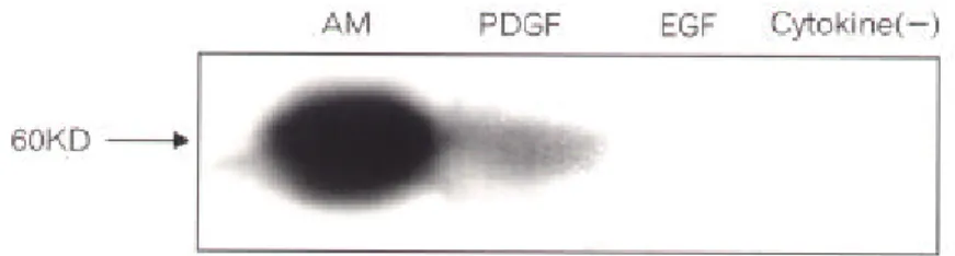
4. PDGF가 신경간세포로부터 TH-ir 신경세포를 분화시키는 것을 tyrosine hydroxylase 에 대한 western blot으로도 확인하였다.
이상의 실험결과로 PDGF는 신경간세포로부터 TH-ir 신경세포의 분화를 유도하는 것을 알 수 있었다. TH-ir 신경세포 분화유도 기전과 TH-ir 신경세포 발현율을 증가시 키는 조건은 추후 연구할 과제로 생각한다.
핵심되는 말: 분화, 신경간세포, dopamine 신경세포, platelet-derived growth factor, epi-dermal growth factor, 신경세포
Platelet-derived growth factor가 dopaminergic
신경세포 분화에 미치는 영향
<지도 연 동 수 부교수> 연세대학교 대학원 의학과손
인
숙
I. 서 론 포유동물의 중추신경계를 구성하고 있는 신경세포와 별아교세포 및 희소돌기아교세포 는 발생초기 뇌실부(ventricular zone)에 있는 신경상피에서 유래한다.1,2 신경상피에서 유래 된 신경간세포(neuronal stem cell)가 중추 신경계의 다양한 형태의 세포로 분화되는 기전은아직 확실히 밝혀지지 않았으나, 세포외기질3
(extracellular matrix)과 세포유착분자,4 그리고 epidermal growth factor (EGF),5 ,6 transforming growth factor-α (TGFα),7 basic fibroblast growth
factor (bFGF)8 등을 포함한 peptide growth factor 등이 관여하는 것으로 알려졌다.9
신경간세포로부터 신경세포의 분화과정에는 세포간 접촉이 중요한 역할을 하는 것으로
알려졌다. 예를 들어 발생기시 신경세포는 radial glial cell과 부착 이동되면서 분화된다.10
또 신경을 절단하여 acetylcholine이나 영양인자의 근육으로의 전달을 차단하면 그 신경이 지배하고 있는 근육이 퇴화되며, 실험적으로 근육을 제거하면 그 근육을 지배하는 운동신 경의 퇴화가 오는데 이는 근육에서도 신경의 분화 및 성장에 영향을 주는 신경영양인자 (neurotrophic factor)가 생성되기 때문인 것으로 알려졌다.1 1 이와 유사하게 중추신경계에서 는 신경세포와 별아교세포가 접촉하고 있으면서 상호간의 생존, 증식, 재생 등에 많은 영 향을 준다. 별아교세포는 신경세포에서 분비되는 신경전달물질을 섭취하여 증식에 영향을 받으면서, 여러 cytokine이나 신경영양물질을 신경세포로 분비하여 신경세포의 생존율과 분화에 영향을 주기도 한다.12 또한 신경세포도 별아교세포의 성상에 많은 영향을 준다. 별 아교세포 배양시 신경세포를 직접 접촉시키면서 동시배양할 때는 별아교세포를 단독배양 할때에 비해 별아교세포 내에 특이적으로 존재하는 세포골격 단백질인 glial fibrillary acidic protein (GFAP)의 함량이 감소한다.13 , 14
신경세포의 분화과정에는 그 밖에도 전술한 대로 세포외기질이 관여하는 것으로 보고되
었다. 흰쥐 시신경은 태아기 14일째에 laminin이 발현된 안포경(optic stalk) 부위에서 처음 자라기 시작한다.15 이 부위의 laminin은 생후 10일, 즉 시신경이 발육하는 기간 동안 계속 존재한다. 그러나 생후 12일 시신경 발육이 완료된 후에는 시신경 주위 혈관에서만 laminin 이 발현되는 것으로 보고되었다. 또 흰쥐에서 시신경 손상후 손상부위 별아교세포 막에 laminin이 발현되며, 발현된 laminin은 신경재생에 관여하는 것으로 알려졌다.16 따라서 laminin은 태아기시 중추신경계 신경세포의 이동, 증식, 분화, 성숙 과정에 관여할 가능성 이 있으며, 또 중추신경계 손상 후 신경재생에 영향을 줄 가능성이 있다. 최근 laminin은 망 막에 존재하는 신경간세포로부터 신경세포로의 분화과정을 선택적으로 촉진한다는 보고 가 있었다.17 또 laminin은 신경간세포로부터 신경세포의 분화과정 뿐 아니라 GABA 신경 세포의 분화를 촉진하는 것이 보고된 바 있다.18 이처럼 신경간세포로부터 신경세포로의 분화과정 그리고 신경세포 subtype으로의 분화과정에는 여러요인이 작용한다. 특히 신경간세포로부터 신경세포와 별아교세포 및 희소돌기아교세포로의 분화율은 배 양액내에 포함되는 cytokine의 종류에 영향을 받는 것으로 보고되었다.19 ,2 0 즉 EGF를 포함 한 배양액으로 배양된 신경간세포는 brain-derived neurotrophic factor (BDNF), platelet-derived growth factor (PDGF)에 의해 신경간세포로부터 신경세포로의 분화율이 증가되며, ciliary neurotrophic factor (CNTF) 및 triiodothyronine (T3)에 의해서는 각각 신경간세포로부
터 별아교세포 및 희소돌기아교세포로의 분화율이 증가된다.2 0 본 연구에서는 신경간세포로부터 분화된 신경세포가 어떤 신경전달 물질을 함유하고 있 는지, 그리고 어떤 조건에서 dopamine 신경세포로 분화되는지 보고자 하였다. II. 재료 및 방법 1. 신경간세포의 일차 배양 신경간세포의 일차배양은 Reynolds 및 Weiss의 방법으로 시행하였다.2 1 즉, 생후 1~2일
된 흰쥐 머리를 절단하고 두개골을 제거한 후, 대뇌를 Ca /Mg -free Hanks balanced
salts solution (HBSS, Gibco, Grand Island, NY, USA)에 넣었다. 수술 현미경 하에서 뇌막을 완전히 제거한 후 이를 10 ml 파이펫을 이용하여 조직 분쇄하였다. 세포를 분리하기 위해
trypsin을 0.13%되도록 첨가한 후 37oC에서 15분간 incubation하였다. Incubation이 끝난 다음
이를 Nitex filter (Tetko, Elmsford, NY, USA) #210 및 #130에 통과시키고, 통과된 세포는
100 g에서 5 분간 원심분리하였다. Trypsin을 제거하기 위해 세포 침전물을 신선한 HBSS
에 재부유시키고, 이를 다시 Nitex filter #130에 통과시킨 다음 원심분리하였다. 최종적인 세포 부유액을 얻기 위해 이 세포 침전물을 EGF가 포함된 무혈청 배양액(serum-free me-dium, SFM)에 재부유시켜 Nitex filter #40에 통과시켰다. 이렇게 얻은 세포 부유액의 세포
수는 hemocytometer를 이용하여 측정하고 6-well 배양판에 2.5 105
cells/ml의 농도로 1 ml
씩 plate하여 배양하였다. 배양 5~7일이 경과한 다음 배양액에 떠있는 신경간세포 세포군 (cell clusters)를 위상차 현미경으로 확인하였다. SFM은 50 nM hydrocortisone (Sigma, St Louis, MO, USA), 100 μM putrescine (Sigma), 30 nM selenium (Sigma), 20 μg/ml transferrin (Sigma), 10 μg/ml insulin (Sigma), 20 ng/ml EGF (Sigma)와 5 mM HEPES (Sigma)를 포함한 Dulbecco's modified Eagle's medium (Gibco) 용액으로 조성하였다.
2. 신경간세포와 신경간세포에서 분화된 신경세포의 특성 연구 배양액에 부유하고 있는 세포군이 신경간세포로 구성되어 있음을 증명하기 위해 다음과 같은 실험을 하였다. 이 세포군을 poly-L-lysine (PLL)으로 처리된 chambered 슬라이드에 재 분주하고 1시간 동안 세포군이 chambered 슬라이드에 부착되기를 기다린 후, 신경간세포에 만 존재하는 단백질인 nestin의 면역화학염색 방법으로 확인하였다. 이때의 세포군에는 분 화된 신경세포와 별아교세포 및 희소돌기아교세포가 없음을 각각 신경세포 미세관(micro-tubule)에 특이적으로 존재하는 tau와 별아교세포에만 존재하는 세포골격단백질인 GFAP 및 희소돌기아교세포 세포막에만 존재하는 galactocerebroside (GC)가 면역화학염색에서 음 성인 것으로 확인하였다. 신경세포와 별아교세포 및 희소돌기아교세포가 신경간세포로부터 분화되는 것은 일차 배양된 신경간세포 세포군을 PLL로 처리된 chambered 슬라이드에서 7일간 EGF가 포함되 지 않는 SFM으로 이차배양한 후 tau, GFAP 및 GC에 대한 면역화학염색으로 확인하였다. 또한 신경간세포로부터 분화된 신경세포의 특성 연구는 다음과 같이 시행하였다. 즉 이 차 배양 7일 후 dopamine 생성의 병목효소인 tyrosine hydroxylase 및 중요 neurotransmitter들 인 γ-aminobutyric acid (GABA), substance P, serotonin등을 신경간세포로부터 분화된 신경 세포가 함유하고 있는지 면역화학염색으로 확인하였다.
3. Tyrosine hydroxylase 신경세포의 분화
어떤 종류의 cytokine이 신경간세포로부터 tyrosine hydroxylase를 발현하는(TH immuno-reactive. TH-ir) 신경세포로 분화시키는지 보기 위해 다음과 같은 실험을 하였다. 즉, 일차 배양된 신경간세포 세포군을 PLL이 처리된 chambered 슬라이드에 재분주하고 배양액 내에 각기 20 ng/ml EGF, 20 ng/ml bFGF (Sigma), 100 ng/ml nerve growth factor (NGF, Sigma), 10 ng/ml BDNF (Sigma) 10 ng/ml CNTF (Sigma), 1 ng/ml glial cell-derived neurotrophic factor (GDNF, Sigma), 10 ng/ml PDGF (Sigma)를 처리하고 7일간 배양하였다. 이때 대조군으로는 cytokine이 첨가되지 않은 배양액에서 이차배양한 것으로 하였다.
이차배양 7일 후 신경간세포로부터 TH-ir 신경세포가 분화되었는지는 tyrosine hydroxylase 에 대한 면역화학염색과 western blot으로 확인하였다.
4. 면역화학염색 방법
면역화학염색은 간접 형광면역염색 방법으로 시행하였다. 배양된 세포를 4% parafor-maldehyde를 포함한 20 mM phosphate buffered saline (PBS, pH 7.4)에 4oC에서 20분간 고정
하였다. 표본을 PBS로 세척한 다음, 95% ethanol로 4o
C에 10 분간 노출시켜 세포막 지방질
을 제거하였다. 표본을 PBS로 세척하고 37o
C에서 30분간 10% 우태아 혈청이 포함된 20 mM PBS (blocking 용액)에 incubation하여 비특이적 항원을 차단하였다. Blocking 용액을 제
거하고 표본을 1차 항원 용액에 37o
C에서 60분간 incubation하였다. 1차 항원 용액을 버리
고 표본을 PBS로 세척한 다음 2차 항원 용액에 37o
C에서 30분간 incubation하였다. 2차 항 원 용액을 버리고 표본을 다시 PBS로 세척한 다음, 50% glycerol과 형광 보존체인 0.05 mg/ ml propyl gallate가 포함된 PBS로 mount하였다. 이를 형광 현미경으로 관찰하고 Kodak Ektachrome 400을 이용하여 사진 기록을 하였다. 세포막에 존재하는 항원에 대한 면역화학 염색을 할 때는 세포막 지방질 제거 과정을 생략하였다.
Rabbit anti tau IgG (Sigma); 1:100, rabbit anti GFAP IgG (Sigma); 1:100, rabbit anti GC IgG (Sigma); 1:100, rabbit anti substance P IgG (Boehringer Mannheim, Indianapolis, IN, USA); 1:200, mouse anti tyrosine hydroxylase IgG (Boehringer Mannheim); 1:500, mouse anti GABA IgG (Boehringer Mannheim); 1:500, mouse anti serotonin IgG (Boehringer Mannheim); 1:10, mouse anti neurofilament IgG (SMI-31, Boehringer Mannheim); 1:1,000.
2차 항체의 종류 및 희석 배율은 다음과 같다. FITC conjugated sheep anti mouse IgG (Boehringer Mannheim); 1:400, rhodamine conjugated goat anti rabbit IgG (Boehringer Mannheim); 1:400.
5. Western blot
Tyrosine hydroxylase에 대한 western blot은 다음과 같은 방법으로 실시하였다. 세포내
tyrosine hydroxylase는 Zhou 등의 방법으로 추출하였다.22
전기영동은 10% SDS-PAGE gel을 이용하였으며, 전기영동한 gel의 단백질은 Towin 등의 방법에 따라 nitrocellulose (NC)막으
로 전이시켰다.2 3
단백질이 전이된 NC막을 3% BSA/TBS 차단용액에 1시간 30분간 흔들면 서 비특이적 항원을 차단하였다. NC막을 TBS로 3회 세척한 후 0.5% BSA/TBS mouse anti-TH IgG를 1:500으로 희석하여 2시간 동안 흔들면서 반응시켰다. TBS로 3회 세척하고 horseradish-peroxidase conjugated goat anti-mouse IgG (Jackson, West Grove, Pennsylvania, USA)을 0.5% BSA/TBS에 1:5000으로 희석하여 1시간 30분간 반응시켰다. 이를 다시 TBS 로 3회 세척한 후 enhanced chemiluminescence (Amersham Pharmacia Biotech, Piscataway, NJ, USA)로 발색시켰다.
III. 결 과
1. 일차배양된 신경간세포의 특성
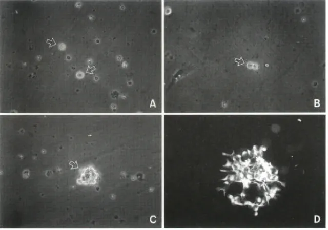
신생 흰쥐 대뇌 세포를 EGF가 포함된 SFM으로 배양했을 때 배양 1일후 대부분의 세포 는 죽고 일부 세포만 살아 있었다. 이 세포들은 위상차 현미경하에서 둥글고 밝게 빛나는 모양이었다(Fig. 1A). 세포들은 배양 2일에 2개의 세포로 분열 및 증식하였으며(Fig. 1B), 배 양 7일에는 50개 이상의 세포로 증식하였다(Fig. 1C). 세포들은 덩어리로 뭉쳐 증식하였으 며, 덩어리에서 떨어져 나온 단일 세포도 일부 있었다.
일차배양된 이 세포들의 특성을 연구하기 위해 다음과 같은 실험을 진행하였다. 즉, 세포 군을 PLL으로 처리된 chambered 슬라이드에 재분주하여 세포군이 슬라이드에 부착하기를
Fig. 1. Light microscopic appearance of EGF-responsive neuronal stem cells from newborn rat forebrains. (A) single neuronal stem cell (arrows) after 1 day in primary culture ( 200). (B) Dividing neuronal stem cells (arrow) after 2 days in primary culture ( 200). (C) A cluster of cells (arrow) after 7 days in primary culture ( 200). (D) Floating cell clusters were replated on poly-L-lysine coated slides and allowed to attach for 1 hr : indirect immunocytochemistry was then performed using anti-nestin antibody. Virtually all cells expressed nestin ( 400).
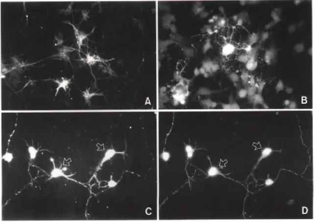
기다린 후 nestin, GFAP, tau, GC에 대한 면역화학염색을 하였다. 거의 모든 세포가 nestin을 발현하였으나(Fig. 1D), GFAP, tau, GC를 발현한 세포는 없었다. 따라서 일차배양된 세포들 은 신경간세포이며, 이 세포군에는 별아교세포, 신경세포, 희소돌기아교세포는 발현되지 않았음을 알 수 있었다. 또한 fibronectin 및 OX-42 항체에 대해 면역화학적 염색에서는 음 성으로 섬유아세포 및 미세아교세포나 대식세포는 존재하지 않았다. Chambered 슬라이드 에 부착한 세포들은 세포군 중심부에서 주변부로 이동하였다. 이동한 세포는 점차 가늘고 긴 돌기를 가진 세포 모양으로 형태가 변하였다. 2. 신경간세포의 분화과정 및 신경간세포에서 유래된 신경세포의 특성 신경간세포의 분화과정을 보기 위해 PLL으로 처리된 chambered 슬라이드에서 신경간세 포를 7일간 EGF가 포함되지 않은 SFM으로 이차 배양하였다. 이차 배양 전 기간을 통하여
Fig. 2. Immunocytochemical characterization of different cellular types derived from EGF-responsive neuronal stem cells. After 1 week in secondary culture, cells were processed for indirect immunocytochemistry using anti GFAP and anti GC antibodies and for double immunocytochemistry using anti tau and anti neurofilament antibodies. (A) GFAP-positive cells with flat, polygonal morphology ( 400). (B) GC-positive cells (×400). (C) Tau-positive cells ( 400). (D) Neurofilament-positive cells ( 400). Each arrow in C and D indicates an indentical cell.
세포들은 계속 증식 및 이동하였다. 세포들이 증식 및 이동하여 이웃한 세포군과 만나게 되는 경우에는 더 이상의 세포 이동 없이 증식만 일어났는데, 이때는 기존 세포층 위로 증 식이 일어나는 양상을 보였다.
별아교세포는 배양 2일에 분화되기 시작하였으며, 배양 7일 후에는 세포군에 있는 세포 중 80% 이상이 별아교세포로 분화되었다(Fig. 2A). 세포군 중심부에서는 다각형 모양의 별 아교세포(polygonal shape astrocyte)가 주로 있었으며, 세포군 주변부에서는 가늘고 긴 돌기 를 가진 별아교세포(process bearing astrocyte)가 있었다. 배양 7일에는 GC를 발현한 세포, 즉 희소돌기아교세포도 발현되었다(Fig. 2B). 희소돌기아교세포는 세포군 중심부에서만 있 었다. Tau를 발현한 세포(Fig. 2C)는 모두 neurofilament (Fig. 2D)도 발현하였다. 따라서 신 경간세포로부터 신경세포도 분화된 것을 알 수 있었다. 분화된 신경세포는 원형의 세포체 와 여러개의 가늘고 긴 돌기를 가지고 있었으며 세포군 중심부에서 관찰할 수 있었다. Fig. 2C와 Fig. 2D는 tau와 neurofilament에 대해 이중면역화학염색한 것이다. 배양 7일 후에도 미세아교세포나 섬유아세포는 없었다.
신경간세포는 EGF가 포함되지 않은 배양액에서 7일간 배양하더라도 배양 전 기간에 걸 쳐 존재하였다. 따라서 본 실험 배양조건에서는 신경간세포는 계속 증식되며, 증식된 세포 중 일부가 신경세포와 별아교세포, 희소돌기아교세포로 분화가 되는 것을 알 수 있었다.
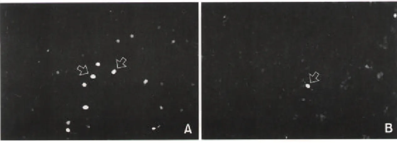
신경간세포로부터 분화된 신경세포가 어떤 신경전달물질을 갖고 있는지 확인하기 위해 tau와 dopamine 생성의 주효소인 tyrosine hydroxylase 및 몇 가지 신경전달물질들에 대한 이 중형광면역화학염색을 하였다. GABA 또는 substance P를 함유하고 있는 세포는(Fig. 3) 모 두 neurofilament도 발현하였다. 그러나 TH-ir 신경세포나, serotonin을 함유하는 신경세포는 없었다.
Fig. 3. Characterization of neuronal cells differentiated from EGF-responsive neuronal stem cells. After 1 week in secondary culture, cells were processed for indirect immunocytochemistry using several antibodies. Arrows indicate GABA-positive cells (A, 200) or substance P-positive cell (B, 200).
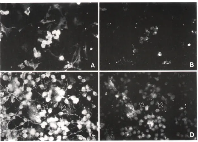
3. PDGF가 신경세포 및 TH-ir 신경세포 분화에 미치는 영향
신경간세포를 chambered 슬라이드에서 PDGF가 포함된 SFM으로 이차배양했을 때(Fig. 4C) cytokine을 포함하지 않은 SFM으로 이차배양했을 때(Fig. 4A) 보다 더 많은 신경세포 를 발현하였다. 또 NGF, BDNF CNTF, GDNF 등이 각각 포함된 SFM으로 신경간세포를 배 양했을 때 신경세포 발현율은 cytokine을 포함하지 않은 SFM으로 이차배양했을 때와 신경 세포 발현율의 차이는 없었으며, EGF나 bFGF를 포함한 SFM으로 배양했을 때는 신경세포 발현율이 오히려 낮게 관찰되었다.
TH-ir 신경세포는 cytokine이 포함되지 않은 배양액(Fig. 4B), 또는 EGF, bFGF, NGF, BDNF CNTF, GDNF가 각각 포함된 배양액으로 신경간세포를 이차배양했을 때는 발현되
Fig. 4. Effects PDGF on neuronal cell differentiation from EGF-responsive neuronal stem cells. Cell clusters were replated and cultured in serum-free medium (SFM, B) and PDGF-containing SFM (C, D). After 1 week in secondary culture, cells were processed for double immunocytochemistry using anti tau (A, C, 400) and anti tyrosine hydroxylase (B, D, 400). There are more tau expressed neuronal cells in culture of PDGF-containing SFM (C) than in culture of SFM (A). Tyrosine hydroxylase immuno-reactive cells are seen in culture of PDGF-containing SFM (D, arrows). Each arrow in C and D indicates an identical cell.
지 않았으나, PDGF가 포함된 배양액으로 이차배양했을 때는 발현되었다(Fig. 4D). 따라서 PDGF는 신경간세포로부터 TH-ir 신경세포의 발현을 유도할 수 있다는 것을 알 수 있었다. TH-ir 신경세포가 tau도 발현하는 것을 이중면역화학염색으로 관찰하여 이 세포가 신경세 포임을 증명하였다(Fig. 4C, D).
4. Western blot
TH-ir 신경세포 분화가 PDGF에 의해 유도되는 것을 western blot으로도 확인하였다(Fig. 5). 대조군으로는 tyrosine hydroxylase를 많이 함유하고 있는 부신수질조직을 이용하였다. PDGF가 포함된 배양액으로 신경간세포를 배양 분화시킨 후 western blot을 했을 때는 부신 수질조직에서 나타나는 tyrosine hydroxylase band와 동일한 위치에서 band가 나타났다. 그 러나 EGF를 포함하거나 또는 cytokine을 포함하지 않았을 때는 band가 나타나지 않았다. 결과에는 나타내지 않았으나 bFGF, NGF, BDNF CNTF, GDNF가 포함된 배양액에서 배양 했을 때도 tyrosine hydroxylase band는 나타나지 않았다.
IV. 고 찰 신경간세포는 태아 뿐 아니라 성숙한 포유류의 중추신경계 즉 대뇌 뇌실의 subependy-mal layer, 선조체, 해마, 그리고 소뇌와 척수에도 존재함이 증명되었다.2 4 이 세포는 배양 시 EGF, bFGF, TGF 에 의해 증식이 일어난다.2 ,7 ,2 1,2 5 성숙한 포유류 중추신경계에 있는 신 경간세포의 역할은 아직 알려지지 않았다. 단지 대뇌 뇌실 subependymal layer에 존재하는 신경간세포는 생체내에서 후각신경 재생에 관여한다는 보고가 있을 뿐이다.2 6 즉, 신경간세 포는 신경세포로의 분화 및 증식이 일어나며 분화된 신경세포는 후각신경 쪽으로 이동되 면서 손상된 후각 신경을 대치한다는 것이다. 본 실험에서는 EGF를 포함한 무혈청 배양액을 이용하여 신생 흰쥐 대뇌에 존재하는 신 경간세포를 배양하였다. 신경간세포는 PLL이 처리된 chambered 슬라이드 배양했을 때 신
Fig. 5. Western blot analysis of tyrosine hydroxylase in neuronal cells dif-ferentiated from neuronal stem cells. AM: adrenal medulla for control. PDGF: cultivated cell in PDGF-containing SFM. EGF: cultivated cells in EGF-containing SFM. cytokine ( ): cultivated cell in SFM without cytokine.
경세포와 별아교세포 및 희소돌기아교세포로 분화되었다. 또 본 실험에서 사용한 여러 cytokine 중 PDGF에 의해서만 신경간세포로부터 신경세포로의 발현율이 증가되었다. 신경 세포 발현율이 증가된 것이 PDGF에 의한 신경세포로의 분화 유도가 촉진되었기 때문인 지, 아니면 이미 분화된 신경세포의 생존율이 증가되었기 때문인지는 본 실험 결과만으로 는 알 수 없다. 그러나 신경간세포를 bFGF를 사용하여 증식시킬때는 PDGF를 처리하더라 도 신경세포의 발현율이 증가되지 않는 것으로 보아2 0 PDGF 자체가 신경세포 분화유도에 일차적으로 작용하지는 않을 것으로 생각된다.
신경간세포로부터 신경세포로의 분화 유도에는 insulin-like growth factor (IGF)-1이 작용 한다는 보고가 있다. 즉, Brooker 등은 신경간세포 배양시 IGF-1에 대한 항체를 처리하면 신경세포 분화가 중단되며, IGF-1을 투여하면 신경세포 분화가 증가하는 것으로 보아 신경
간세포의 내재성 IGF-1이 신경세포로의 분화 유도 물질일 가능성을 제시한 바 있다.2 7
본 실험 결과에서 신경간세포로부터 GABA와 substance P 신경세포는 분화되였으나 TH-ir 신경세포나 serotonin 신경세포로는 분화되지 않았다. TH-ir 신경세포나 serotonin 신 경세포가 분화되지 않은 이유는 본 실험에서 이용한 신경간세포 배양 조건이 TH-ir 또는 serotonin 신경세포를 발현시키지 못하는 조건이었기 때문일 가능성이 있다. 성숙한 흰쥐 대뇌에 존재하는 신경간세포와 태아 흰쥐 대뇌에 존재하는 신경간세포가 특성이 일치하는 신경간세포인지, 즉 한 종류의 신경간세포인지는 아직 확실히 밝혀지지 않았다.2 8,2 9 성숙한 포유류와 태아기에 존재하는 신경간세포는 EGF에 의한 증식 및 분화율이 서로 다르 며,30 ,3 1,32 중뇌에서 분리한 신경간세포는 같은 배양 조건에서 선조체에서 분리한 신경간세 포와는 달리 비록 적은 수이지만 TH-ir 신경세포로 분화하는 것으로 보아 이 세포들은 신 경간세포라기 보다는 계열이 다른 신경아세포(committed stem cell, 또는 progenitor cell)의
형태로 존재할 것이라는 보고도 있다.3 3 ,34
그러나 배양한 신경간세포에 retrovirus-mediated recombinant gene을 transfection 시킨 후 이를 대뇌피질, 망막, optic tectum에 이식했을때 하 나의 세포로부터 다양한 종류의 신경세포와 아교세포로 분화되는 것으로 보아 이세포는 신경간세포이며, 어떤 세포로 분화하는가는 세포간 접촉과 각종 성장물질, 또는 성장 억제 물질 등 주위환경에 따라 달라질 것이라는 주장도 있다.35 ,36 ,37 PDGF는 신경간세포로부터 TH-ir 신경세포를 분화시킨 반면, EGF, bFGF, NGF, BDNF, CNTF, GDNF를 단독 처리 했을 때는 TH-ir 신경세포를 분화시키지 못하였다. 이는 PDGF 가 전술한대로 신경세포의 생존율을 증가시키는 작용뿐 아니라, TH-ir 신경세포의 분화 유 도 작용을 갖고 있기 때문으로 생각한다. Cytokine이나 세포외기질 등에 의한 신경세포의 생존율을 증가시키는 작용과 특정 신경전달물질을 함유하는 신경세포로의 분화 작용은 서 로 다른 별개의 기전이 작용할 가능성이 있다. 이는 GDNF와 낮은 농도의 BDNF는 이미 분화된 TH-ir 신경세포의 생존율은 증가시키는 것은 잘 알려져 있으나,38 ,39 PDGF와는 달리 GDNF나 BDNF는 TH-ir 신경세포의 분화를 유도하지 못하였기 때문이다. 11
TH-ir 신경세포 분화를 유도하는 cytokine으로는 본 실험에서 증명한 PDGF 외에
inter-leukin (IL)-1이 있다.4 0 ,4 1 Ling 등은 태아 흰쥐 중뇌 신경간세포를 배양할 때 IL-1이 TH-ir 신
경세포로의 분화를 유도하며, 배양액에 IL-11 또는 leukemia inhibitory factor을 첨가하거나, 중뇌조직 세포막 또는 선조체 배양액을 첨가하면 분화된 신경세포로부터 dopamine이 생성
되며, 형태학적으로 성숙한 TH-ir 신경세포의 수도 증가하는 것을 보고한 바 있다.4 1
또 TH-ir 신경세포 분화 유도에는 cytokine 외에 세포간 접촉이 관여한다는 보고도 있다. 즉, 태아 흰쥐 중뇌 조직을 신경관 기저(floor plate neural tube) 조직과 동시 배양할 때 두 조직 이 접촉하지 않았을 때는 확산물질에 의해 choline 신경세포만 분화되나 두 조직이 접촉되
어 있을 때는 TH-ir 신경세포도 분화된다.4 2
Cytokine이 아닌 세포간 접촉에 의한 신호전달 체계의 영향으로 TH-ir 신경세포 분화가 유도할 수 있으며, dopamine 신경세포로 분화를 유도하는 신호전달체계를 활성화하는 방법은 여러 경로가 있을 것으로 생각한다.
TH-ir 신경세포의 분화 유도 기전은 아직 확실히 알려져 있지 않으나 sonic hedgehog
(SHH) 신호전달체계가 관여할 것이라는 보고가 있다.4 3 SHH는 45kDa의 전구단백질로 생
성된 후 자가단백분해과정을 거쳐 20 kDa 미만의 SHH-N (amino-terminal cleavage product) 과 SHH-C (carboxy-terminal cleavage product)이 생성되며, SHH-N은 세포막에 결합된 후 신
경세포의 분화 및 증식에 관여한다.4 4 ,4 5
흰쥐 태아기(태아기 9일) 중뇌 조직 배양시 SHH-N 을 첨가하면 TH-ir 신경세포의 분화율이 증가하는 것으로 보고되었다. SHH는 발생기 척삭 (notochord)에서 발현하며, 척삭의 기저판세포(floor plate cell)의 발생을 유도하여 신경관 (neural tube)을 형성하게 하는데,4 6 ,4 7 cyclic AMP-dependent protein kinase A (PKA)와는 길항
작용이 있어 PKA 활성도가 증가하면 SHH의 작용이 억제된다.4 8 ,4 9
Dopamine뿐 아니라 choline, GABA 등 특정신경전달물질을 함유하는 신경세포로 분화를 유도하는 보고도 있다. 신경간세포로부터 choline 신경세포로 분화는 미세아교세포-대식세 포 배양조건액에 의해 유도되며, 배양조건액에 NGF를 넣으면 choline 신경세포의 수가 증 가할 뿐 아니라 choline acetyltransferase의 활성도도 15배 정도 증가한다.5 0 그러나 배양조건 액내의 어떤 물질이 choline 신경세포로 분화 유도를 하는지는 아직 모른다. 또 세포외기질 물질인 laminin은 망막에 있는 신경간세포로부터 특이적으로 GABA 신경세포의 분화를 유 도하거나,17 증식률을 증가시킨다.18 따라서 특정 신경전달물질을 함유하는 신경세포로의 분화 유도 신호전달체계를 활성화하는 데는 여러 경로가 있을 것으로 생각한다. 이상의 실험결과로 PDGF는 신경간세포로부터 TH-ir 신경세포의 분화를 유도하는 것을 알 수 있었다. TH-ir 신경세포 분화유도 기전과 TH-ir 신경세포 발현율을 증가시키는 조건 은 추후 연구할 과제로 생각한다. 12
V. 결 론 신경간세포로부터 분화된 신경세포가 어떤 신경전달 물질을 함유하고 있는지, 그리고 어떤 cytokine이 신경간세포로부터 dopamine 신경세포를 분화시킬 수 있는지 알아보고자 하였으며 다음과 같은 결과를 얻었다. 1. EGF를 포함한 무혈청 배양액으로 신경간세포를 배양했을 때 신경간세포로부터 별아 교세포, 희소돌기아교세포, 신경세포가 모두 발현되었다.
2. 이때 분화된 신경세포 중 일부는 GABA 신경세포와 substance P 신경세포였으며, tyro-sine hydroxylase를 발현한(tyrotyro-sine hydroxylase immuno-reactive, TH-ir) 신경세포나 serotonin 신경세포 등은 관찰되지 않았다.
3. PDGF는 신경간세포로부터 TH-ir 신경세포의 분화를 유도하였다. 그러나 EGF, bFGF, NGF, BDNF CNTF, GDNF는 TH-ir 신경세포의 분화를 유도하지 못하였다.
4. PDGF가 신경간세포로부터 TH-ir 신경세포를 분화시키는 것을 tyrosine hydroxylase에 대한 western blot으로도 확인하였다.
이상의 실험결과로 PDGF는 신경간세포로부터 TH-ir 신경세포의 분화를 유도하는 것을 알 수 있었다. TH-ir 신경세포 분화유도 기전과 TH-ir 신경세포 발현율을 증가시키는 조건 은 추후 연구할 과제로 생각한다.
참 고 문 헌
1. Jacobson M. Developmental Neurobiology. 3rd ed. New York: Plenum Press; 1991;311-58. 2. Davis AA, Temple S. A self-renewing multipotential stem cell in embryonic rat cerebral cortex.
Nature 1994;372:263-6.
3. Gordon-Weeks PR, Golding JP, Clarke HD, Tonge D. A study of the expression of laminin in the spinal cord of the frog during development and regeneration. Exp Physiol 1992;77: 681-91.
4. Masuda-Nakagawa LM, Wiedemann C. The role of matrix molecules in regeneration of leech CNS. J Neurobiol 1992;23:551-67.
5. Murphy M, Drago J, Bartlett PF. Fibroblast growth factor stimulates the proliferation and differentiation of neural precursor cells in vitro. J Neurosci Res 1990;25:463-75.
6. Mytileneou CT, Park H, Shen J. Epidermal growth factor induced survival and proliferation of neuronal precursor cells from embryonic rat mesencephalon. Neurosic Lett 1992;135:62-6. 7. Reynolds BA, Tesslaff W, Weiss S. A multipotent EGF-responsive striatal embryonic progenitor
cell produces neurons and astrocytes. J Neurosci 1992;12:4565-74.
8. McLoon Sc, McLoon LK, Palm SL, Furcht LT. Transient expression of laminin in the optic nerve of the developing rat. J Neurosci 1988;8:1981-90.
9. Cattaneo E, Mckay R. Proliferation and differentiation of neuronal stem cells regulated by nerve growth factor. Nature 1990;347:762-5.
10. Sephel GC, Burrous BA, Kleinman HK. Laminin; Neural activity and binding proteins. Dev Neurosci 1989;11:313-31.
11. Davis HL. Trophic action of nerve extract on denervated skeletal muscle in vivo. Exp Neurol 1983;70:383-7.
12. Heinicke EA, Davis HL. Soluble proteins of rat muscle; Effects of denervation and nerve extract. Brain Res 1985;343:176-81.
13. Schwartz JP, Sheng JG, Mitsuo K. Trophic factor production by reactive astrocytes in injured brain. Ann N Y Acad Sci 1993;679:226-35.
14. Bae HM, Park JS, Yeon DS. Effects of glial-neuronal cell co-culture on GFAP expression of astrocytes. Korean J Physiol Pharmacol 1997;1:285-96.
15. Liesi P, Kaakkola S, Dahl D, Vaheri A. Laminin is induced in astrocytes of adult brain by injury. EMBO J 1984;3:683-6.
16. Nurcombe V. Laminin in neural development. Pharmacol Ther. 1992;56:247-64.
17. Frade JM, Martinez-Morales JR, Rodriguez-Tebar A. Lmninin-1 selectively stimulates neuron generation from cultured retinal neuroepithelial cells. Exp Cell Res 1996;22:140-9.
18. Park DS, Park JS, Yeon DS. The effects of laminin on the characteristics and differentiation of neuronal cell from epidermal growth factor-responsive neuroepithelial cells. Yonsei Med J
1998;39(2):130-40.
19. Barres BA, Lazar MA, Raff MC. A novel role for thyroid hormone, glucocorticoides and retinoic acid in timing oligodendrocyte development. Development 1994;120:1097-108. 20. Johe KK, Hazel TG, Muller T, Dugich-Djordjevic M, McKay RDG. Single factors direct the
differentiation of stem cells from the fetal and adult central nervous system. Genes Develop-ment 1996;10:3129-40.
21. Reynolds Ba, Weiss S. Generation of neurons and astrocytes from isolated cells of the adult mammalian central nervous system. Science 1992;255:1707-10.
22. Bradfod JH, Zhou J, Stern GM. Induction of dopaminergic neurotransmitter phenotype in rat embryonic cerebrocortex by the synergistic action of neurotrophins and dopamine. Eur J Neurosci 1996;8:2328-39.
23. Towbin H, Staehelm T, Gordon J. Electrophoretic transfer of proteins from polyacrylamide gels to nitrocellulose sheets. Procedure and some applications. Proc Natl Acad Sci USA 1979; 78:4350-6.
24. Eriksson PS, Perfilieva E, Bjork-Eriksson T, Alborn AM, Nordborg C, Peterson DA, Gage FH. Neurogenesis in the adult human hippocampus. Nature Med 1998;4:1313-7.
25. Von Visger JR, Yeon DS, Oh TH, Markelonis GJ. Differentiation and maturation of astrocytes derived from neuroepithelial progenitor cells in culture. Exp Neurol 1994;128:34-40. 26. Pasterkamp RJ, Dewinter F, Holtmaat AJ, Verhaagrn J. Evidence for a role of the
chemore-pellent semaphorin III and its receptor neurophin-I in the regeneration of primary olfactory axons. J Neurosci 1998;18:9962-76.
27. Brooker GJF, Kalloniatis M, Russo UC, Murphy M, Werther GA, Bartlett PE. Endogenous IGF-1 regulates the neuronal differentiation of adult stem cells. J Neurosci Res 2000;59:332-41. 28. Tropepe V, Graig CG, Morshead CM, vander Kooy D. Transforming growth factor null and senescent mice show decreased neural progenitor cell proliferation in the forebrain sube-pendyma. J Neurosci 1997;17:7850-9.
29. Yokoyama M, Morrison RS, Black IB, Dreyfus CF. Septal neuron cholinergic and GABAergic functions: Differential regulation by basic fibroblast growth factor and epidermal growth factor. Brain Res 1994;78:201-8.
30. Arsenijevic Y, Weiss S. Insulin-like growth factor-1 is a differentiation factor for postmitotic CNS stem cell-derived neuronal precursors: distinct actions from those of brain-derived neurotrophic factor. J Neurosci 1998;18:2118-28.
31. Santa-Olalla J, Covarrubias L. Epidermal growth factor, transforming growth factor-alpha, and basic fibroblast growth factor differentially influence neural precursor cells of mouse embryonic mesencephalon. J Neurosci Res 1995;42:172-83.
32. Ptak LA, Hart KR, Lin D, Carvey PM. Isolation and manipulation of rostral mesencephalic tegmental progenitor cells from rat. Cell Transplant 1995;4:335-42.
33. Luskin MB. Neuronal cell lineage in the vertebrate central nervous system. FASEB J 1994;8:722-30.
34. Parnavelas JG, Barfield JA, Franke E, Luskin MB. Seperate progenitor cells give rise to pyramidal and nonpyramidal neurons in the rat telencephalon. Cereb Cortex 1991;1:463-8. 35. Galileo DS, Gray GE, Owens GC, Majors I, Sanes JR. Neurons and glia arise from a common
progenitor in chicken optic tectum: Demonstration with two retroviruses and cell type-specific antibodies. Proc Natl Acad Sci USA 1990;87:458-62.
36. Walsh C, Cepko CL. Clonally related cortical cells show several migration patterns. Science 1988;241:1342-5.
37. Bartlett P, Brooker G, Faux C, Dutton R, Murphy M, Turnkey A, et al. Regulation of neuronal stem cell differentiation in the forebrain. Immunol Cell Biol 1998;76:414-8.
38. Leu-Fen H, Doherfy DH, Lile JD, Bektesh S, Collins F. GDNF: A glial cell line-derived neurotrophic factor for midbrain dopaminergic neurons. Science 1993;260:1130-2.
39. Zigova T, Pencea V, Wiegand ST, Luskin MB. Intraventricular administration of BDNF increases the number of newly generated neurons in the adult olfactory bulb. Mol Cell Neurosci
1998;11:234-45.
40. Potter ED, Ling ZD, Carvey PM. Cytokine-induced conversion of mesencephalic derived progenitor cells into dopamine neurons. Cell Tissue Res 1999;296:235-46.
41. Ling ZD, Potter ED, Lipton JW, Carvey PM. Differentiation of mesencephalic progenitor cells into dopaminergic neurons by cytokines. Exp Neurol 1998;149:411-23.
42. Hynes M, Porter JA, Chang C, Chang D, Tessier-Lavigne M, Beachy PA, et al. Induction of midbrain dopaminergic neurons by sonic hedgehog. Neuron 1995;15:35-44.
43. Davis S, Gale NW, Aldrich TH, Maisonpierre PC, Lhotak V, Pawson T, Goldfarb M, Yanco-poulos GD. Ligands for EPH-related receptor tyrosine kinases that require membrane attach-ment or clustering for acitivity. Science 1994;266:816-9.
44. Bumcrot DA, Takada R, McMahon AP. Proteolytic processing yields two secreted forms of sonic hedgehog. Mol Cell Biol 1995;15:2294-303.
45. Porter JA, Ekker SC, Young KE, von Kessler DP, Lee JJ, Moses D, Beachy PA. The product of hedgehog autoproteolytic cleavage active in local and long-range signalling. Nature 1995; 374:363-6.
46. Roelink H, Porter JA, Chiang C, Tanabe Y, Chang DT, Beachy PA, Jessell TM. Floor plate and motor neuron induction by different concentrations of the amino-terminal cleavage product of sonic hedgehog autoproteolysis. Cell 1995;81:445-55.
47. Marti E, Bumcrot DA, Takada R, McMahon AP. Requirement of 19K sonic hedgehog for induction of distinct ventral cell types in CNS explants. Nature 1995;375:322-4.
48. Jiang J, Struhl G. Protein kinase A and hedgehog signaling in Drosophila limb development. Cell 1995;80:563-72.
49. Fan CM, Porter JA, Chiang C, Chang DT, Beachy PA, Tessier-Lavigne M. Long-range sclero-tome induction by sonic hedgehog: direct role of the amino terminal cleavage product of modulation by the cyclic AMP signalling pathway. Cell 1995;81:457-65.
50. Jonakait GM, Wen Y, Wan Y, N L. Macrophage cell-conditioned medium promotes cho-linergic differentiation of undifferentiated progenitors and synergizes with nerve growth factor action in the developing basal forebrain. Exp Neurol 2000;161:285-96.
A bstract
Effects of platelet-derived growth factor on dopaminergic
neuron differentiation from neuronal stem cells in vitro
In Sook Sohn
Dep artment of Medicine
Brain Korea 21 Proj ect f or Medical Sciences
(Directed by Asssociate Professor Dong-Soo Yeon)
It is generally acknowledged that extrinsic factors such as cytokines, cell-to-cell contact and extracellular matrix components are important in generating cellular diversities observed in the central nervous system. Johe et al. reported that platelet-derived growth factor (PDGF) enhanced neuronal differentiation and that cilliary neurotrophic factor acted on stem cells to generate astrocytes. This study was undertaken to investigate which cytokine can differentiate dopaminergic neurons from neuronal stem cells obtained from fetal rat fore-brains.
Results are summarized as belows.
1. Neuronal stem cells cultured in EGF-containing serum-free medium subsequently dif-ferentiate into neurons, astrocytes, and oligodendrocytes.
2. Differentiated neuronal cells were immuno-reactive for γ-aminobutyric acid or sub-stance P, but not for serotonin and tyrosine hydroxylase.
3. PDGF converted neuronal stem cells into tyrosine hydroxylase immuno-reactive (TH-ir) cells, but epidermal growth factor, basic fibroblast growth factor, nerve growth factor, brain-derived neurotrophic factor, cilliary neurotropfic factor, or glial cell-derived neuro-trophic factor did not.
4. PDGF-induced TH-ir cells from neuronal stem cells were confirmed using western blot. These results suggest that PDGF may be a important regulator in differentiation of TH-ir cells from neuronal stem cells, and cytokines can affect neurotransmitter phenotype of the neurons generated from neuronal stem cells.
Key Words: differentiation, neuronal stem cell, dopaminergic neuron, platelet-derived growth factor, epidermal growth factor, neuron