Copyright © 2020 by Asian-Australasian Journal of Animal Sciences
This is an open-access article distributed under the terms of the Creative Commons Attribution License
Asian-Australas J Anim Sci
Vol. 33, No. 12:1957-1964 December 2020 https://doi.org/10.5713/ajas.19.0842 pISSN 1011-2367 eISSN 1976-5517
Comparison of overfed Xupu and Landes geese in performance,
fatty acid composition, enzymes and gene expression related to
lipid metabolism
Xu Liu1,2,3, Peng Li1,2,3, Changqing He1,2,3, Xiangyong Qu1,2,3,*, and Songchang Guo1,2,3,*
Objective: The aim of this study was to compare overfeeding performance, fatty acid com position, blood chemistry, enzymes and genes expression overfed Xupu and Landes geese.
Methods: Sixty male Xupu geese (80 d) and Landes geese (80 d) were selected. After a period of oneweek of preoverfeeding, Xupu and Landes geese were overfed three meals of 550 and 350 g/d, respectively, of a highcarbohydrate diet in the first week of the overfeeding period. The next week, geese were given four meals of 1,200 and 850 g/d, respectively, over 8 to 14 d. Finally, geese were given five meals of 1,600 and 1,350 g/d, respectively, for the last two weeks.
Results: After overfeeding for 28 d: Compared with Landes geese, Xupu geese liver weight and livertobody weight ratio decreased (p<0.05), while final weight, slaughter weight, total weight gain, abdominal fat weight, and feedtoliver weight ratio increased (p<0.05). The levels of elaidic acid (C18:1t9), oleic acid (C18:1n9), eicosenoic acid, and arachidonic acid in the liver of Xupu geese significantly increased (p<0.05), and the levels of myristic acid and stearic acid significantly decreased (p<0.05), while methyleicosanoate acid significantly increased (p<0.05). Xupu geese had higher plasma concentrations of triglyceride and very low density lipoprotein cholesterol (p<0.05), and decreased activities of alanine aminotrans ferase, aspartate aminotransferase, and lipase (LPS) (p<0.05). Landes geese had higher LPS activity (p<0.05), but lower cholinesterase activity (p<0.05) when compared with Xupu geese. The mRNA expression levels of fatty acid dehydrogenase (FADS) gene, elongase of long chain fatty acid 1 (ELOVL1) gene, ELOVL5, and acylCo A: cholesterol acyltransferase 2 (ACAT2) gene were significantly upregulated (p<0.05) in Landes goose when compared with Xupu geese.
Conclusion: This study demonstrates that the liver production performance of Landes geese was better than that of Xupu geese to some extent, which may be closely related to LPS activity, as well as the expression of FADS, ELOVL1, ELOVL5, and ACAT2.
Keywords: Xupu Geese; Landes Geese; Liver; Serum Parameters; Fatty Acid Composition; Genes Expression
INTRODUCTION
With the occurrence of African swine fever in China, the consumption of safer poultry meat has increased dramatically [1,2]. When compared with other poultry meat, goose meat has beneficial characteristics including high protein and low fat content [3]. Moreover, goose liver has a high capacity of fat accumulation and is used for the production of “foie gras” in poultry production [4,5]. Unlike mammalian fatty liver, main components of foie gras are unsaturated fatty acids (UFA), which have been shown to help protect against car diovascular and cerebrovascular disease in humans [6,7].
* Corresponding Author: Xiangyong Qu Tel: +86-13874856185, Fax: +86-0731-84618532, E-mail: [email protected] Songchang Guo Tel: +86-15074836991, Fax: +86-0731-84618176, E-mail: [email protected]
1 College of Animal Science and Technology, Hunan
Agricultural University, Changsha, Hunan 410128, China
2 Hunan Engineering Research Center of Poultry
Production Safety, Hunan 410128, China
3 Hunan Co-Innovation Center of Animal Production
Safety, Changsha, Hunan 410128, China
ORCID Xu Liu https://orcid.org/0000-0001-6254-6322 Peng Li https://orcid.org/0000-0002-2037-5868 Changqing He https://orcid.org/0000-0001-9906-3906 Xiangyong Qu https://orcid.org/0000-0002-2775-1942 Songchang Guo https://orcid.org/0000-0001-6067-3475 Submitted Oct 30, 2019; Revised Dec 11, 2019; Accepted Jan 10, 2020
The Landes goose (Anser anser) originated in the south western Landes province of France. Landes geese have a beneficial liver capacity in that it can increase 5 to 10fold in size over the course of a shortterm overfeeding proce dure. As a result, the Landes geese have become the world’s most famous breed for producing fatty liver products [8]. The Xupu goose (Anser cygnoides domesticus) is an indige nous breed of western Hunan Province in China and has been included in the list of National Livestock and Poultry Genetic Resources Protection in China [9,10]. When com pared with other domestic goose breeds, the Xupu goose has the best capacity for fat accumulation in the liver. To this end, Fournier et al [11] showed that the difference of liver size between the two breeds of geese may be closely related to heredity. This difference may also be attributable to a wide range of other factors, including sex, nutrition, housing den sity, and housing environment.
At present, many studies have been conducted on Landes geese relative to Xupu geese; as a result, comparative studies between the two breeds remain scarce. Here, we sought to investigate overfeeding performance, plasma biochemistry indices, fatty acid composition, enzymes and gene expression related to lipid metabolism between Xupu and Landes geese. These data not only form the basis for the study regarding the mechanisms of liver fat deposition, but also provided a theoretical reference for the breeding of fatty liver geese.
MATERIALS AND METHODS
Experimental design, diets, and animal management
All experiment were approved by the Institutional Animal Care and Use Committee of the Hunan Agricultural University (Hunan, China). All methods and procedures were performed in accordance with the approved guidelines and provided by the regional Animal Ethics Committee.
Sixty male Xupu geese (4,947.00±377.54 g) and sixty male Landes geese (4,751.00±244.73 g) were selected for this study. All geese were maintained under the same feeding and man agement conditions. Xupu geese were provided by the Hunan Hongyu Xupu Goose Industry Development Co., Ltd. (Huai hua City, Hunan Province, P. R. China) and Landes geese were provided by the Hunan Fugoose Industry Development Co., Ltd. (Chenzhou City, Hunan Province, P. R. China). At 80 d of age, a period of oneweek of preoverfeeding began. During this time, food intake was progressively increased to enlarge the volume of the digestive tract and to initiate metabolic adaptation to overfeeding. At the end of the preoverfeeding period, all geese were forcefed with a carbohydrate diet con sisted of 98% boiled maize, 1.0% plant oil, 0.5% salt, and 0.5% multivitamin (multivitamin provided per kilogram of diet: vitamin A, 80,000,000 IU; vitamin D, 6,000,000 IU; vitamin E, 40,000 IU; vitamin B1, 12,000 mg; vitamin B2, 50,000 mg;
vitamin B6, 500 mg; vitamin B12, 2,000 mg; vitamin K3, 3,000
mg; vitamin C, 16,000 mg; pantothenic acid, 3,000 mg; folic acid, 2,000 mg; nicotinic acid, 4,000 mg; biotin, 2,000 mg; methionine, 10,000 mg; lysine, 8,000 mg; tryptophan, 800 mg; arginine, 1,800 mg; serine, 8,000 mg; alanine, 18,000 mg). In total, this intake amounted to 3,370 kcal/kg, with a compasition of 90 g of protein/kg and 4.5 g of fat/kg. Xupu geese, having a greater capacity for overfeeding ingestion, were fed by the operator to the maximum of their ingestion potential. The feed intake of Xupu and Landes geese were different because of their different body weights. Xupu and Landes geese were overfed three meals of 550 and 350 g/d, respectively, of a highcarbohydrate diet in the first week of the overfeeding period. The next week, geese were given four meals of 1,200 and 850 g/d, respectively, over 814 d. Finally, geese were given five meals of 1,600 and 1,350 g/d, respectively, for the last two weeks.
This overfeeding experiment was conducted from July to August in 2018. The overfeeding room had a constant tem perature (28°C to 34°C) and humidity (65% to 70%) range. All geese were divided into two groups according to the va rieties and reared on the ground (4 m×6 m). Each goose was labeled with a ring on its right foot. An overfeeding machine was used in our study, and operated by the same person each time overfeeding occurred. During the overfeeding period, all goose had ad libitum access to water.
Sample collection
Initial weight of each geese was recorded before the first meal of overfeeding. After 28 d of overfeeding, all geese were food deprived overnight for 12 h. During this time, geese had ad
libitum access to water. On the following morning, geese were
weighted and blood samples were taken by puncture of the occipital venous sinus. Blood sampling were maintained at room temperature for 1 h, and plasma was obtained by cen trifugation at 3,000×g for 20 min at 4°C. After blood sampling, the goose were killed by exsanguination and each individual liver was quickly removed and weighed. Liver sample were immediately taken from the ventromedial portion of the main lobe (right lobe) of eight Xupu geese and eight Landes geese with similar body weight gain. Liver samples were immedi ately frozen in liquid nitrogen and stored at –80°C until later analysis of enzyme activities and mRNA levels. Initial weight, final weight, slaughter weight, liver weight were recorded daily by each goose to calculate the total weight gain, body weight gain rate, livertobody weight ratio, and feedtoliver weight ratio. The indicator is calculated as follows:
Total weight gain = Mf – Mi
Livertobody weight ratio = Ml / Ms ×100%
Feedtoliver weight ratio = MF / Ml
Mf, final weight; Mi, initial weight; Ms, slaughter weight;
Ml, liver weight; MF, total feed consumption of each goose
during overfeeding.
Fatty acid composition
The fatty acid composition of the liver was determined ac cording to a previously described method [12]. Briefly, total lipids were extracted from the liver tissue using petroleum ether/anhydrous diethyl ether (1:1, v/v). Methyl esters of the lipids were prepared using saponification with a solu tion of KOH: methanol (4 mol:1 L). The organic layer was aspirated for fatty acid analysis using an Agilent 7890N gas chromatography equipped with a flame ionization detector (Agilent Technologies, Santa Clara, CA, USA) and a CPSil 88 fused silica open tube capillary column (100 m×0.25 nm; Agilent Technologies, USA). The gas chromatograph tem perature program was as follows: Initial temperature of 140°C for 5 min, temperature increase of 3°C/min to 220°C, 1 min temperature hold at 220°C, and then holding temperature at 220°C for additional 40 min. The injector and detector temperatures were maintained at 240°C and 260°C, respec tively. Hydrogen was used as the carrier gas at a flow rate of 40 mL/min. Individual fatty acid peaks were identified by comparing their retention times with those of the standards (Cat#: 189191AMP; Sigma Chemicals, St. Louis, MO, USA). The results were expressed as grams per 100 g of total iden tified fatty acids.
Plasma biochemistry
Concentrations of triglyceride (TG), cholesterol (TC), low density lipoprotein cholesterol (LDLC), very low density lipoprotein cholesterol (VLDLC), and high density lipopro tein cholesterol (HDLC) in plasma were measured using a Mindray automatic analyzer (BS300; Shenzhen Mindray BioMedical Electronics Co., Ltd, Shenzhen, Guangdong, China) using a commercially available kits (Shenzhen Min dray BioMedical Electronics Co., Ltd., China) according to the manufacturer’s instructions. Aspartate aminotrans ferase (AST, C0101) and alanine aminotransferase (ALT, C0092) were determined using corresponding, commercially, available diagnostic kits (Nanjing Jiancheng Bioengineering Institute, Nanjing, China) using a microplate reader (Multi skan GO; Thermo Fisher Scientific, Waltham, CT, USA) according to the instructions of the manufacturer.
Lipid metabolism enzymes activities of liver and plasma
Approximately 0.5 g of liver sample was used to prepare the
tissue homogenate. Tissues were diluted in 1:9 (w/v) using icecold 154 mmol/L sodium chloride solution, and homog enized using an UltraTurrax homogenizer (T10BS25, IKA, BadenWurttemberg, Germany). Resulting homogenates were then centrifuged at 3,500×g at 4°C for 10 min.
The supernatant and plasma were used to determine the activities of cholinesterase (CHE), lipase (LPS), lipoportein lipase (LPL), hepaticlipase (HL), and content of nonesterified free fatty acids (NEFA). All activities and content were deter mined according to corresponding, commercially available diagnostic kits (Nanjing Jiancheng Bioengineering Institute, China) according to the manufacturer’s instructions using a microplate reader (Multiskan GO; Thermo Fisher Scien tific, USA). Protein concentration in the supernatant of the liver homogenate was measured by using a protein assay kit (A0452; Nanjing Jiancheng Institute of Bioengineering, China).
Total RNA extraction, reverse transcription, and quantitative real-time polymerase chain reaction
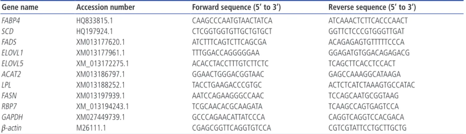
Total RNA was isolated from liver tissues using a TaKaRa Min BEST Universal RNA Extraction Kit (Takara, Osaka, Japan) according to the manufacturer’s protocol. The concentration and integrity of RNA were determined using a NanoDrop 2,000 Spectrophotometer (Thermo Scientific, Hudson, NH, USA) and 1% agarose gel electrophoresis, respectively. Only RNA specimens with an A260/A280 ratio of 1.8 to 2.0 and an A260/A230 ratio ≥2.0 were used for subsequent analyses. Total RNA from each sample was reverse transcribed into cDNA using the PrimeScript RT reagent Kit with gDNA Eraser kit (Takara, Japan), and cDNA was then diluted 1:10 with nucleasefree water before being used for quantitative realtime polymerase chain reaction (PCR). The primer pairs for the amplification of adipocyte fatty acid binding protein (FABP4), stearoylCoA desaturase (SCD), fatty acid dehydro genase (FADS), elongase of longchain fatty acid 1 (ELOVL1), acylCo A: cholesterol acyltransferase 2 (ACAT2), LPL, fatty acid synthase (FASN), glyceraldehyde3phosphate dehydro genase (GAPDH), and betaactin (β-actin) gene were designed from GeneBank sequences using Primer Premier 5.0 and obtained from Shanghai ShengGong Biological Company (Shanghai, China), as shown in Table 1.
Quantitative realtime PCR (qPCR) was performed using SYBR Green Master Mix (Vazyme Biotech, Nanjing, China) in CFX96 Touch RealTime PCR Detection System (BioRad Laboratories, Hercules, CA, USA). The PCR systems con sisted of 2 μL of diluted cDNA template (1:9), 12.5 μL of SYBR Premix Ex Taq II, 1 μL PCR Forward Primer (10 μmol/L), 1 μL PCR Reverse Primer (10 μmol/L) and 8.5 µL of sterilized distilled water. The PCR programs was as fol lows: 95°C for 30 s, followed by 35 cycles of denaturation at 95°C for 5 s and 60°C for 30 s. Dissociation curves of the
products were generated by increasing the temperature of samples incrementally from 55°C to 95°C as the final step of the PCR. GAPDH and β-actin genes were used as the dual in ternal standard for normalizing transcript abundance of mRNA expression. The relative expression levels of target genes were calculated by the 2–ΔΔCt method as described by
Livak and Schmittgen [13].
Statistical analyses
All data were analyzed using SPSS 21.0 (2015, IBMSPSS Inc., Chicago, IL, USA). Variability of all the data is expressed as standard error of the mean (SEM). Differences between mean values were compared using independent samples ttest, and considered significant at p<0.05.
RESULTS
Overfeeding performance
The results of overfeeding performance of Xupu and Landes geese are presented in Table 2 and 3. When compared with the Landes geese, final weight, total weight gain, slaughter weight, abdominal fat weight and feedtoliver weight ratio of Xupu geese significantly increased (p<0.05). In Xupu geese, the liver weight and livertobody weight ratio both decreased
(p<0.05). There were no significant differences in the initial weight and body weight gain rate between Xupu and Landes geese (p>0.05).
Fatty acid composition
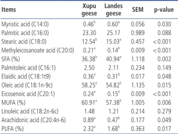
The fatty acid composition in liver of Xupu and Landes geese are shown in Table 4. Analyses of liver fatty acids showed that the major fatty acids were palmitic acid (C16:0), stearic acid (C18:0), palmitoleic acid (C16:1), oleic acid (C18:1n9), and linoleic acid (C18:2n6c). Oleic acid (C18:1n9) was the most abundant fatty acid, accounting for 58.25% and 54.82% of total fatty acid in livers of Xupu and Landes geese, respec
Table 1. The primers used for gene cloning, quantitative real-time polymerase chain reaction
Gene name Accession number Forward sequence (5’ to 3’) Reverse sequence (5’ to 3’)
FABP4 HQ833815.1 CAAGCCCAATGTAACTATCA ATCAAACTCTTCACCCAACT
SCD HQ197924.1 CTCGGTGGTGTTGCTGTGCT GGTTCTCCCGTGGGTTGAT
FADS XM013177620.1 ATCTTTCAGTCTTCAGCGA ACAGAGAGTGTTTTTCCCA
ELOVL1 XM013177961.1 TTTGGACCAGGGGGAA GGAGATGTGGACAGAGACG
ELOVL5 XM_013172275.1 ACACCTACCTTTGTCTTCTC TCAGCTTCACCTCCACT
ACAT2 XM013186797.1 GGAACTGGGACGGTAAC GAGCCAAAGGCATAAGA
LPL XM013188252.1 TACCTGAAGACCCGTGC ACTCTCATCTAAAGTGCCATAC
FASN XM013197939.1 AATCCAGAAGGGCCAAC TCCAGCAATGCGGTAAG
RBP7 XM_013194243.1 TCGCAACACGCAAGATA TCAAGCCAGTGAGTCCA
GAPDH XM027449739.1 GCCCAGAACATTATCCCA CAGGTCAGGTCCACGACA
β-actin M26111.1 CGAGCGGTTCAGGTGTCCA CGTCGTATTCCTGCTTGCTG
FABP4, adipocyte fatty acid binding protein; SCD, stearoyl-CoA desaturase; FADS, fatty acid dehydrogenase; ELOVL1, elongase of long-chain fatty acid 1; ELOVL5, elongase of long-chain fatty acid 5; ACAT2, cholesterol acyltransferase 2; LPL, lipoprotein lipase; FASN, fatty acid synthase; RBP7, retinol binding protein 7; GAPDH, glyceralde-hyde-3-phosphate dehydrogenase; β-actin, beta-actin.
Table 2. Comparison of performance parameters between overfed Xupu and
Landes geese
Items geeseXupu Landes geese SEM p-value Initial weight (g) 4,947.00 4,851.00 100.606 0.120 Final weight (g) 8,843.00a 8,031.00b 194.983 < 0.001
Total weight gain (g) 3,896.00a
3,280.00b
93.726 0.001 Body weight gain rate (%) 78.74 74.14 3.017 0.139
Data are means of 8 geese per group. SEM, standard error mean.
a,b Means within a row with different superscripts differ significantly (p < 0.05).
Table 3. Comparison of slaughter traits between overfed Xupu and Landes geese
Items Xupu geese Landes geese SEM p-value
Slaughter weight (g) 8,154.75a 7,188.25b 208.733 < 0.001
Liver weight (g) 609.08b 938.40a 58.886 < 0.001
Abdominal fat weight (g) 557.25a
360.45b
32.814 < 0.001
Liver-to-body weight ratio (%) 7.47b 13.07a 0.731 < 0.001
Feed-to-liver weight ratio (kg · kg) 44.44a 22.82b 3.083 < 0.001
Data are means of 8 geese per group. SEM, standard error mean.
tively. When compared with the Landes geese, the levels of myristic acid (C14:0) and stearic acid (C18:0) decreased (p< 0.05) in the livers of Xupu geese, contrastingly, levels of methyleicosanoate acid (C20:0) increased (p<0.05). In the livers of Xupu geese, the levels of elaidic acid (C18:1t9), oleic acid (C18:1n9), eicosenoic acid (C20:1), and arachidonic acid (C20:4n6) significantly increased (p<0.05). However, no significant differences were found in palmitic acid (C16:0), palmitoleic acid (C16:1), or linoleic acid (C18:2n6) between either Xupu or Landes geese (p>0.05).
Blood chemistry
As is shown in Table 5, the results of blood chemistry analyses for Xupu and Landes geese revealed that there were no signifi
cant differences in either TC or LDLC (p>0.05). However, plasma levels of TG and VLDLC were significantly higher (p<0.05) in Xupu geese when compared with Landes geese. When compared with Landes geese, Xupu geese had a sig nificant decrease (p<0.05) in HDLC content, as well as ALT and AST activities.
Lipid metabolism-related enzymes activities
The results of lipid metabolismrelated enzymes activities in the plasma and liver of Xupu and Landes geese are presented in Table 6. When compared with the Landes geese, LPS ac tivity in the plasma and liver of Xupu geese both significantly decreased (p<0.05). However, a significant enhancement (p<0.05) of CHE activity in the liver of Xupu geese was ob served. There were no significant differences in either plasma or liver LPL or HL activities or in the NEFA content between Xupu and Landes geese (p>0.05).
Gene expression
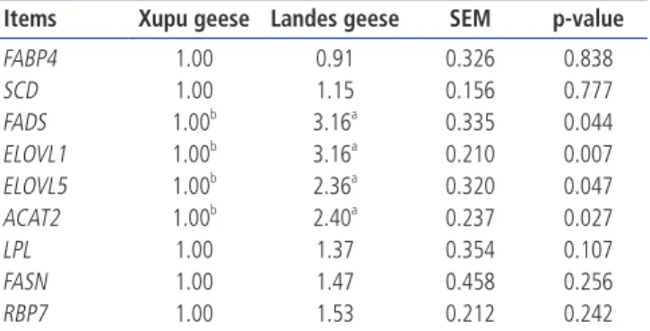
The lipid metabolismrelated gene mRNA expression in livers of Xupu and Landes geese are shown in Table 7. The mRNA expression of FADS, ELOVL1, ELOVL5, and ACAT2 (p<0.05) in liver of Xupu goose were significantly downregulated than in liver of Landes goose. There were no significant differences in the mRNA expression of FABP4, SCD, LPL, FASN, and retinol binding protein 7 in liver between Xupu and Landes geese (p<0.05).
DISCUSSION
In our study, we found that the livers of Xupu geese were
Table 4. Comparison of fatty acid composition between overfed Xupu and
Landes geese
Items geeseXupu Landes geese SEM p-value Myristic acid (C14:0) 0.46b 0.60a 0.056 0.030 Palmitic acid (C16:0) 23.30 25.17 0.989 0.088 Stearic acid (C18:0) 12.54b 15.03a 0.457 < 0.001 Methyleicosanoate acid (C20:0) 0.21a 0.14b 0.009 < 0.001 SFA (%) 36.38b 40.94a 1.118 0.002 Palmitoleic acid (C16:1) 2.50 2.11 0.234 0.149 Elaidic acid (C18:1t9) 0.36a 0.31b 0.017 0.048 Oleic acid (C18:1n-9c) 58.25a 54.82b 1.135 0.015 Eicosenoic acid (C20:1) 0.24a 0.15b 0.009 < 0.001 MUFA (%) 60.91a 57.38b 1.005 0.006 Linoleic acid (C18:2n-6c) 1.48 1.21 0.214 0.279 Arachidonic acid (C20:4n-6) 0.89a 0.47b 0.177 0.049 PUFA (%) 2.32a 1.68b 0.363 0.017
Data are means of 8 geese per group.
SEM, standard error mean; SFA, saturated fatty acids; MUFA, monounsaturated fatty acids; PUFA, polyunsaturated fatty acids.
a,b Means within a row with different superscripts differ significantly (p < 0.05).
Table 5. Comparison of plasma biochemical metabolites between overfed Xupu
and Landes geese
Items geeseXupu Landes geese SEM p-value TG (mmol/L) 6.04a 4.33b 0.378 0.001 TC (mmol/L) 10.42 11.51 0.659 0.125 LDL-C (mmol/L) 3.13 3.47 0.280 0.245 VLDL-C (mmol/L) 0.92a 0.52b 0.151 0.026 HDL-C (mmol/L) 6.12b 7.24a 0.353 0.009 ALT (U/L) 50.97b 94.68a 5.181 < 0.001 AST (U/L) 87.73b 234.98a 7.199 < 0.001
Data are means of 8 geese per group.
SEM, standard error mean; TG, triglyceride; TC, cholesterol; LDL-C, low density lipoprotein cholesterol; VLDL-C, very low density lipoprotein cholesterol; HDL-C, high density lipoprotein cholesterol; ALT, alanine aminotransferase; AST, aspartate aminotransferase.
a,b Means within a row with different superscripts differ significantly (p < 0.05).
Table 6. Comparison of enzyme activities in blood and liver between overfed
Xupu and Landes geese
Items geeseXupu Landes geese SEM p-value Plasma CHE (U/mL) 330.12 342.50 64.194 0.851 LPS (U/L) 16.40b 27.81a 3.854 0.034 LPL (U/mL) 5.58 5.84 0.447 0.567 HL (U/mL) 5.62 5.62 0.650 0.995 LPL+HL (U/mL) 10.67 11.45 0.873 0.385 NEFA (U/mL) 490.97 470.77 93.500 0.833 Liver CHE (U/mgprot) 26.77a 7.41b 7.038 0.022 LPS (U/gprot) 66.70b 139.77a 21.311 0.027 LPL (U/mgprot) 0.46 0.56 0.137 0.505 HL (U/mgprot) 0.45 0.45 0.265 0.990 LPL+HL (U/mgprot) 0.84 1.01 0.296 0.562 NEFA (U/gprot) 66.48 56.04 14.670 0.497
Data are means of 8 geese per group.
SEM, standard error mean; CHE, cholinesterase; LPS, lipase; LPL, lipoporteinlipase; HL, hepaticlipase; NEFA, nonesterified free fatty acids.
smaller and the feedtoliver weight ratio of Xupu geese were higher than those of Landes geese. Since Xupu geese had a larger body shape, the index of livertobody weight ratio re vealed an opposite result. The abdominal fat weight of Xupu geese was higher than that of Landes geese. Possible reasons for this result include that a large amount of fat was trans ferred from the liver to extrahepatic (e.g., abdominal adipose tissue) in Xupu geese. The above results confirmed that Landes geese are the best breed globally to produce fatty liver products. Overfeeding with a carbohydraterich diet results in high
de novo lipogenesis. The present study was the first time to
compare the fatty acid composition of the fatty liver in Landes and Xupu geese. There were both similarities and differences between the two breeds. The main products of fatty acid syn thesis were 16:0, 18:0, and above all 18:1. This fatty acid in particular accounted for more than 90% of the hepatic TG fatty acid content, and the proportions of 18:1 was more than 50% in both breeds. These results were consistent with the fatty liver content found previously in both geese and duck [14,15]. Relative to Landes geese, Xupu geese had an increase in the proportions of 18:1, but a decrease in the proportions of 18:0 in their fatty liver. This finding was in agreement with those found by both Cazeils et al [16] and Hatsugai et al [17]. Simultaneously, the proportions of monounsaturated fatty acids and polyunsaturated fatty acids (PUFA) in the fatty liver of Xupu geese were significantly higher than those in Landes geese. Moreover, the proportion of saturated fatty acids in the fatty liver of Xupu geese was significantly lower than that of Landes geese. These results were consistent with the pre vious study, which used 21 days of feeding for both Xupu and the Landes geese [18]. Given these findings, it is clear that the performance of overfeeding production and fat deposition in Xupu geese were inferior to that of Landes geese. However,
the proportions of UFA in fatty liver of Xupu geese was signifi cantly higher than that of Landes geese, which may indicate that Xupu fatty liver is better for human health.
Fatty liver occurs in geese or duck when fat synthesis ex ceeds fat secretion. In response to overfeeding, de novo hepatic lipogenesis is dramatically increased, TG do not fully enter the secretion pathway, and a large proportion of TG remains stored in the liver [11,19]. During overfeeding, part of the newly synthesized TG in the liver are incorporated into he patic lipoprotein, mainly VLDL which can be secreted into blood and used (or stored) in extrahepatic tissues. We found that the plasma contents of TG and VLDL of Xupu geese were higher than those of Landes geese. These results were in agreement with previous studies, which showed higher VLDL concentration in Poland geese when compared with Landes geese, even though the liver weight of the former was less than the latter [11]. Xu et al [20] found that the plasma concentrations of TG and VLDL were higher in Sichuan white geese than in Landes geese. In the present study, overfeed ing with highenergy corn diet for 28 d induced elevations in the concentration of plasma HDL, which was in accor dance with findings of previous studies in broiler chickens and geese [21,22]. It clear the transfer of fat from the liver to the extrahepatic tissue is one of the reasons why the liver weight of Xupu geese was smaller, but their abdominal fat heavier than that of Landes geese. These results suggested that the mechanism behind geese fatty liver formation is mainly attributable to an imbalance between the storage and secretion (as plasma lipoproteins) of newly synthesized endogenous lipids and exogenous lipids in the cytoplasm. When overfeeding using a highenergy corn diet, plasma ALT and AST are mainly from the liver, resulting in higher ALT and AST activities. Our study found that the plasma activities of ALT and AST in Xupu geese were lower than those of Landes geese, indicating that Landes geese had a higher degree of liver damage due to longterm overfeeding. The results in this experiment were in agreement with those of Zhu et al [21], which showed that longterm overfeeding induced liver cell inflammation. Therefore, we supported the opinion that goose hepatic adaptation to overfeeding is of notable importance. Our study found that the activity of LPS in the plasma and liver of Landes geese increased relative to that of Xupu geese. Pancreatic lipase plays an important role in fat absorption. Kobayashi et al [23] showed that lipase activity was high by using the higher fat content of diets. Krogdahl [24] also reported that when given a highfat diet, the lipase activity of birds was higher than that in birds fed a lowfat diet. Much past work has found that the activity of LPL was positively correlated with the weight of fatty liver in goose or duck, moreover, that LPL was useful in the se lection of Landes geese breeders with a higher susceptibility to liver steatosis [6,25,26]. Despite this, we found no differ
Table 7. Comparison of lipid metabolism related gene expressions in liver
between overfed Xupu and Landes geese
Items Xupu geese Landes geese SEM p-value
FABP4 1.00 0.91 0.326 0.838 SCD 1.00 1.15 0.156 0.777 FADS 1.00b 3.16a 0.335 0.044 ELOVL1 1.00b 3.16a 0.210 0.007 ELOVL5 1.00b 2.36a 0.320 0.047 ACAT2 1.00b 2.40a 0.237 0.027 LPL 1.00 1.37 0.354 0.107 FASN 1.00 1.47 0.458 0.256 RBP7 1.00 1.53 0.212 0.242
Data are means of 8 geese per group.
SEM, standard error mean; FABP4, adipocyte fatty acid binding protein; SCD, stea-royl-CoA desaturase; FADS, fatty acid dehydrogenase; ELOVL1, elongase of long-chain fatty acid 1; ELOVL5, elongase of long-long-chain fatty acid 5; ACAT2, cholesterol acyltransferase 2; LPL, lipoprotein lipase; FASN, fatty acid synthase; RBP7, retinol binding protein 7.
ence in the activity of LPL in either plasma or liver between Xupu and Landes geese, suggesting further study is needed. Collectively, our previous study [18] and current data in dicate the development of severe steatosis was closely related to the genes involved in lipid synthesis, packaging, secretion, transportation, deposition or metabolism, including FADS,
ELOVL1, ELOVL5, and ACAT2 [27,28]. It is well known that FADS gene plays a key role in the synthesis of longchain poly
unsaturated fatty acid (LCPUFAs) and the metabolism of essential fatty acids [2933]. ELOVL1 plays an important role in the elongation of superlongchain saturated fatty acids and superlongchain monosaturated fatty acids. ELOVL5 is mainly responsible for the elongation of 18carbon fatty acids. ACAT2 catalyzes the conjugation of cholesterol and longchain fatty acids to form cholesterol esters, and plays an important role in the absorption, storage, transport, and apolipoprotein metabolism of cholesterol. Given this, we observed that the expression of FADS, ELOVL1, ELOVL5, and ACAT2 in the liver of Landes geese increased significantly relative to Xupu geese. Osman et al [34] showed that FADS expression levels were gradually increased after overfeeding, moreover, that the induction of FADS promoted the generation of LCPUFAs in goose fatty liver. This increase appeared to be well coordi nated with the size of fatty liver in Landes geese, which suggests that the FADS, ELOVL1, ELOVL5, and ACAT2 genes are im portant to the development of goose fatty liver.
In conclusion, the results of the present study showed that the overfeeding performance of Xupu geese was inferior to that of Landes geese, which may be related to the activity of LPS, and the expression of FADS, ELOVL1, ELOVL5, and
ACAT2. However, the the proportions of UFA in Xupu fatty
liver were significantly higher than those of Landes geese. Taken together, there results provide new insights into the cultivation of highquality fatty liver geese.
CONFLICT OF INTEREST
We certify that there is no conflict of interest with any financial organization regarding the material discussed in the manu script.
ACKNOWLEDGMENTS
This study was supported by the Key Project of Hunan Edu cation Department (Nature) (18A089) and the Key Research and Development programs in Hunan—Agricultural Tech nology Innovation projects (2016NK2106). We would like to thank BPG Editing for the English language editing.
REFERENCES
1. Galindo I, Alonso C. African swine fever virus: a review. Viruses
2017;9:103. https://doi.org/10.3390/v9050103
2. Costard S, Mur L, Lubroth J, SanchezVizcaino JM, Pfeiffera DU. Epidemiology of African swine fever virus. Virus Res 2013;173:1917. https://doi.org/10.1016/j.virusres.2012.10. 030
3. Su SY, Dodson MV, Li XB, Li QF, Wang HW, Xie Z. The effects of dietary betaine supplementation on fatty liver performance, serum parameters, histological changes, methylation status and the mRNA expression level of Spot14α in Landes goose fatty liver. Comp Biochem Physiol Part A Mol Integr Physiol 2009;154:30814. https://doi.org/10.1016/j.cbpa.2009.05.124 4. Liu L, Zhao X, Wang Q, et al. Prosteatotic and protective
components in a unique model of fatty liver: gut microbiota and suppressed complement system. Sci Rep 2016;6:31763. https://doi.org/10.1038/srep31763
5. Geng TY, Yang B, Li FY, et al. Identification of protective com ponents that prevent the exacerbation of goose fatty liver: characterization, expression and regulation of adipo nectin receptors. Comp Biochem Physiol B Biochem Mol Biol 2016;194:328. https://doi.org/10.1016/j.cbpb.2016.01. 006
6. Lu LZ, Chen Y, Wang Z, et al. The goose genome sequence leads to insights into the evolution of waterfowl and suscepti bility to fatty liver. Genome Biol 2015;16:89. https://doi.org/ 10.1186/s130590150652y
7. De Souza RJ, Mente A, Maroleanu A, et al. Intake of saturated and trans unsaturated fatty acids and risk of all cause mortality, cardiovascular disease, and type 2 diabetes: systematic review and metaanalysis of observational studies. BMJ 2015;351: h3978. https://doi.org/10.1136/bmj.h3978
8. Mourot J, Guy G, Lagarrigue S, Peiniau P, Hermier D. Role of hepatic lipogenesis in the susceptibility to fatty liver in the goose (Anser anser). Comp Biochem Physiol B Biochem Mol Biol 2000;126:817. https://doi.org/10.1016/S03050491 (00)001711
9. Lin Q, Cao R, Jiang GT, et al. The complete mitochondrial genome of the Xupu goose. Mitochondrial DNA A 2016;27: 10101. https://doi.org/10.3109/19401736.2014.926528 10. Dai QZ, Lin Q, Jiang GT. Phylogenetic studies of four Anser
cygnoides (Anserini: Anserinae) in Hunan province of China
based on complete mitochondrial DNA sequences. Mitochon drial DNA A DNA Mapp Seq Anal 2016;27:24645. https:// doi.org/10.3109/19401736.2015.1033699
11. Fournier E, Peresson R, Guy G, Hermier D. Relationships between storage and secretion of hepatic lipids in two breeds of geese with different susceptibility to liver steatosis. Poult Sci 1997;76:599607. https://doi.org/10.1093/ps/76.4.599 12. Li FN, Duan YH, Li YH, et al. Effects of dietary n6:n3 PUFA
ratio on fatty acid composition, free amino acid profile and gene expression of transporters in finishing pigs. Br J Nutr 2015;113:73948. https://doi.org/10.1017/S0007114514004346 13. Livak KJ, Schmittgen TD. Analysis of relative gene expression
data using realtime quantitative PCR and the 2ΔΔCt method.
Methods 2001;25:4028. https://doi.org/10.1006/meth.2001 14. Hermier D, Salichon MR, Guy G, Peresson R. Differential
channelling of liver lipids in relation to susceptibility to hepatic steatosis in the goose. Poult Sci 1999;78:1398406. https://doi. org/10.1093/ps/78.10.1398
15. Molee W, Bouillieroudot M, Auvergne A, Babilé R. Changes in lipid composition of hepatocyte plasma membrane induced by overfeeding in duck. Comp Biochem Physiol B, Biochem Mol Biol 2005;141:43744. https://doi.org/10.1016/j.cbpc.2005. 05.007
16. Cazeils JL, BouillierOudot M, Auvergne A, Candau M, Babile R. Lipid composition of hepatocyte plasma membranes from geese overfed with corn. Lipids 1999;34:93742. https://doi. org/10.1007/s117459990443z
17. Hatsugai K, Ohkohchi N, Fukumori T, Akamatsu Y. Satomi S. Mechanism of primary graft nonfunction in a rat model for fatty liver transplantation. Transpl Int 2000;13:S58390. https://doi.org/10.1007/s001470050408
18. Liu X, Liu YW, He CQ, et al. Comparison of nutritional com ponents and serum biochemical indices in fatty liver between Xupu geese and Landes geese. J Anim Nutr (in chinese) 2019; 31:6228. https://doi.org/10.3969/j.issn.1006267x.2019.02. 018
19. Hermier D, Saadoun A, Salichon MR, Sellier N, Rousselot Paillet D, Chapman MJ. Plasma lipoproteins and liver lipids in two breeds of geese with different susceptibility to hepatic steatosis: changes induced by development and forcefeeding. Lipids 1991;26:3319. https://doi.org/10.1007/BF02537194 20. Xu HY, Wang Y, Han CC, et al. Estimation of lipoprotein
lipase activity (LPL) and other biochemical changes in two breeds of overfeeding geese. AsianAustralas J Anim Sci 2010; 23:12218. https://doi.org/10.5713/ajas.2010.10013
21. Zhu LH, Meng H, Duan XJ, Xu GQ, Zhang J, Gong DQ. Gene expression profile in the liver tissue of geese after overfeeding. Poult Sci 2011;90:10717. https://doi.org/10.3382/ps.2009 00616
22. Peebles ED, Cheaney JD, Brake JD, Boyle CR, Latour MA, McDaniel CD. Effects of added lard fed to broiler chickens during the starter phase. 2. Serum lipids. Poult Sci 1997;76: 164854. https://doi.org/10.1093/ps/76.12.1648
23. Kobayashi S, Terashima Y, Itoh H. Effects of dietary chitosan on fat deposition and lipase activity in digesta in broiler chickens. Br Poult Sci 2002;43:2703. https://doi.org/10.1080/0007166
0120121490
24. Krogdahl A. Digestion and absorption of lipids in poultry. J Nutr 1985;115:67585. https://doi.org/10.1093/jn/115.5.675 25. Davail S, Guy G, Andre J, Hermier D, HooParis R. Metabolism
in two breeds of geese with moderate or large overfeeding induced liversteatosis. Comp Biochem Physiol Part A Mol Integr Physiol 2000;126:919. https://doi.org/10.1016/S1095 6433(00)001902
26. André JM, Guy G, Gontierlatonnelle K, et al. Influence of lipoproteinlipase activity on plasma triacylglycerol concen tration and lipid storage in three genotypes of ducks. Comp Biochem Physiol Part A Mol Integr Physiol 2007;148:899 902. https://doi.org/10.1016/j.cbpa.2007.09.006
27. Sassa T, Ohno Y, Suzuki S, et al. Impaired epidermal perme ability barrier in mice lacking Elovl1, the gene responsible for verylongchain fatty acid production. Mol Cell Biol 2013; 33:278796. https://doi.org/10.1128/MCB.0019213
28. Shikama A, Shinozaki H, Takeuchi Y, et al. Identification of human ELOVL5 enhancer regions controlled by SREBP. Biochem Biophys Res Commun 2015;465:85763. https:// doi.org/10.1016/j.bbrc.2015.08.101
29. Burdge GC, Calder PC. Conversion of alphalinolenic acid to longerchain polyunsaturated fatty acids in human adults. Reprod Nutr Dev 2005;45:58197. https://doi.org/10.1051/ rnd:2005047
30. Koletzko B. Fatty acids and early human growth. Am J Clin Nutr 2001;73:6712. https://doi.org/10.1093/ajcn/73.4.671 31. MoltoPuigmarti C, Plat J, Mensink RP, et al. FADS1 FADS2
gene variants modify the association between fish intake and the docosahexaenoic acid proportions in human milk. Am J Clin Nutr 2010;91:136876. https://doi.org/10.3945/ ajcn.2009.28789
32. Arterburn LM, Hall EB, Oken H. Distribution, interconver sion, and dose response of n3 fatty acids in humans. Am J Clin Nutr 2006;83:1467S76S. https://doi.org/10.1093/ajcn/ 83.6.1467S
33. Park WJ, Kothapalli KS, Reardon HT, Lawrence P, Qian SB, Thomas Brenna J. A novel FADS1 isoform potentiates FADS2 mediated production of eicosanoid precursor fatty acids. J Lipid Res 2012;53:150212. https://doi.org/10.1194/jlr.M025312 34. Osman RH, Liu L, Xia LL, et al. Fads1 and 2 are promoted
to meet instant need for longchain polyunsaturated fatty acids in goose fatty liver. Mol Cell Biochem 2016;418:103 17. https://doi.org/10.1007/s1101001627377