Copyright ⓒ Korean Academy of Dental Technology
This is an Open Access article distributed under the terms of the Creative Commons Attribution Non-Commercial License (http://creativecommons.org/licenses/by-nc/4.0) which permits unrestricted non-commercial use, distribution, and reproduction in any medium, provided the original work is properly cited.
INTRODUCTION
치아가 상실된 경우 과거에는 단순히 저작과 발음기능의 회복과 같 은 기능적인 측면이 주된 관심사였지만, 최근에는 환자들의 심미적 요 구 증가와 심미보철 시스템의 개발로 전부도재관의 사용이 증가하고 있으며, 적합한 기계적 성질을 가지기 위하여 여러 소재들이 개발되 었다[1]. 2000년대 초반부터 치과용 지르코니아를 이용한 보철물이 소개가 되었는데, 치과용 지르코니아는 산화 지르코니움(Zirconium oxide, ZrO2)의 총칭으로 화학적 안정성과 체적안정성을 갖는 다형 (polymorphic)구조로 되어있으며, 상전이 시 발생하는 체적확장에 의 해 균열의 진행을 억제하여 기존의 도재에 비해 높은 굴곡 및 파절강도 를 갖고 있다[2]. 또한 우수한 생체적합성과 구조적 안정성으로 전치 및 구치부 고정성 보철물, 포스트, 임플란트 지대주, 교정용 브라켓 등 활 용범위가 다양하다[3,4]. 지르코니아는 자연치아와 같은 색상과 투명도를 정교하게 재현하는 데는 한계가 있어 일반적으로 지르코니아 위에 도재를 축성하여 심미 성의 한계를 보완하고 있으며, 최근 지르코니아 표면에 다양한 색상을 첨가하여 지르코니아의 심미성을 개선하고자 하는 노력을 기울이고 있 다[5]. 하지만, 이러한 지르코니아 하부구조를 포함하는 전부도재관이 라 하더라도 내/외부요인에 의해 지르코니아 하부구조와 베니어 세라Purpose: This study aimed to compare the shear bond strength between zirconia cores and veneer ceramics as per the sand blasting and liner treatments.
Methods: The following 4 groups of zirconia-veneering ceramic specimens were prepared: (1) Group I, untreated; (2) Group II, with 110 μm aluminium oxide (Al2O3) sandblasting; (3)
Group III, with liner (IPS e.max ZirLiner; Ivoclar Vivadent); and (4) Group IV, with 110 μm Al2O3 sand blasting and liner. Surface roughness was measured for all the prepared
speci-mens, and the surface morphology was observed using a scanning electron microscope. All the samples (n=40) were fixed with measuring jigs, and shear bond strengths were ob-tained using a universal testing machine with a crosshead speed of 0.5 mm/min. The shear bond strength data were analyzed using one-way analysis of variance and t-test. The post hoc comparison was performed using the Tukey’s test (α=0.05).
Results: A significant difference in the surface roughness was observed between the speci-mens of groups I and II (p<0.05). Surface treatment with liner and sandblasting showed higher shear bond strength between zirconia core and veneering ceramic (p<0.05).
Conclusion: The sand blasting and liner treatment increased the shear bond strength be-tween zirconia core and veneering ceramic.
Key Words: Liner, Sand blasting, Shear bond strength, Veneering ceramic, Zirconia
Article Info Received February 24, 2021 Revised March 15, 2021 Accepted March 17, 2021 Corresponding Author Jae-Hong Kim
Department of Dental Laboratory Science, College of Health Science, Catholic Univer-sity of Pusan, 57 Oryundae-ro, Geumjeong-gu, Busan 46252, Korea
E-mail: [email protected]
https://orcid.org/0000-0002-2679-8802
https://doi.org/10.14347/jtd.2021.43.1.6 pISSN: 1229-3954 eISSN: 2288-5218
J Tech Dent 2021;43(1):6-12
Original Article
샌드블라스팅 처리와 라이너가 지르코니아와 전장도재의 전단결합
강도에 미치는 영향
김기백
1, 김재홍
21대전보건대학교 치기공학과, 2부산가톨릭대학교 보건과학대학 치기공학과
Effect of sandblasting and liner on shear bond strength of veneering ceramic to
zirconia
Ki-Baek Kim
1, Jae-Hong Kim
21Department of Dental Lab Science, Daejeon Health Institute of Technology, Daejeon, Korea
2Department of Dental Laboratory Science, College of Health Science, Catholic University of Pusan, Busan, Korea
JOURNAL OF TECHNOLOGIC DENTISTRY
Ki-Baek Kim, Jae-Hong Kim: Effect of surface treatments on the zirconia-to-veneering ceramic shear bond strength 믹 사이의 계면 탈락 또는 응력 집중에 의한 파괴가 발생된다는 보고가 있다[6,7]. 이와 같은 실패는 세라믹의 두께 차이[8], 소결과 냉각에 따 른 열 수축 및 팽창[9], 그리고 지르코니아의 부적절한 디자인에 따른 잔류응력[10] 등의 요인에 영향을 받는 것으로 알려져 있다. 최근 지르코니아와 전장도재 사이의 결합력을 개선하기 위한 많은 연구가 이루어지고 있으며[11-13], 이 연구들은 지르코니아와 전장도 재의 결합 메커니즘을 최적화하기 위해 다양한 물리/화학적 표면처리 방법을 적용하였다. 선행연구에 의하면 가장 빈번하게 사용되는 물리 적 처리 방법은 표면에 거칠기를 형성하는 방법으로 압축된 공기를 이 용하여 알루미나 입자를 분사하는 방법인 air-particle abrasion 또는 sandblasting를 활용하는 것이며[14], 화학적 처리 방법은 산부식 처 리방법과 liner 사용이 지르코니아와 전장도재와의 결합력 개선에 기 여할 수 있음이 보고되었다[15,16]. 기존 발표된 선행연구들은 다양한 조건에서 결합력을 관찰한 결과로 직접적인 비교 관찰 연구는 부족한 것으로 판단되어, 지르코니아 소재의 이중구조 전부도재관 제작 시 실 제 임상과정에서 시행되는 표면처리방법에 따른 전단결합강도의 차이 를 직접적으로 비교하는 것이 필요하다고 생각된다. 이에 본 연구에서는 지르코니아를 완전 소결한 후 임상에서의 진행 과정을 모사하여 표면 연마를 시행하고 지르코니아 표면에 물리적 표 면처리인 sandblasting과 화학적 결합을 위한 liner 처리를 실시한 후 두 재료 간 전단결합강도에 미치는 영향을 비교분석하고, 이를 통해 어 떠한 표면 처리방법이 지르코니아와 전단도재의 접착을 향상시킬 수 있는지 알아보고자 하였다.
MATERIALS AND METHODS
1. 실험 재료
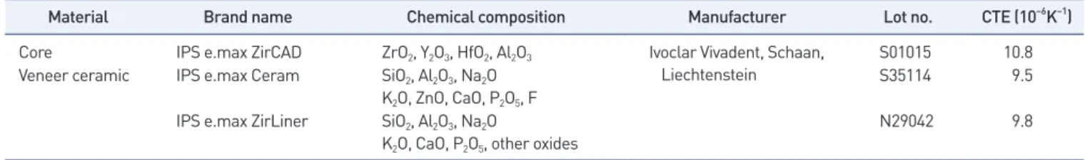
본 연구에 사용된 재료는 Table 1에 제시하였다. 코어(core)는 지르 코니아 블록(IPS e.max ZirCAD; Ivoclar Vivadent, Schaan, Liech-tenstein), veneering ceramic은 장석류 도재(IPS e.max Ceram; Ivoclar Vivadent), liner는 IPS e.max ZirLiner (Ivoclar Vivadent) 를 사용하였다.
2. 시편 제작
소결 후 최종 시편의 크기(가로 13.5 mm, 세로 13.5 mm, 높이 3.5
mm)를 정한 후, 소결 과정의 수축률 20%로 고려하여 CAD software (exocad; exocad GmbH, Darmstadt, Germany)로 디자인하여 CAM software (hyperDENT; FOLLOW-ME! Technology Group, Munich, Germany)로 가공데이터를 얻은 후 절삭장비(Trione; DIO Implant, Busan, Korea)를 사용하여 가공하였다. 모든 시편은 치과 기공 임상에서의 방법과 같이 Stone point (Abrasive stones GC#13; R&D Dental, Seoul, Korea)를 사용하여 35,000 rpm 속도로 좌우 왕복 50회에 걸쳐 연마를 시행하였다. 연마 후 소결로(Zirkonofen 600/V2; Zirkonzahn GmbH, Gais, Italy)에 넣은 후 소성 스케줄에 따라 소결하여 최종 시편을 얻었다. 준비된 시편은 증류수에 침수시켜 초음파 세척기로 10분간 세척 후 스팀 클리너로 세척하여 실온에서 자 연건조 시켰다(Fig. 1). 완전 소결된 지르코니아 코어마다 동일한 접촉면과 크기를 갖도록 하기 위하여 최종시편과 유사한 형태의 실리콘 주형을 제작하였다. 각 각의 코어 위에 같은 형태의 도재를 축성하였다. 전장도재를 도재용 브러쉬로 축성하고 도재전용 퍼니스에 1차 소성을 한 후 소성으로 인 한 수축을 보상하기 위해 수축된 양만큼 다시 축성하여 2차 소성을 진 행하였다. 소성이 끝난 시편은 전체 두께와 크기를 확인하여 오차범위 안으로 제작이 되었는지 확인하였다. 지르코니아 전용 전장도재(IPS e.max Ceram Dentin; Ivoclar Vivadent)로 축성하였으며, 소성 전 20개의 시편은 liner (IPS e.max ZirLiner; Ivoclar Vivadent)를 도 포하여 소성하였다. 도재전용 퍼니스(Programat P300/G2; Ivoclar Vivadent)로 제조사의 소성 스케줄에 맞게 진행하였다(Table 2).
Table 1.
Table 1. Composition of the ceramic materials used in this study
Material Brand name Chemical composition Manufacturer Lot no. CTE (10–6K–1)
Core IPS e.max ZirCAD ZrO2, Y2O3, HfO2, Al2O3 Ivoclar Vivadent, Schaan,
Liechtenstein
S01015 10.8
Veneer ceramic IPS e.max Ceram SiO2, Al2O3, Na2O
K2O, ZnO, CaO, P2O5, F
S35114 9.5
IPS e.max ZirLiner SiO2, Al2O3, Na2O
K2O, CaO, P2O5, other oxides
N29042 9.8
CTE: coefficient of thermal expansion.
Figure 1. Specimen of sintered zirconia.
Figure 1.
8 https://doi.org/10.14347/jtd.2021.43.1.6 JOURNAL OF TECHNOLOGIC DENTISTRY
J
TD
3. 표면 처리
모든 시편들은 연마 과정을 거친 후 무작위로 네 그룹으로 나누어 실 험을 진행하였다(n=10). Group I은 아무 처리도 하지 않은 대조군,Group II는 110 μm의 alumina oxide (Al2O3)로 4기압 하에 10초간
sandblasting한 뒤, 10분간 초음파세척 후 건조하였다. Group III는 sandblasting 처리 없이 시편에 liner(IPS e.max ZirLiner; Ivoclar Vivadent)를 제조사의 지시에 따라 도포한 후 소결하였다. Group IV는 Sandblasting 한 후 제조사의 지시에 따라 liner를 도포하였다 (Table 3).
4. 표면 형상 및 거칠기 평가
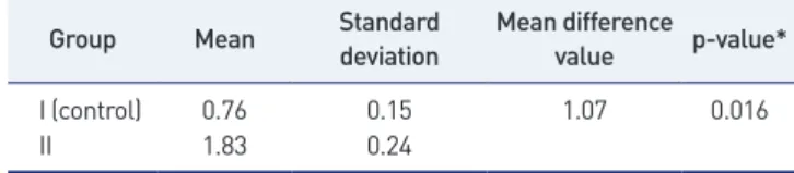
물리적 표면처리인 sandblasting에 의한 표면 거칠기를 비교 관찰하 기 위해 3D surface profilometer (Contour GT-X3 base; Bruker, Bremen, Germany)를 이용하여 시편 표면의 거칠기를 평가하였다. 시편의 중앙 부위에서 3.0 mm의 거리를 시편 당 5회 측정하여 각 평 균 거칠기 결과 값을 기록하였다.
5. 전단결합강도 측정 및 파절면 관찰
지름 30 mm, 높이 22 mm 원통 형태에 교정용 아크릴릭 레진(Or-tho-Jet; Lang Dental, Wheeling, IL, USA)으로 포매하여 전단결합 강도 측정용 지그(zig)에 고정될 수 있도록 하였다. 힘의 하중이 지르코 니아 코어와 도재 사이의 접착면에 동일한 방향으로 전달되도록 전단 결합강도 측정용 지그에 고정하여 지르코니아 코어에서 전장도재의 파 절이 발생할 때까지 실험을 시행하였으며, 만능시험기(Instron model 3366; Instron, Nowood, MA, USA)로 crosshead 속도 0.5 mm/ min의 조건에서 최대하중값을 측정하였다(Fig. 2). 파절된 계면의 상 태는 전단결합강도를 측정한 후 시편에서 파절된 부위를 백금코팅 처 리하여 scanning electron microscope (JSM-6510; Jeol, Tokyo, Japan)를 이용하여 관찰하였다.
6. 통계분석
전단결합강도의 기술적 통계는 평균과 표준편차로 나타내었다. 측
정된 값들에 대한 동질성 검정 Shapiro–Wilk test로 정규성 검정을 수
행한 결과, 네 그룹 모두 정규분포로 검증되었다. 이와 같은 결과를 토 대로 모수적 통계방법인 일원분산분석(one-way ANOVA)과 t-test를 실시하였으며, 다중비교 검정(Tukey’s multiple comparison test)으 로 사후분석을 시행하였다. 모든 통계분석은 IBM SPSS 22.0 for win-dows (IBM, Armonk, NY, USA)를 이용하였으며, 통계적 판단을 위 한 제 1종 오류의 수준은 0.05로 하였다.
RESULTS
1. 물리적 표면처리에 따른 표면거칠기
지르코니아 코어의 sandblasting 처리 전과 후의 표면조도 측정결과 는 Table 4와 같다. 표면조도는 Group I이 0.76 μm, Group II는 1.83 μm로 통계적으로 유의한 차이를 나타냈다(p<0.05). 3D optical im-age analyzer와 Scanning Electron Microscope (SEM)을 이용한 표 면관찰에서 Group I은 절삭흔 형태가 그대로 노출된 표면형상을 보였 으며, Group II는 알루미나 입자의 충돌에 의해 bur의 절삭흔은 모두
Table 2.
Table 2. Firing schedule
Brand name Start ℃/℉ Dry time ℃/min Final℃/℉ Hold time Vacuum start℃/℉ Vacuum release℃/℉
IPS e.max ZirLiner 403/757 4 min 40 960/1760 1 min 450/842 959/1758
IPS e.max Ceram 403/757 4 min 40 750/1382 1 min 450/842 749/1380
Table 3.
Table 3. Specimen groups prepared in this study Group
(n=10)
Surface treatment Abrasive stone
point polishing Sandblasting Liner
I ○ ❌ ❌
II ○ ○ ❌
III ○ ❌ ○
IV ○ ○ ○
Figure 2. Schematic diagram of the shear bond strength test sep-up on.Figure 2.Figure 2. Schematic diagram of the shear bond strength test sep-up on. Table 4.
Table 4. Roughness values experimental groups (unit: μμm)
Group Mean Standard deviation Mean difference value p-value*
I (control) 0.76 0.15 1.07 0.016
II 1.83 0.24
Ki-Baek Kim, Jae-Hong Kim: Effect of surface treatments on the zirconia-to-veneering ceramic shear bond strength
소실되었으나 시편의 표면에 다른 물리적 거칠기가 생성되었다(Fig. 3, 4).
2. 전단결합강도
각 실험군의 평균 전단결합강도와 표준편차는 Table 5와 같다. Group I 대조군에서는 11.91±1.19 MPa, Group II 실험군은 15.38 ±1.13 MPa, Group III 실험군은 15.53±1.29 MPa, Group IV 실험 군은 20.32±1.93 MPa로 나타났다. 각 실험군 간의 전단결합강도 차 이를 검정하기 위해 일원분산분석을 시행한 결과 각 실험군 별로 전단 결합강도에 유의한 차이가 있었다(p<0.05). 각 집단 간의 유의한 차이 를 자세히 검증하기 위해 다중비교 검정 실시 결과, Group II 실험군과 Group III 실험군 간에만 유의한 차이를 보이지 않았고, 나머지 집단 간에는 유의한 차이를 보였다(p<0.05).
3. 파절면 관찰
전단결합강도 실험에 사용된 시편에서 지르코니아와 전장도재가 접 착되어 있던 면을 중심으로 파절양상을 SEM으로 관찰하였다. 대조군 인 Group I 시편에서는 완전히 박리된 상태인 접착성 파절양상(adhe-sive failure)을 보였으며, Group II, III 시편에서는 완전 박리되지 않 고 전장도재가 일부 남아있는 복합 파절양상(mixed failure)을 나타내 었다. 반면에 Group IV 시편에서는 liner와 전장도재 간의 응집성 파 절양상(cohesive failure)을 보였다(Fig. 5).DISCUSSION
최근 들어 심미보철의 적용범위가 넓어지면서 전치부를 전부도재관 으로 수복하거나, 심지어 구치부까지 수복하는 경우가 증가하고 있고, 이에 따라 강도가 개선된 전부도재 수복용 소재가 등장하고 있다[17]. 더불어 CAD/CAM (computer-aided design/computer-aided manufacturing) 기술의 발달은 높은 기계적 강도와 생체 적합성을 갖 는 전부도재관의 사용을 더욱 증가시킬 수 있는 가능성을 열어주고 있 다. 새로운 전부도재관 시스템의 코어-전장도재의 결합강도와 실패양 상에 대한 정보는 전부도재관의 임상 적용 시 내구성을 예측하는데 도
Figure 3. 3D-optical image of specimens after sandblasting treatment. (A) Group I, (B) group II.
Figure 3.
Figure 3. 3D-optical image of specimens after sandblasting treatment. (A) Group I, (B) group II.
Figure 4. Scanning electron micrographic images of the zirconia surface after
Figure 4.
Figure 4. Scanning electron micrographic images of the zirconia surface after sand-blasting treatment. (A) Group I, (B) group II. Table 5.
Table 5. Mean of shear bond strength and standard deviation (SD) (unit: MPa)
Specimen Group I Group II Group III Group IV
1 11.96 14.51 14.01 21.87 2 13.43 13.21 13.89 19.43 3 10.67 17.88 17.68 22.96 4 11.81 15.61 15.00 20.62 5 10.18 17.41 17.21 24.15 6 12.36 15.22 15.28 21.67 7 11.23 16.67 16.32 19.35 8 12.32 14.36 14.52 21.77 9 11.22 17.01 17.30 19.26 10 10.95 16.18 16.95 21.13 Mean 11.91a 15.38b 15.53b 20.32c SD 1.19 1.13 1.29 1.93
a,b,cData with the different letters are significantly different at 0.05
10 https://doi.org/10.14347/jtd.2021.43.1.6 JOURNAL OF TECHNOLOGIC DENTISTRY
J
TD
움을 준다[18]. 최근 지르코니아와 전장도재 사이의 결합력을 개선하기 위한 많은 연구가 이루어지고 있으며, 이들 연구는 지르코니아와 전장 도재의 결합 메커니즘을 최적화하기 위해 다양한 물리, 화학적 표면처 리방법을 적용하였다[19,20]. 선행연구를 토대로 지르코니아 전부도재 관의 표면처리 방법에 따른 변화를 직접적으로 비교하고자 하였다. 따 라서 본 연구에서는 지르코니아의 표면에 알루미나 sandblasting에 의 한 물리적 표면처리를 실시하였고, 표면에 liner를 도포한 후 소결하여 화학적 표면처리를 실시하였다. 지르코니아 코어의 물리적 처리에 의한 표면 거칠기 경향은 Group II 군이 1.83 μm로 Group I 군의 0.76 μm보다 높아 통계적으로 유의 한 차이를 나타냈다(p<0.05). 3D optical image analyzer와 SEM을 이용한 표면관찰에서도 다른 양상을 관찰 할 수 있었다. 이는 Wegner 등[21]의 연구 결과와 같이 Alumina Oxide를 사용한 sandblasting은 고강도 도재의 표면 거칠기를 만드는 가장 효과적인 방법이라 보고한 적이 있다[21]. 이는 지르코니아 표면에 하는 sandblasting이 표면에 너지, 접착을 위한 표면적, 젖음성 등을 향상 시킨다고 보고했던 선행 연구와 일치하는 것으로 해석된다[12]. 지르코니아에 대한 전장도재의 접착력을 향상시키기 위해 화학적 처 리방법도 연구되어 있는데, 가장 대표적인 방법이 liner를 도포하는 것 이다. Liner의 적용에 대해 Aboushelib 등[22]은 코어를 차폐(mask-ing)하기 위해 사용하는 것으로 전장도재의 젖음성을 증가시키고 결합 강도를 강하게 한다는 연구결과를 보고하였으나, 반면에 liner 적용이 결합강도를 약하게 만든다는 연구결과도 보고되었다[23]. 따라서 본 연 구에서는 sandblasting의 물리적 표면처리와 liner의 화학적 표면처리 를 적용한 시편들의 전단결합강도를 분석하였다. 연구결과, 물리적 및 화학적 처리를 모두 시행한 Group IV에서 가장 높은 전단결합강도를 나타냈고, 다음으로는 Group III, Group II 그리고 Group I 순으로 나 타났다. 이와 같은 결과를 토대로 물리적 표면처리나 화학적 표면처리 를 단일하게 적용하는 것으로는 지르코니아와 전장도재간의 결합강도 를 월등히 향상시키는데 영향을 미칠 수 없으며, 물리적 및 화학적 표 면처리를 동시에 시행하였을 때 지르코니아와 전장도재 간의 전단결합 강도가 유의하게 증가한 것을 확인할 수 있었다(p<0.05). 본 연구에 사 용된 liner는 유리 세라믹 기질로, 지르코니아 코어와 균일하며 강한 화 학적 결합을 유도하여 선행 연구들과 유사한 결과[22,24]를 보였다고 생각 된다. 또한 sandblasting에 의한 표면처리는 지르코니아 표면에 일정한 깊이의 균열을 유발함으로써 지르코니아 표면에 거칠기를 형성 하여 그 결과로 지르코니아 코어와 전장도재 간의 결합강도를 증가 시 킨 것으로 판단된다[25]. 전단결합강도 비교 후 파절양상을 관찰한 결과 sandblasting과 liner를 함께 처리한 Group IV에서 전장도재의 파절에 의한 양상이 응 집성 파절양상으로 관찰되었다. 이러한 파절양상은 결합강도와 관련된 것으로 응집성 파절양상이 전단결합강도와 연계되어 있는 것으로 생각 되며, 이는 지르코니아 코어와 전장도재 사이의 결합력이 전장도재의 파절강도보다 높았기 때문이라고 해석할 수 있다. 지르코니아의 접착력을 향상시키기 위해 다양한 기계적, 화학적 처 리 방법들이 계속 연구되고 있다. 치과영역에서 지르코니아는 계속적 으로 영역이 확대되고 있기 때문에 지르코니아의 접착력의 향상은 앞 으로 더 중요할 것이다. 본 연구를 임상적 결과로 확대 해석하기에는 다소 무리가 있지만, 임상에서 지르코니아와 전장도재의 결합력을 증 가시키고자 한다면 sandblasting과 liner를 동시에 수행할 수 있을 것Figure 5. Scanning electron microscope micrograph of specimen after shear bond
strength test. (A) Group I, (B) group II, (C) group III, (D) group IV.
Figure 5.
Figure 5. Scanning electron microscope micrograph of specimen after shear bond strength test. (A) Group I, (B) group II, (C) group III, (D) group IV.
Ki-Baek Kim, Jae-Hong Kim: Effect of surface treatments on the zirconia-to-veneering ceramic shear bond strength 이다. 그러나 이 실험에서 사용한 시편은 임상적인 치과 수복물의 형태 를 반영하지 못하며, 오랜 기간 구강 내 수분에 노출되었을 때의 상황 과도 다르다는 한계점이 있다. 이를 보완하기 위해서는 치과 수복물과 유사한 형태의 시편을 제작하고 구강 내 환경을 재현한 수분환경 하에 기계적 강도를 측정하는 연구가 뒷받침 되어야 할 것으로 생각 된다.
CONCLUSIONS
본 연구는 sandblasting (물리적 표면처리)과 liner (화학적 표면처 리) 사용이 지르코니아와 전장도재의 전단결합강도에 미치는 영향을 알아보고자 하였고, 제한적이지만 다음과 같은 결론을 얻었다. 1. 지르코니아 코어의 물리적 표면처리는 표면 거칠기에 통계적으로 유의한 영향을 주었다(p<0.05). 2. 단독적인 물리적 표면처리와 화학적 표면처리는 전장도재의 결합 력에 큰 영향을 끼치지 못하며, sandblasting 처리와 liner를 함께 적 용하는 것이 지르코니아 코어와 전장도재의 접착력에 효과적인 요인으 로 작용함을 확인하였다(p<0.05). 3. 파절면 상태는 전단결합강도가 높은 Group IV에서만 응집성 파 절양상(cohesive failure)을 보였다.CONFLICT OF INTEREST
No potential conflict of interest relevant to this article was reported.
ORCID
Ki-Baek Kim, https://orcid.org/0000-0003-3821-9764 Jae-Hong Kim, https://orcid.org/0000-0002-2679-8802
REFERENCES
1. Blatz MB, Sadan A, Kern M. Resin-ceramic bonding: a re-view of the literature. J Prosthet Dent. 2003;89:268-274. 2. Piconi C, Maccauro G. Zirconia as a ceramic biomaterial.
Biomaterials. 1999;20:1-25.
3. Luthardt RG, Holzhüter M, Sandkuhl O, Herold V, Schnapp JD, Kuhlisch E, et al. Reliability and properties of ground Y-TZP-zirconia ceramics. J Dent Res. 2002;81: 487-491.
4. Yildirim M, Fischer H, Marx R, Edelhoff D. In vivo frac-ture resistance of implant-supported all-ceramic restora-tions. J Prosthet Dent. 2003;90:325-331.
5. Ban S, Suzuki T, Yoshihara K, Sasaki K, Kawai T, Kono H. Effect of coloring on mechanical properties of dental zirconia. J Med Biol Eng. 2014;34:24-29.
6. Ozcan M, Vallittu PK. Effect of surface conditioning methods on the bond strength of luting cement to ceram-ics. Dent Mater. 2003;19:725-731.
7. Zhang Y, Lawn BR, Rekow ED, Thompson VP. Effect of sandblasting on the long-term performance of dental ceramics. J Biomed Mater Res B Appl Biomater. 2004;71: 381-386.
8. Mainjot AK, Schajer GS, Vanheusden AJ, Sadoun MJ. Influence of cooling rate on residual stress profile in ve-neering ceramic: measurement by hole-drilling. Dent Mater. 2011;27:906-914.
9. Al-Amleh B, Lyons K, Swain M. Clinical trials in zirconia: a systematic review. J Oral Rehabil. 2010;37:641-652. 10. Fukushima KA, Sadoun MJ, Cesar PF, Mainjot AK.
Re-sidual stress profiles in veneering ceramic on Y-TZP, alumina and ZTA frameworks: measurement by hole-drilling. Dent Mater. 2014;30:105-111.
11. Kern M, Thompson VP. Sandblasting and silica coating of a glass-infiltrated alumina ceramic: volume loss, mor-phology, and changes in the surface composition. J Pros-thet Dent. 1994;71:453-461.
12. Della Bona A, Borba M, Benetti P, Cecchetti D. Effect of surface treatments on the bond strength of a zirconia-reinforced ceramic to composite resin. Braz Oral Res. 2007;21:10-15.
13. Uo M, Sjögren G, Sundh A, Goto M, Watari F, Bergman M. Effect of surface condition of dental zirconia ceramic (Denzir) on bonding. Dent Mater J. 2006;25:626-631. 14. Kirmali O, Akin H, Ozdemir AK. Shear bond strength of
veneering ceramic to zirconia core after different surface treatments. Photomed Laser Surg. 2013;31:261-268. 15. Smielak B, Klimek L. Effect of hydrofluoric acid
concen-tration and etching duration on select surface roughness parameters for zirconia. J Prosthet Dent. 2015;113:596-602.
16. Jian YT, Tang TY, Swain MV, Wang XD, Zhao K. Effect of core ceramic grinding on fracture behaviour of bilayered zirconia veneering ceramic systems under two loading schemes. Dent Mater. 2016;32:1453-1463.
den-12 https://doi.org/10.14347/jtd.2021.43.1.6
JOURNAL OF TECHNOLOGIC DENTISTRY
J
TD
tistry: historical roots and current perspectives. J Prosthet Dent. 1996;75:18-32.
18. Guess PC, Kulis A, Witkowski S, Wolkewitz M, Zhang Y, Strub JR. Shear bond strengths between different zirconia cores and veneering ceramics and their susceptibility to thermocycling. Dent Mater. 2008;24:1556-1567.
19. Fischer J, Grohmann P, Stawarczyk B. Effect of zirconia surface treatments on the shear strength of zirconia/ve-neering ceramic composites. Dent Mater J. 2008;27:448-454.
20. Piascik JR, Swift EJ, Braswell K, Stoner BR. Surface fluori-nation of zirconia: adhesive bond strength comparison to commercial primers. Dent Mater. 2012;28:604-608. 21. Wegner SM, Kern M. Long-term resin bond strength to
zirconia ceramic. J Adhes Dent. 2000;2:139-147.
22. Aboushelib MN, Kleverlaan CJ, Feilzer AJ. Microtensile bond strength of different components of core veneered all-ceramic restorations. Part II: zirconia veneering ce-ramics. Dent Mater. 2006;22:857-863.
23. Tinschert J, Natt G, Mautsch W, Spiekermann H, Anusav-ice KJ. Marginal fit of alumina-and zirconia-based fixed partial dentures produced by a CAD/CAM system. Oper Dent. 2001;26:367-374.
24. Aboushelib MN, de Jager N, Kleverlaan CJ, Feilzer AJ. Microtensile bond strength of different components of core veneered all-ceramic restorations. Dent Mater. 2005;21:984-991.
25. Peterson IM, Pajares A, Lawn BR, Thompson VP, Rekow ED. Mechanical characterization of dental ceramics by Hertzian contacts. J Dent Res. 1998;77:589-602.