IGFBP-3 suppresses VEGF expression and tumor angiogenesis
in head and neck squamous cell carcinoma
Seung-Hyun Oh*, Woo-Young Kim*, Ok-Hee Lee*, Ju-Hee Kang, Jong-Kyu Woo, Jai-Hyun Kim, Bonnie Glisson, and Ho-Young Lee
College of Pharmacy, Gachon University of Medicine and Science, Incheon, Republic of Korea (SHO), College of Pharmacy, Sookmyung Women’s University, Seoul, 140-742 Republic of Korea (WYK), National Cancer Center, Goyang-si, Gyeonggi-do 410-769, Republic of Korea (JHKang), Department of Food and Nutrition, Chung-Ang University, Ansung, Gyeonggi-do 456-756, Republic of Korea (JHKang), Department of Thoracic/Head and Neck Medical Oncology, The University of Texas MD Anderson Cancer Center, 1515 Holcombe Boulevard, Houston, Texas 77030 USA (WYK, OHL, JHKim., BG) and College of Pharmacy, Seoul National University, Seoul 151-742, Republic of Korea (JKW, HYL)
Abstract
Angiogenesis, the process by which new blood vessels are recruited to existing ones, is essential for tumor development. Insulin-like growth factor (IGF) binding protein-3 (IGFBP-3), which modulates bioavailability of IGF, has been studied for its potential role in angiogenesis during tissue regeneration and cancer development. In this study, we assessed the role of IGFBP-3 in tumor angiogenesis in head and neck squamous cell carcinoma (HNSCC) and human umbilical endothelial cells (HUVECs) using adenoviral (Ad-BP3) and recombinant (rBP3) IGFBP-3. Utilizing an in vivo orthotopic tongue tumor model, we confirmed that both Ad-BP3 and rBP3 suppress the growth of UMSCC38 HNSCC cells in vivo. Ad-BP3 inhibited vascularization in tongue tumors and chorio-allantoic membrane, and suppressed angiogenesis-stimulating activities in UMSCC38 cells. In HUVECs, Ad-BP3 decreased migration, invasion, and tube formation. rBP3 also suppressed production of VEGF in HUVECs and UMSCC38 cells. IGFBP-3-GGG, a mutant IGFBP-3 with loss of IGF binding capacity, suppressed VEGF production. In addition, we found that IGFBP-3 suppressed VEGF expression, even in mouse embryonic fibroblasts from an IGF-1R-null mouse. Finally, we demonstrated that IGFBP-3-GGG inhibits tumor angiogenesis and growth to the same degree as wild-type IGFBP-3. Taken together, these results support the hypothesis that IGFBP-3 has antiangiogenic activity in HNSCC, at least in part due to IGF-independent suppression of VEGF production from vascular endothelial cells and cancer cells.
Introduction
Angiogenesis is an orchestrated process in which new blood vessels are formed from preexisting vasculature. Angiogenesis is essential not only for normal development and tissue regeneration of vertebrates but also for the growth, invasion, and metastasis of
tumors.1 Through this process, a solid tumor that has grown larger than 2–3 mm in diameter
recruits new blood vessels to gain the necessary oxygen and nutrition supply. Angiogenesis is regulated by many external signaling factors, including pro angiogenic and antiangiogenic
Correspondence to : Ho-Young Lee, PhD, College of Pharmacy, Seoul National University, 599 Gwanak-ro, Gwanak-gu, Seoul 151-742, Republic of Korea; Telephone: +82-2-880-9277; Fax: +82-2-872-1795; [email protected].
*These authors contributed equally to this work.
NIH Public Access
Author Manuscript
Cancer Sci
. Author manuscript; available in PMC 2013 July 01.Published in final edited form as:
Cancer Sci. 2012 July ; 103(7): 1259–1266. doi:10.1111/j.1349-7006.2012.02301.x.
$watermark-text
$watermark-text
factors.2 Therefore, the intervention of these signaling factors to block tumor angiogenesis has been proposed as a therapeutic approach against a variety of solid tumors. The most widely studied pro-angiogenic factor is vascular endothelial growth factor (VEGF); agents that target this molecule, including monoclonal antibodies and small-molecule kinase
inhibitors, have been used in both monotherapy and combination therapy.3
The expression of VEGF is regulated by hypoxia-induced factor-1 (HIF-1),4 a transcription
factor that is induced by insulin-like growth factor (IGF) 1 and 2.5 Therefore, it was
postulated that IGFs promote tumor angiogenesis,6 at least in part through the induction of
HIF-1 and VEGF. In fact, the blocking of IGF signaling inhibited tumor angiogenesis.7–8
IGF-binding protein 3 (IGFBP-3) is the major binding protein of IGF in serum
IGFBP-3inhibits the bioactivity of IGF by sequestering it and inhibiting this ligand from binding to its receptor, IGF-1R. Through the inhibition of IGF, IGFBP-3 has a variety of
antitumor effects, including anti-angiogenic activity.9 IGFBP-3 has also been suggested to
have IGF-independent anti-angiogenic antitumor activity.9–13 However, in contrast to these
findings, IGFBP-3 has been shown to enhance the stability of IGFs. Several reports have demonstrated that IGFBP-3 has pro-angiogenic effects during normal development and
tumor angiogenesis.9, 14–16 These controversial findings prompted us to perform an in-depth
study of the effect of IGFBP-3 on tumor angiogenesis and growth in HNSCC. Utilizing in vitro, ex vivo and in vivo model systems with recombinant IGFBP-3 and adenoviral IGFBP-3, we observed that HNSCC tumor angiogenesis and growth were inhibited by overexpression of IGFBP-3. We found that IGFBP-3 was able to suppress VEGF expression in vascular endothelial and HNSCC cells independent of its binding affinity to IGF. This mechanism may explain how IGFBP-3 suppresses tumor angiogenesis.
Materials and Methods
Cell culture, animals, and other reagents
Head and neck squamous cancer cell (HNSCC) cell lines, UMSCC38, SQCC/Y1, TR146, and human umbilical vein endothelial cells (HUVECs; Lonza; Charles City, IA) lines were
cultured as previously described.17–18 Female athymic nude mice (6 weeks old) and chick
eggs were purchased from Harlan-Sprague Dawley (Indianapolis, IN) and Charles River Laboratories (Wilmington, MA), respectively. Adenoviruses expressing wild type IGFBP-3 (Ad-BP3) or mutant IGFBP-3 (Ad-BP3-GGG) were established using the plasmids encoding
Flag-BP3-wt and Flag-BP3-GGG.19 The empty virus (Ad-EV) was used as a negative
control. Bovine serum albumin, gelatin, and
3-(4,5-dimethylthiazol-2-yl)-2,5-diphenyltetrazolium bromide (MTT) were obtained from Sigma-Aldrich (St. Louis, MO). IGF-I and Amicon Ultra-4 concentrator units were purchased from R&D Systems (Minneapolis, MN) and Millipore (Billerica, MA) respectively. Recombinant human IGFBP-3 (rBP3) was a gift from Insmed. (Glen Allen, VA) Antibodies for IGFBP-3 VEGF and actin were purchased from Diagnostic Systems Laboratories (Webster, TX), Cell Signaling Technology (Danvers, MA) and Santa Cruz Biotechnology (Santa Cruz, CA) respectively.
Orthotopic tongue tumor model and immunohistochemical analysis
After the orthotopic tongue tumor volume reached ~30–50mm3 (day0), mice (n = 5) were
given intratumoral injections of 1 × 10¹⁰ virus particles (1sh, 5th, and 9th days per 3–5 days;
either Ad-EV, Ad-BP3, or Ad-BP3-GGG) as described elsewhere.13, 17, 20 To treat the
tumor bearing mice with rBP3, the recombinant protein (10 mg/kg in 50μl PBS) was
subcutaneously injected once a week for 3 weeks. For CD31 staining, frozen tumor tissue sections were stained with anti-CD31 antibody from BD PharMingen (San Diego, CA;
$watermark-text
$watermark-text
1:100 dilution). The number of tumor vessels per high-power field was counted by blinded observer.
Chorio-allantoic membrane (CAM) assay
The CAM assay was conducted by using 4.5-day-old chick embryos as previously
described.21 This experiment was repeated 3 times with >20 eggs for HNSCC cells in vitro.
A cover slip loaded with vehicle or 1 × 10¹⁰ particles of Ad-EV or Ad-BP3 with angiogenic
factors, including IGF-1 (100ng), bFGF (100ng), or VEGF (100ng), was applied onto the surface of the CAM. After 2 days of incubation, a fat emulsion was injected into the CAM to allow visualization and quantification of the blood vessels.
Migration, invasion, and tube formation assay
The in vitro migration, invasion, and tube formation assays were performed using either uninfected HUVECs or those infected with Ad-EV or Ad-BP3 as described
elsewhere.17, 22–23 Briefly, HUVECs were uninfected (con) or infected with BP3 or
Ad-EV (10 and 50 pfu/cell) for 1 day, and the number of living cells was counted using 0.4% trypan blue. Only cells with viability ≥95% were used for further experiments. For the migration assay and invasion assays, we used 24-well cell culture chamber inserts (6.5 mm
diameter, 8 μm pore size; Corning Costar, Cambridge, MA) coated with a 0.1 mg/mL
collagen type IV (Trevigen, Gaithersburg, MD) and Matrigel (BD Labware, Bedford, MA), respectively. For the tube formation assay, HUVECs treated with rBP3, conditioned medium (CM) or infected with Ad-EV or Ad-BP3 were seeded onto presolidified Matrigels. After 8 h, the cells were fixed and photographed by microscopy. Three branch points were considered to indicate positive tube formation and scored by a blinded observer. Each experiment was conducted at least twice with three sets per experiment. The average values
of representative results were graphed.17, 23
Cell proliferation assay
To test the effects of IGFBP-3 to the growth factor supported HUVEC cell proliferation and
survival, HUVECs (1×10⁴) were incubated in endothelial growth medium (EGM; Lonza,
Walkersville, MD) supplemented with bFGF, VEGF or IGF. The HUVEC cells were infected with EV or Ad BP3 and cultured for three days and then followed by MTT assay. Six replicate wells were used for each analysis, and at least three independent experiments were performed.
Chick aortic arch and Mouse Matrigel plug assays
The chick aortic arch assay was performed as described previously.24 Briefly, the aortic ring
arches of 14-day-old chick embryos were dissected and placed on a 10-μL Matrigel drop in
48-well plates, and EBM and CM were added. Average sprouting was measured with Axiovision 4.3 software (Carl Zeiss, Thornwood, NY) after the plates had been
photographed under a stereomicroscope (Carl Zeiss) and scored by a blinded observer. Each condition was replicated in six wells, and the experiment was repeated three times. The in
vivo mouse Matrigel plug assay was performed as described elsewhere 22–23 using
UMSCC38 cells preinfected with Ad-BP3, Ad-BP3-GGG, or Ad-EV (50 pfu/cell). Each treatment group included 7–10 mice. After 7 days, the Matrigel plugs were excised and the level of vessel formation was evaluated by determining hemoglobin content as previously
described.24
RT-PCR analysis
RNA was purified from UMSCC38, HUVECs, and R− cells (mouse embryonic fibroblasts
from an IGF-1R-null mouse, gifted from Dr. Basergga) that had been infected with Ad-EV
$watermark-text
$watermark-text
or Ad-BP3 or treated with rBP3 for 2 days. Semiquantitative RT-PCR products were separated on agarose gels and visualized by ethidium bromide staining under ultraviolet
transillumination. The primer sequences were as follows: (sense) 5′-CCATG AACTT
TCTGC TGTCT T-3′ and (antisense) 5′-ATCGC ATCAG GGGCA CACAG-3′ for human
VEGF; (sense) 5′-GGACC CTGGC TTTAC TGC-3′ and (antisense)5′-CGGGC TTGGC
GATTT AG-3′ for mouse VEGF: 5′-GGTG AAGGT CGGTG TGAAC GGATT T-3′
(sense) and5′-AATGC CAAAG TTGTC ATGGA TGACC-3′ for human GAPDH :
5′-TAAGG GCATC CTGGG CTACA CT -3′ (sense) and 5′-TTACT CCTTG GAGGA
GGCCA TGTAG G -3′ for mouse GAPDH.
Statistical analysis
The data acquired from the MTT assay were analyzed using ANOVA in GraPhad Prism (La Jolla, CA) or student t-test. All means from multiple replicates (3 to 20 samples) and 95% CIs were calculated using SAS software (release 8.02; SAS Institute, Cary, NC). Statistical significance of tumor growth was analyzed by ANOVA using SAS. In all statistical analyses, two-sided P values of <0.05 were considered statistically significant.
Results
IGFBP-3 inhibits in vivo tumor growth
We have previously demonstrated that delivery of IGFBP-3 via a recombinant adenovirus (Ad-BP3) is effective in inhibiting the growth of NSCLC cells in vitro and in vivo by
inducing apoptosis.13 In the current study, we tested the effects of Ad-BP3 on the growth of
UMSCC38 HNSCC orthotopic tongue tumors established in athymic nude mice. We have
previously shown that Ad-BP3 induces expression of IGFBP-3 in the tumor 13. We further
confirmed that the virus induced expression of IGFBP3 protein in HUVECs and secretion of the protein from the infected cells (supplement Figure 1). Injection of Ad-BP3 into the tumors completely suppressed the growth of these tumors without significant effects on the body weights of the mice (Fig. 1a). Because the levels of adenovirus-expressing IGFBP-3 can be much higher than the levels observed under real-life conditions, we confirmed the antitumor effect of IGFBP-3 using recombinant proteins (rBP3). Growth of UMSCC38-derived orthotopic tongue tumors was also inhibited significantly by treatment with rBP3 (Fig. 1b).
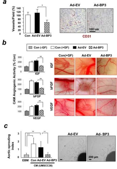
IGFBP-3 suppresses angiogenic activities in HNSCC and vascular endothelial cells
We determined Ad-BP3’s effect on angiogenesis in UMSCC38-derived tumors. Anti-CD31 staining of the Ad-BP3-treated tumors showed significantly decreased micro vessel density compared with those injected with Ad-EV (Fig. 2a). We clarified whether IGFBP-3’s effects on tumor angiogenesis was caused by the direct effect of IGFBP-3 on vascular endothelial cells. The CAM assay revealed that Ad-BP3 (50 pfu/egg) significantly inhibited IGF-, basic fibroblast growth factor (bFGF)-, and VEGF-stimulated neovascularization in chicken embryos with no signs of thrombosis, hemorrhage, or egg lethality (Fig. 2b). To test whether IGFBP-3’s inhibition of tumor angiogenesis was also caused by the suppression of
angiogenic potential in cancer cells, we examined the angiogenic effect of conditioned medium (CM) from UMSCC38 cells pre-treated with the recombinant viruses. The pro-angiogenic effect of the cancer cells was inhibited by pretreatment with Ad-BP3, as demonstrated by diminished ex vivo chick aortic sprout growth (Fig. 2c). These findings suggest that overexpression of IGFBP-3 inhibits the angiogenic potential of vascular endothelial and cancer cells.
$watermark-text
$watermark-text
IGFBP-3 inhibits migration, invasion, and tube formation of vascular endothelial cells
To investigate the mechanisms underlying antiangiogenic activities of IGFBP-3, we examined the effects of IGFBP-3 on vascular endothelial cells. We found that the migration, invasion and tube formation of HUVECs were significantly inhibited by Ad-BP3 (Fig. 3a), whereas no detectable cyototoxic activity of Ad-BP3 was observed in the same condition (data not shown). We next tested the effects of IGFBP-3 on proliferation of HUVECs. The treatment of HUVECs with 5 to 50 pfu/cell of Ad-BP3 for 3 days significantly suppressed cell proliferation induced by IGF, bFGF, or VEGF (Fig. 3b).
IGFBP-3 suppresses VEGF expression in vascular endothelial and HNSCC cells
Because one of the most potent and widely investigated activators of angiogenesis is VEGF, we asked whether Ad-BP3 inhibited angiogenesis by modulating VEGF expression. Semi-quantitative RT-PCR analysis revealed that Ad-BP3 treatment in fact decreased the mRNA levels of VEGF in UMSCC38 cells and HUVECs (Fig. 4a). The CM from Ad-BP3-treated UMSCC38 cells also showed a dose-dependent decrease in the levels of VEGF protein (Fig. 4b). However, the same doses of Ad-EV induced an increased expression of VEGF,
probably trough the activation of AKT by the RGD motif of capsid.25 Because the
adenoviral system induces large amounts of IGFBP-3 expression, we also tested the effects
of rBP3 at doses of 0.1 to 10 μg/ml; these doses are below, within, and above the
physiological range of IGFBP-3.26 rBP3 at doses greater than 5 μg/ml markedly decreased
VEGF expression in three HNSQCC cell lines, UMSCC38, SQCC/Y1 and TR146, and HUVEC (Fig. 4c). The same amount of rBP3 also decreased the tube formation of HUVEC (Fig. 4d, left). In addition, CM from UMSCC38 cells that had been incubated in serum- and
drug-free medium for 1 day after a 2-day treatment with rBP3 at doses greater than 5 μg/ml
stimulated tube formation of HUVECs significantly less than CM from vehicle-treated cells (control) (Fig. 4d, right). However, we were unable to observe detectable changes in these angiogenic parameters in UMSCC38 cells and HUVECs treated with rBP3 at doses of 0.1–1
μg/ml. These findings suggest that the antiangiogenic effects of IGFBP-3 that we observed
were physiological and pharmacological effects of IGFBP-3.
IGFBP-3 suppresses the expression of VEGF through IGF-independent mechanisms
IGF-1 is known to induce VEGF synthesis at the levels of transcription and translation.27
We investigated whether the effect of IGFBP-3 on VEGF expression is IGF-dependent. To this end, we tested the effects of a mutated adenoviral IGFBP-3 (Ad-BP3-GGG). This mutant has glycine substitutions at three residues that are critical for IGF-1 binding (i.e.,
Ile→Gly56, Leu→Gly80, and Leu→Gly81).19 We found that Ad-BP3-GGG also suppressed
VEGF production in a dose-dependent manner (Fig. 5a). To confirm the IGF-independent
inhibitory effects of Ad-BP3 on VEGF mRNA expression, we utilized R− cells, mouse
embryonic fibroblast cells that do not have IGF-1 receptor (Fig. 5b). IGFBP-3-expressing virus efficiently inhibited the expression of VEGF in the R- cells, strongly suggesting that the action of IGFBP-3 on the suppression of VEGF expression is through
IGF-1-independent mechanisms.
IGFBP-3 inhibits tumor growth and angiogenesis through an IGF-independent mechanism
We finally investigated whether the IGF-independent anti-angiogenic activities of IGFBP-3 can restrict tumor growth in vivo using Ad-BP3 and Ad-BP3-GGG. We found that the antiangiogenic and antitumor activities of Ad-BP3-GGG were similar to those of Ad-BP3. Matrigel plugs containing Ad-BP3-GGG–infected cells and plugs containing Ad-BP3– infected cells had significantly less hemoglobin levels compared with the plugs containing Ad-EV–infected cells (p< 0.01; Fig. 6a). Furthermore, Ad-BP3 and Ad-BP3-GGG both induced significant regression of UMSCC38 orthotopic tongue tumors to a similar degree
$watermark-text
$watermark-text
when injected intratumorally (Fig. 6b). Collectively, these results demonstrate that IGFBP-3 has IGF-independent anti-angiogenic/antitumor activity in HNSCC.
Discussion
In this report, we have demonstrated the anti-angiogenic antitumor effects of IGFBP-3 in HNSCC: 1) inhibition of endothelial cell migration and invasion, 2) IGF-independent inhibition of VEGF production in vascular endothelial and cancer cells, and 3) IGF-independent antiangiogenic and anti-tumor growth in vivo.
IGFBP-3, one of the most abundant proteins in serum, has been known to regulate the IGF axis, which is essential for survival and metabolism of cancer cells in a hypoxic and
energy-limited environment.28 IGFBP-3 has also been shown to inhibit vascular endothelial cell
survival 18, 29 and induce normalization of tumor vasculature.30 Based on these functions,
IGFBP-3 has been suggested to have anti-angiogenic properties. In support of this notion, IGFBP-3-mediated antitumor activities in a variety of cancer types have been found to
involve the suppression of angiogenesis.17–18, 29 In the current study, we have shown that
overexpression of IGFBP-3 also has antiangiogenic and antitumor activities in HNSCC. Nevertheless, further research is required to understand mechanisms by which IGFBP-3 regulates angiogenesis.
While investigating the mechanisms that mediate the anti-angiogenic actions of IGFBP-3, we found that IGFBP-3 regulates the expression of VEGF, a potent pro-angiogenic factor. The suppressed expression of this potent pro-angiogenic factor in cancer and vascular endothelial cells may contribute to the inhibition of the blood supply in HNSCC. Based on conventional wisdom, which states that 1) IGFBP-3 regulates cell growth by sequestering free IGF, 2) IGF-1R/PI3K/AKT signaling plays an important role in angiogenesis by
contributing to the expression of a number of pro-angiogenic factors, including VEGF,31–33
and 3) more than 80% of circulating IGF binds to IGFBP-3, it is possible that the observed inhibitory effects of IGFBP-3 on VEGF expression could have been conducted solely through dependent mechanisms. However, a number of studies have indicated
IGF-independent antitumor activities of IGFBP-3.19, 34 Recently, we have demonstrated that
several angiogenic factors, including bFGF and PDGF, are regulated by IGFBP-3 through IGF-independent mechanisms. These previous findings and our current in vitro and in vivo data showed that 1) IGFBP-3 inhibited angiogenesis induced by bFGF and VEGF, as well as IGF, 2) both Ad-BP3 and rBP3 suppressed VEGF expression in UMSCC38 cells, HUVECs,
and R− cells, and 3) wild-type IGFBP-3 and mutant IGFBP-3 lacking IGF-binding affinity
both revealed similar levels of inhibitory effects on VEGF expression, tumor angiogenesis and growth of UMSCC38 HNSCC tumors; these findings suggest that the ability of IGFBP-3 to regulate tumor angiogenesis and growth are mediated through IGF-independent mechanisms.
However, recent reports have suggested that the effect of IGFBP-3 on angiogenesis is more complicated than what is described here. IGFBP-3 has been shown to promote vascular
regrowth after vascular destruction in vivo.15 IGFBP-3 has also been shown to direct
endothelial progenitor cells to differentiate into endothelial cells and to increase cell
migration and capillary tube formation,14 especially after vascular injury.16 Findings
reported by Granata et al. showed that IGFBP-3 induced angiogenesis in vitro through the
expression of pro-angiogenic genes, such as VEGF,9 in human endothelial cells, and that
IGF signaling was essential for the pro-angiogenic activity of IGFBP-3. Therefore, it is interesting that the same molecule, IGFBP-3, shows opposing activities in regulating angiogenesis under very similar experimental conditions. It has been shown that circulating
levels of IGFBP-3 in adults are approximately 2–7 μg/ml26; the amount of recombinant
$watermark-text
$watermark-text
IGFBP-3 protein used in this previous study was 1 μg/ml.9 However, we noted in our study
that at doses of 0.1 to 1 μg/ml, recombinant IGFBP-3 had no detectable effect on VEGF
expression or tube formation, whereas at doses greater than 5 μg/ml, rBP3 inhibited both
VEGF expression and tube formation well. Therefore, it is likely that treatment with IGFBP-3 at greater than the physiological concentration inhibits VEGF expression and angiogenesis in vascular endothelial and HNSCC cells, leading to the suppression of tumor growth that we observed in this study. These data suggest that IGFBP-3 treatment is a promising approach for antiangiogenic antitumor therapy in HNSCC.
In summary, our data suggest that IGF-independent actions of IGFBP-3 contribute to its antiangiogenic antitumor activity in HNSCC by suppressing VEGF expression. However, it is possible that other secreted factors with specific effects on tumor angiogenesis may have been modulated by IGFBP-3 treatment. Our findings clearly showed the ability of IGFBP-3 to inhibit VEGF, a potent angiogenic factor, and to suppress tumor angiogenesis and growth. However, given the possibility that 1) IGFBP-3 could work as both an inducer and
suppressor of VEGF expression,5, 35–37 2) reduced tumor vascularization following
antiangiogenic therapy could induce hypoxia and, thus, promote the spread of cancer cells
toward a more oxygenated environment,38 and 3) antiangiogenic therapies could induce
acquired resistance through other growth factor-induced angiogenesis,39 extensive research,
including studies of how IGFBP-3 regulates angiogenesis, is warranted to assess the therapeutic effect of IGFBP-3 before performing clinical trials.
Supplementary Material
Refer to Web version on PubMed Central for supplementary material.
Acknowledgments
This work was supported by a grant from the National Research Foundation of Korea (NRF), Ministry of Education, Science and Technology (MEST), Republic of Korea (No. 2011-0017639), the Global Core Research Center (GCRC) grant (No. 2011-0035681) from NRF, MEST, Republic of Korea, National Institutes of Health grant R01 CA100816, the Converging Research Center Program (No. 2011-K000975) from MEST, Republic of Korea (H-Y. Lee).
References
1. van Moorselaar RJ, Voest EE. Angiogenesis in prostate cancer: its role in disease progression and possible therapeutic approaches. Mol Cell Endocrinol. 2002; 197:239–50. [PubMed: 12431818] 2. Folkman J. Tumor angiogenesis: therapeutic implications. N Engl J Med. 1971; 285:1182–6.
[PubMed: 4938153]
3. Kim WY, Lee HY. Brain angiogenesis in developmental and pathological processes: mechanism and therapeutic intervention in brain tumors. FEBS J. 2009; 276:4653–64. [PubMed: 19664069] 4. Ryan HE, Lo J, Johnson RS. HIF-1 alpha is required for solid tumor formation and embryonic
vascularization. EMBO J. 1998; 17:3005–15. [PubMed: 9606183]
5. Feldser D, Agani F, Iyer NV, Pak B, Ferreira G, Semenza GL. Reciprocal positive regulation of hypoxia-inducible factor 1alpha and insulin-like growth factor 2. Cancer Res. 1999; 59:3915–8. [PubMed: 10463582]
6. Shigematsu S, Yamauchi K, Nakajima K, Iijima S, Aizawa T, Hashizume K. IGF-1 regulates migration and angiogenesis of human endothelial cells. Endocr J. 1999; 46 (Suppl):S59–62. [PubMed: 12054122]
7. Menu E, Jernberg-Wiklund H, De Raeve H, et al. Targeting the IGF-1R using picropodophyllin in the therapeutical 5T2MM mouse model of multiple myeloma: beneficial effects on tumor growth, angiogenesis, bone disease and survival. Int J Cancer. 2007; 121:1857–61. [PubMed: 17546599]
$watermark-text
$watermark-text
8. Moser C, Schachtschneider P, Lang SA, et al. Inhibition of insulin-like growth factor-I receptor (IGF-IR) using NVP-AEW541, a small molecule kinase inhibitor, reduces orthotopic pancreatic cancer growth and angiogenesis. Eur J Cancer. 2008; 44:1577–86. [PubMed: 18445520] 9. Granata R, Trovato L, Lupia E, et al. Insulin-like growth factor binding protein-3 induces
angiogenesis through IGF-I- and SphK1-dependent mechanisms. J Thromb Haemost. 2007; 5:835– 45. [PubMed: 17388800]
10. Huang SS, Ling TY, Tseng WF, et al. Cellular growth inhibition by IGFBP-3 and TGF-beta1 requires LRP-1. FASEB J. 2003; 17:2068–81. [PubMed: 14597676]
11. Bhattacharyya N, Pechhold K, Shahjee H, et al. Nonsecreted insulin-like growth factor binding protein-3 (IGFBP-3) can induce apoptosis in human prostate cancer cells by IGF-independent mechanisms without being concentrated in the nucleus. J Biol Chem. 2006; 281:24588–601. [PubMed: 16793770]
12. Ikonen M, Liu B, Hashimoto Y, et al. Interaction between the Alzheimer’s survival peptide humanin and insulin-like growth factor-binding protein 3 regulates cell survival and apoptosis. Proc Natl Acad Sci U S A. 2003; 100:13042–7. [PubMed: 14561895]
13. Lee HY, Chun KH, Liu B, et al. Insulin-like growth factor binding protein-3 inhibits the growth of non-small cell lung cancer. Cancer Res. 2002; 62:3530–7. [PubMed: 12068000]
14. Chang KH, Chan-Ling T, McFarland EL, et al. IGF binding protein-3 regulates hematopoietic stem cell and endothelial precursor cell function during vascular development. Proc Natl Acad Sci U S A. 2007; 104:10595–600. [PubMed: 17567755]
15. Lofqvist C, Chen J, Connor KM, et al. IGFBP3 suppresses retinopathy through suppression of oxygen-induced vessel loss and promotion of vascular regrowth. Proc Natl Acad Sci U S A. 2007; 104:10589–94. [PubMed: 17567756]
16. Kielczewski JL, Jarajapu YP, McFarland EL, et al. Insulin-like growth factor binding protein-3 mediates vascular repair by enhancing nitric oxide generation. Circ Res. 2009; 105:897–905. [PubMed: 19762684]
17. Oh SH, Kim WY, Kim JH, et al. Identification of insulin-like growth factor binding protein-3 as a farnesyl transferase inhibitor SCH66336-induced negative regulator of angiogenesis in head and neck squamous cell carcinoma. Clin Cancer Res. 2006; 12:653–61. [PubMed: 16428512] 18. Liu B, Lee KW, Anzo M, et al. Insulin-like growth factor-binding protein-3 inhibition of prostate
cancer growth involves suppression of angiogenesis. Oncogene. 2007; 26:1811–9. [PubMed: 16983336]
19. Silha JV, Gui Y, Mishra S, Leckstrom A, Cohen P, Murphy LJ. Overexpression of gly56/gly80/ gly81-mutant insulin-like growth factor-binding protein-3 in transgenic mice. Endocrinology. 2005; 146:1523–31. [PubMed: 15550509]
20. Lee HY, Moon H, Chun KH, et al. Effects of insulin-like growth factor binding protein-3 and farnesyltransferase inhibitor SCH66336 on Akt expression and apoptosis in non-small-cell lung cancer cells. J Natl Cancer Inst. 2004; 96:1536–48. [PubMed: 15494604]
21. Kim MS, Lee YM, Moon EJ, Kim SE, Lee JJ, Kim KW. Anti-angiogenic activity of torilin, a sesquiterpene compound isolated from Torilis japonica. Int J Cancer. 2000; 87:269–75. [PubMed: 10861486]
22. Kim MS, Kwon HJ, Lee YM, et al. Histone deacetylases induce angiogenesis by negative regulation of tumor suppressor genes. Nat Med. 2001; 7:437–43. [PubMed: 11283670] 23. Oh SH, Woo JK, Yazici YD, et al. Structural basis for depletion of heat shock protein 90 client
proteins by deguelin. J Natl Cancer Inst. 2007; 99:949–61. [PubMed: 17565155]
24. Oh SH, Woo JK, Jin Q, et al. Identification of novel antiangiogenic anticancer activities of deguelin targeting hypoxia-inducible factor-1 alpha. Int J Cancer. 2008; 122:5–14. [PubMed: 17764071]
25. Philpott NJ, Nociari M, Elkon KB, Falck-Pedersen E. Adenovirus-induced maturation of dendritic cells through a PI3 kinase-mediated TNF-alpha induction pathway. Proc Natl Acad Sci U S A. 2004; 101:6200–5. [PubMed: 15071185]
26. Platz EA, Pollak MN, Rimm EB, et al. Racial variation in insulin-like growth factor-1 and binding protein-3 concentrations in middle-aged men. Cancer Epidemiol Biomarkers Prev. 1999; 8:1107– 10. [PubMed: 10613344]
$watermark-text
$watermark-text
27. Punglia RS, Lu M, Hsu J, et al. Regulation of vascular endothelial growth factor expression by insulin-like growth factor I. Diabetes. 1997; 46:1619–26. [PubMed: 9313759]
28. Tapanainen PJ, Bang P, Muller HL, Wilson K, Rosenfeld RG. Hypoxia-induced changes in insulin-like growth factors and their binding proteins in pregnant rats. Horm Res. 1997; 48:227– 34. [PubMed: 9362393]
29. Franklin SL, Ferry RJ Jr, Cohen P. Rapid insulin-like growth factor (IGF)-independent effects of IGF binding protein-3 on endothelial cell survival. J Clin Endocrinol Metab. 2003; 88:900–7. [PubMed: 12574231]
30. Delafontaine P, Song YH, Li Y. Expression, regulation, and function of IGF-1, IGF-1R, and IGF-1 binding proteins in blood vessels. Arterioscler Thromb Vasc Biol. 2004; 24:435–44. [PubMed: 14604834]
31. Frost P, Shi Y, Hoang B, Lichtenstein A. AKT activity regulates the ability of mTOR inhibitors to prevent angiogenesis and VEGF expression in multiple myeloma cells. Oncogene. 2007; 26:2255– 62. [PubMed: 17016437]
32. Li W, Tan D, Zhang Z, Liang JJ, Brown RE. Activation of Akt-mTOR-p70S6K pathway in angiogenesis in hepatocellular carcinoma. Oncol Rep. 2008; 20:713–9. [PubMed: 18813808] 33. Xue Q, Nagy JA, Manseau EJ, Phung TL, Dvorak HF, Benjamin LE. Rapamycin inhibition of the
Akt/mTOR pathway blocks select stages of VEGF-A164-driven angiogenesis, in part by blocking S6Kinase. Arterioscler Thromb Vasc Biol. 2009; 29:1172–8. [PubMed: 19443844]
34. Firth SM, Baxter RC. Cellular actions of the insulin-like growth factor binding proteins. Endocr Rev. 2002; 23:824–54. [PubMed: 12466191]
35. Nakamura E, Abreu-e-Lima P, Awakura Y, et al. Clusterin is a secreted marker for a hypoxia-inducible factor-independent function of the von Hippel-Lindau tumor suppressor protein. Am J Pathol. 2006; 168:574–84. [PubMed: 16436671]
36. Niu X, Zhang T, Liao L, et al. The von Hippel-Lindau tumor suppressor protein regulates gene expression and tumor growth through histone demethylase JARID1C. Oncogene.
37. Slomiany MG, Rosenzweig SA. Autocrine effects of IGF-I-induced VEGF and IGFBP-3 secretion in retinal pigment epithelial cell line ARPE-19. Am J Physiol Cell Physiol. 2004; 287:C746–53. [PubMed: 15140752]
38. Pennacchietti S, Michieli P, Galluzzo M, Mazzone M, Giordano S, Comoglio PM. Hypoxia promotes invasive growth by transcriptional activation of the met protooncogene. Cancer Cell. 2003; 3:347–61. [PubMed: 12726861]
39. Bergers G, Hanahan D. Modes of resistance to anti-angiogenic therapy. Nat Rev Cancer. 2008; 8:592–603. [PubMed: 18650835]
$watermark-text
$watermark-text
Figure 1. IGFBP-3 suppresses tumor growth of HNSCCs in vivo
UMSCC38-derived orthotopic tongue tumor growth was examined after treatment with Ad-BP3 or Ad-EV (a) or PBS or recombinant IGFBP-3 (10 mg/kg) (b). The relative tumor volume (the starting tumor volume was set to 100%) was expressed as the mean (%) from 5 mice in each group ± SEM. The body weight of mice was also measured. *, p< 0.05; **, p< 0.01. vs. controlAd-EV injected tumors..
$watermark-text
$watermark-text
Figure 2. IGFBP-3 suppresses tumor-associated or growth factor–induced angiogenesis in vivo and in vitro.
(a) Microvessel density of UMSCC38 orthotopic tongue tumors 10 days after injection with Ad-BP3 or Ad-EV. The number of CD31-immunoreactive vessels per field was counted after immunohistochemical analysis. n=5. (b) Chorio-allantoic membrane (CAM) assay for angiogenesis in chicken embryos (n=20) treated with vehicle (Con), Ad-EV or Ad-BP3 in the presence of 50 ng/mL of IGF, bFGF, or VEGF. The blood vessels were scored blindly and relative scores to the control are shown as %. Independent experiments were repeated three times; the values are presented as the mean ± SEM. (c) Effect of CM from HNSCC lines infected with Ad-EV or Ad-BP3 on chick aortic sprouting. n=5. The results represent the mean ± SEM. *, p< 0.05; **, p< 0.01; ***, p< 0.001 vs. the control group.
$watermark-text
$watermark-text
Figure 3. IGFBP-3 inhibits angiogenesis by acting directly on vascular endothelial cells
(a) Inhibitory effects of IGFBP-3 on migration, invasion, and tube formation of HUVECs. (b) Proliferation of HUVECs supplemented with pro-angiogenic growth factors was inhibited by IGFBP-3. Ad-BP3–infected HUVEC culture was supplemented with the indicated growth factors, and the MTT assay was performed 3 days later. Data are presented as the mean ± SD with eight replicates per experiment. Control cells were not treated with the virus. n=5. **, p< 0.01; ***, p< 0.001.
$watermark-text
$watermark-text
Figure 4. IGFBP-3 suppresses expression of VEGF in UMSCC38 HNSCC cells and HUVECs
Semi-quantitative RT-PCR (a) and Western blot analysis (b) on VEGF in the mRNAs and conditioned medium from UMSCC38 HNSCCs. (c) Semi-quantitative RT-PCR analysis of VEGF and GAPDH mRNA expression in UMSCC38 cells and HUVECs treated with the indicated doses of rBP3. (d) Tube formation of HUVECs was inhibited by treatment with rBP3 (left). Angiogenic activity in conditioned medium from UMSCC38 cells after rBP3 treatment was tested by tube formation (right). The control was set to 100%, bars, SDs. Recombinant human IGFBP-3, rBP3. *, p< 0.05; ***, p< 0.001.
$watermark-text
$watermark-text
Figure 5. IGFBP-3 suppresses expression of VEGF through IGF-independent mechanisms
(a) Western blot of UMSCC38 lysates and conditioned medium (CM) after infection with indicated doses of Ad-EV or Ad-BP3-GGG. (b) Semiquantitative RT-PCR analysis of
VEGF and IGFBP-3 mRNA expression in R- cells infected with indicated doses of Ad-EV
or Ad-BP3.
$watermark-text
$watermark-text
Figure 6. IGF-independent inhibition of tumor growth and angiogenesis is mediated byIGFBP-3
(a) IGF-independent tumor angiogenesis inhibition by IGFBP-3. Matrigel plug assay was
done using UMSCC38 cells pretreated with viruses. Hemoglobin amounts (μg per 100 mg
of dissected Matrigel tumors) were measured. Results are the mean from six tumors (except control; Matrigel only, n = 3; ± SEM). **; p<0.01. (b) UMSCC38-derived orthotopic tongue tumor growth was measured 7 and 11 days after intratumoral injection of Ad-BP3, Ad-BP3-GGG, Ad-EV or PBS. The starting tumor volume was set to 100% (± SE). **; p<0.01; n = 5 for each group.