532
Young-Ok Jung1, Inje Kim2,
Suho Kim2, Chang-Hee Suh3,
Han Jung Park3, Won Park4,
Seoung Ryul Kwon4, Jae Cheon Jeong4,
Yun Jong Lee5, Hee Jung Ryu5,
Young Bae Park5, Jisoo Lee6,
You-Hyun Lee6, Young Il Seo2,
Won Tae Chung7, Seung-Jae Hong8,
Yeon-Sik Hong9, Han Joo Baek10,
Hyo Jin Choi10, Hyo-Jong Kang11,
Chan-Hee Lee12, Sang-Hyon Kim13,
and Hyun Ah Kim2
Division of Rheumatology, Department of Internal Medicine1, Kangnam Sacred Heart Hospital, Hallym University College of Medicine, Seoul; Division of Rheumatology, Department of Internal Medicine2, Sacred Heart Hospital, Hallym University College of Medicine, Anyang; Division of Allergy-Rheumatology, Department of Internal Medicine3, Ajou University School of Medicine, Suwon; Division of Rheumatology, Department of Internal Medicine4, Inha University Hospital, Incheon; Division of Rheumatology, Department of Internal Medicine5, Seoul National University Bundang Hospital, Seoul National University College of Medicine, Seongnam; Division of Rheumatology, Department of Internal Medicine6, Ewha Womans University Mokdong Hospital, Seoul; Division of Rheumatology, Department of Internal Medicine7, Dong-A University College of Medicine, Busan; Division of Rheumatology, Department of Internal Medicine8, KyungHee University Medical Center, KyungHee University, Seoul; Division of Rheumatology, Department of Internal Medicine9, Our Lady of Mercy Hospital, Catholic University of Korea, College of Medicine, Incheon; Division of Rheumatology, Department of Internal Medicine10, Gachon University of Medicine and Science, Gil Medical center, Incheon; Division of Rheumatology, Department of Internal Medicine11, Pundang Jesaeng General Hospital, Seongnam; Division of
Rheumatology, Department of Internal Medicine12, NHIC Ilsan Hospital, Goyang; Division of Rheumatology, Department of Internal Medicine13, Keimyung University Dongsan Medical Center, Daegu, Korea
Address for Correspondence
Hyun Ah Kim, M.D.
Division of Rheumatology, Department of Internal Medicine, Hallym University Sacred Heart Hospital, 896 Pyeongchon-dong, Dongan-gu, Anyang 431-070, Korea
Tel : +82.31-380-1726, Fax : +82.31-386-2269 E-mail : [email protected]
This study was supported by a grant from the Hallym University Medical Center (01-2007-18).
J Korean Med Sci 2010; 25: 532-5 ISSN 1011-8934
DOI: 10.3346/jkms.2010.25.4.532
Clinical and Radiographic Features of Adult-onset Ankylosing Spondylitis
in Korean Patients: Comparisons between Males and Females
The objective of this study was to investigate clinical and radiographic features and gender differences in Korean patients with adult-onset ankylosing spondylitis. Multi-center cross-sectional studies were conducted in the rheumatology clinics of 13 Kore-an tertiary referral hospitals. All patients had a confirmed diagnosis of Kore-ankylosing spondylitis according to the modified New York criteria. Clinical, laboratory, and radio-graphic features were evaluated and disease activities were assessed using the Bath ankylosing spondylitis disease activity index. Five hundred and five patients were recruited. The male to female ratio was 6.1:1. Average age at symptom onset was 25.4±8.9 yr and average disease duration was 9.6±6.8 yr. Males manifested symp-toms at a significantly earlier age. HLA-B27 was more frequently positive in males. Hips were more commonly affected in males, and knees in females. When spinal mobility was measured using tragus-to-wall distance and the modified Schober’s test, females had significantly better results. Radiographic spinal changes, includ-ing bamboo spine and syndesmophytes, were more common in males after adjust-ment of confounding factors. In conclusion, we observed significant gender differ-ences in radiographic spinal involvement as well as other clinical manifestations among Korea patients with adult-onset ankylosing spondylitis. These findings may influence the timing of the diagnosis and the choice of treatment.
Key Words : Spondylitis, Ankylosing
Received : 17 April 2009 Accepted : 23 July 2009
ⓒ 2010 The Korean Academy of Medical Sciences.
This is an Open Access article distributed under the terms of the Creative Commons Attribution Non-Commercial License (http://creativecommons.org/licenses/by-nc/3.0) which permits unrestricted non-commercial use, distribution, and reproduction in any medium, provided the original work is properly cited.
Clinical and Radiographic Features in Korean Ankylosing Spondlylitis Patients 533
INTRODUCTION
Ankylosing spondylitis (AS) is a human leukocyte anti-gen (HLA)-B27-associated chronic inflammatory disease that predominantly affects the spine and sacroiliac joints. AS occurs worldwide, but its prevalence ranges from 0 to 1.9%, which implies ethnic and geographic differences (1). Historically, AS has been considered to overwhelmingly affect males, but recent studies have shown that a significant number of AS patients are females, with a male to female ratio approach-ing 2-3:1 (2, 3). Furthermore, the disease pattern also varies by gender, and the spine and pelvis are more commonly affect-ed in males (4). The documentaffect-ed clinical features of AS have been largely derived from studies on Caucasian patients, and studies on Asians are relatively scarce. In particular, gender-based comparisons of clinical manifestations have not been reported in Asia. In the present study, we compared gender differences in the clinical and radiographic features of AS in 505 patients treated at 13 tertiary Korean hospitals.
MATERIALS AND METHODS
Patients
Patients were enrolled at the rheumatology clinics of 13 ter-tiary hospitals between February and December 2006. Pati-ents were included if they met the modified New York crite-ria for AS (5) and were willing to participate in our registry study of AS. The study hospitals were located in four different Korean provinces. Patients with juvenile onset of AS (an age at onset of <15 yr), and patients with a history of inflamma-tory bowel disease or psoriasis were excluded. This study was approved by the ethics committees at all participating hos-pitals and informed consent was obtained from all patients. Methods
All participants completed questionnaires, which includ-ed questions on demographics, such as age, sex, includ-educational attainment, occupation, exercise, family history of AS, and co-morbidities. Heights (cm) and body weights (kg) were measured to the nearest 0.1 cm and 0.1 kg, respectively, with subjects barefoot and wearing light clothing, to calculate body mass indices (BMI). We also collected clinical data, such as, disease duration, age at disease onset, manifesting symptoms, initial joint symptoms, previous site of joint and enthesis involvement, and history of extra-articular manifestations. Disease activities were evaluated using a validated Korean version of Bath Ankylosing Spondylitis Disease Activity Index (BASDAI) (6). We performed complete joint and enthesis examinations and physical measurements on all patients at study entry. Lumbar spinal mobility was measured using Macrae’s modification of Schober’s technique (7).
Measure-ments were taken of tragus-to-wall distance and chest expan-sion at the level of the 4th anterior rib. Joint involvement was defined either if a joint showed swelling, tenderness, or a limited range of motion during physical examination or if the patient reported having arthralgia/arthritis in a joint dur-ing clinical data collection. Entheses were examined usdur-ing direct pressure over standard sites, such as the calcaneal inser-tions of the Achilles tendon, plantar fascia, tibial tuberosi-ties, costosternal junction, iliac crest, and greater trochanter. Enthesitis was defined as either the presence of entheseal ten-derness during physical examination or the history of pain at the above entheseal areas. The laboratory evaluations per-formed included; complete blood cell count, Westergren ery-throcyte sedimentation rate (ESR), C-reactive protein (CRP), HLA-B27, rheumatoid factor (RF), and antinuclear antibody (ANA). The following radiographs were obtained in each case; the pelvis (anteroposterior view), cervical, thoracic, and lum-bar spine (anteroposterior and lateral views). Radiographs of the spine were assessed for the formation of syndesmophytes and for bamboo spine. Bamboo spine was defined as ankylo-sis involving all three spinal levels. Patients with bamboo spine was not included in the counting of spinal syndesmo-phytes. Radiographs were read by academically based rheuma-tologists experienced at reading spine radiographs.
Statistics
All data are presented as means and standard deviation (SD) or as percentages. Unadjusted male-female comparisons were performed using the Student’s t-test for normally distributed continuous measures, and using the chi-square test or Fish-er’s exact test for categorical measures. The odds ratios and 95% confidence interval for bamboo spine was estimated after adjusting age, BMI, duration of disease and HLA-B27 posi-tivity. P value of <0.05 was regarded as significant.
RESULTS
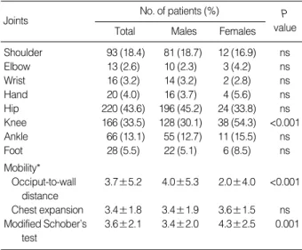
In total, 434 males and 71 females with AS were studied. Males manifested symptoms at a significantly earlier age, and had longer disease duration at the time of study entry (Table 1). Disease activity as measured using BASDAI, ESR and CRP was not significantly different between genders. HLA-B27 positivity was significantly higher among males (94.8% vs. 87.3%; male vs. females, P=0.016). Males experienced joint pain, including back pain, as the first manifestation of AS more often than females (94.7% vs. 85.9%, male vs. females,
P=0.005), while females more frequently experienced uveitis
as the first manifestation compared to males (3.2% vs. 9.9%, male vs. females, P=0.009). The most common joint symp-tom was back pain (63.4% vs. 54.9%, male vs. females). Ex-cluding the spine, the hips and knees were the most common-ly affected joints; knee involvement was significantcommon-ly more
534 Y.-O. Jung, S. Kim, C.-H. Suh, et al.
frequent in females (30.1% vs. 54.3%, male vs. females, P< 0.001), and hip involvement tended to be more frequent in males (45.2% vs. 33.8%, male vs. females, P=0.074) (Table 2). When spinal mobility was measured using tragus-to-wall distance and the modified Schober’s test, females had signif-icantly better results; however, chest expansion was not sig-nificantly different. In our study subjects, Achilles tendini-tis was the most common form of enthesitendini-tis, followed by plan-tar fasciitis. Planplan-tar fasciitis was significantly more frequent in females (6.0% vs. 15.5%, males vs. females, P=0.004), as was uveitis (19.4% vs. 32.4%, males vs. females, P=0.013). The pattern of spinal involvement manifesting as
syndesmo-phytes or bamboo spine is shown in Table 3. Bamboo spine and syndesmophytes in the thoracic spine was significantly more common among male patients. Male patients had sig-nificantly increased odds ratio for bamboo spine compared to females after adjustment of age, BMI, duration of disease and HLA-B27 positivity (unadjusted odds ratio [OR]2.24
[95% confidence interval [CI]1.34-3.74], adjusted OR 1.90
[95% CI 1.13-3.39]).
DISCUSSION
In this study, we aimed to investigate the clinical and radio-graphic features of 505 Korean patients with adult-onset AS and to identify gender differences. It was observed that males manifested symptoms at a significantly earlier age, had a greater HLA-B27 positive rate, and had more extensive radiograph-ic spinal involvement. On the other hand, females had more uveitis.
As compared with previous reports of male-to-female ratios of 1.9:1 to 3:1 (2, 8-9), we found a ratio of 6.1:1 in the pre-sent study, which concurs with previous Korean and Chinese reports on patients with AS (10, 11). However, we do not know whether this higher male to female ratio is a specific feature among Asian subjects. The majority of Asian studies have been conducted at single centers, usually referral hospi-tals, and thus, patients with more severe symptoms may have been selectively recruited. Although most of the study cen-ters involved in the present study were university affiliated tertiary hospitals, the study included hospitals located in 4 different Korean provinces (Seoul, Pusan, Daegu, Kyunggi), and it is one of the largest studies conducted in Asia, and thus, it might be more representative than previous Asian studies. Contrary to a previous report that revealed longer diagnostic delays among female patients (12), no difference in gender-associated diagnostic delays was observed in the present study (4.5 yr vs. 3.8 yr; male vs. female, respectively). Thus, the larger male to female ratio observed in the present study is unlikely to be entirely due to the under-diagnosis of female patients. A larger study involving patients from a community cohort or nationwide patient registry is warranted to determine gen-der differences in AS prevalence more precisely in Korea.
Although the overall frequency of HLA-B27 positivity of
BMI, body mass index; BASDAI, bath ankylosing spondylitis disease activity index; ns, non-significant.
Parameters Total (n=505) Males (n=434) Females (n=71) P value Age (yr) 35.0±10.0 34.9±10.1 35.4±9.6 ns
Age at diagnosis (yr) 30.0±9.7 29.8±9.7 31.5±9.7 ns
Age at symptom 25.4±8.9 25.0±8.9 27.7±8.8 0.015 onset (yr) Duration of 9.6±6.8 9.9±7.0 7.7±5.4 0.004 disease (yr) Education level (%) ≤6 yr 7 (1.4) 7 (1.7) 0 6-12 yr 171 (35.2) 148 (35.5) 23 (33.3) ns ≥12 yr 308 (63.4) 262 (62.8) 46 (66.7) ns BMI (kg/m2) 22.8±3.1 23.0±3.0 21.5±3.3 <0.001 Family history of 64 (12.7) 55 (12.9) 9 (12.7) ns AS (%) HLA-B27-positivity: 465 (93.8) 403 (94.8) 62 (87.3) 0.016 n (%) BASDAI 4.4±2.3 4.5±2.4 4.2±2.0 ns
Table 1. Baseline characteristics of AS patients
*cm, mean±SD.
Joints
Total Males Females
No. of patients (%) P value Shoulder 93 (18.4) 81 (18.7) 12 (16.9) ns Elbow 13 (2.6) 10 (2.3) 3 (4.2) ns Wrist 16 (3.2) 14 (3.2) 2 (2.8) ns Hand 20 (4.0) 16 (3.7) 4 (5.6) ns Hip 220 (43.6) 196 (45.2) 24 (33.8) ns Knee 166 (33.5) 128 (30.1) 38 (54.3) <0.001 Ankle 66 (13.1) 55 (12.7) 11 (15.5) ns Foot 28 (5.5) 22 (5.1) 6 (8.5) ns Mobility* Occiput-to-wall 3.7±5.2 4.0±5.3 2.0±4.0 <0.001 distance Chest expansion 3.4±1.8 3.4±1.9 3.6±1.5 ns Modified Schober’s 3.6±2.1 3.4±2.0 4.3±2.5 0.001 test
Table 2. Peripheral joint involvements and spinal mobility of male and female AS patients
Radiographic
findings Total Males Females
No. of patients (%) P value Lumbar syndesmophyte 123 (24.4) 106 (24.4) 12 (24.5) ns Thoracic syndesmophyte 61 (12.8) 59 (13.6) 2 (2.8) 0.027 Cervical syndesmophyte 13 (2.6) 10 (2.3) 3 (4.2) ns Bamboo spine 104 (20.6) 96 (22.1) 8 (11.3) 0.023 Normal 266 (52.7) 220 (50.7) 46 (64.8) 0.042
Table 3. Patterns of radiographic spinal involvement in AS pati-ents
Clinical and Radiographic Features in Korean Ankylosing Spondlylitis Patients 535
our subjects was similar to those reported previously, females had significantly lower positivity compared to males in our study patients, which is contradictory to previous reports including that from Korea (10). Whether this discrepancy stems from the limited sample size or true difference among Korean subjects also needs to be investigated in a larger pati-ent registry.
The findings of the present study concur with those of pre-vious studies regarding the common involvements of joints of the lower extremities, particularly the hips, knees and ankles, in AS (10, 13), and although the knee joint was significant-ly more involved in females and the hip joint in males, peri-pheral joints including those of the ankle, foot, wrist and elbow were similarly involved in both genders. Furthermore, in line with a previous Korean report, our patients tended to have higher prevalences of shoulder, hip and peripheral joint involvement than Caucasians (1).
Ankylosis is regarded as the end point of the inflammato-ry process in AS. Sacroiliac joint involvement and spinal anky-losis have been reported to be more frequent among male patients (14). And the present study also shows a higher pro-portion of males had spinal involvement, which manifested as spinal syndesmophytes and bamboo spine. It is presumed that the better mobility of our female AS patients, despite similar disease activities (as measured by BASDAI) and acute phase reactant levels, were the result of reduced spinal involve-ment. It would be interesting to determine whether this dif-ference in spinal involvement and mobility is related to bet-ter functional outcomes in female patients.
The underlying pathogenetic mechanism that results in gender-associated differences in terms of the manifestations of AS is unknown. Recently, different haplotype combina-tions in the ankylosis homologue (ANKH) gene were report-ed in males and females with AS (15). Further genetic stud-ies may identify more genes that contribute to observed eth-nic and gender differences and the severity of AS.
The limitations of this study include its cross-sectional design and possible selection bias due to the recruitment of patients from rheumatology clinics in tertiary referral hospi-tals. In addition, due to limited resources, no standardized reading scheme was used for spinal radiographs, such as the Stoke ankylosing spondylitis spinal scoring system, and only limited analysis of spine involvement extent was performed. The reading was performed by as many as 13 readers, and because of the logistics, we could not obtain inter-reader vari-ability. However, limited inter-reader variability analysis involving 2 readers and 45 subjects showed that the kappa statistics were good for our simple scheme evaluating only the presence or absence of syndesmophytes and bamboo spine. Joint examination findings were recorded at the time of recruit-ment, and thus, previous involvement may not have been included, which possibly caused peripheral joint involvement to be underestimated.
In conclusion, we have examined the clinical
characteris-tics of a large number of Korean patients with adult-onset AS. We observed significant gender differences in radiograph-ic spinal involvement as well as other clinradiograph-ical manifestations among our subjects. These findings may influence the tim-ing of the diagnosis and the choice of treatment.
REFERENCES
1. Baek HJ, Shin KC, Lee YJ, Kang SW, Lee EB, Yoo CD, Song YW.
Clinical features of adult-onset ankylosing spondylitis in Korean patients: patients with peripheral joint disease (PJD) have less severe spinal disease course than those without PJD. Rheumatology (Oxford) 2004; 43: 1526-31.
2. Gran JT, Husby G. The epidemiology of ankylosing spondylitis. Semin
Arthritis Rheum 1993; 22: 319-34.
3. Will R, Edmunds L, Elswood J, Calin A. Is there sexual inequality
in ankylosing spondylitis? A study of 498 women and 1202 men. J Rheumatol 1990; 17: 1649-52.
4. Resnick D, Dwosh IL, Goergen TG, Shapiro RF, Utsinger PD, Wies-ner KB, Bryan BL. Clinical and radiographic abnormalities in
anky-losing spondylitis: A comparison of men and women. Radiology 1976; 119: 293-7.
5. Goie The HS, Steven MM, van der Linden SM, Cats A. Evaluation
of diagnostic criteria for ankylosing spondylitis: a comparison of the Rome, New York and modified New York criteria in patients with a positive clinical history screening test for ankylosing spondylitis. Br J Rheumatol 1985; 24: 242-9.
6. Calin A, Nakache JP, Gueguen A, Zeidler H, Mielants H, Dougados M. Defining disease activity in ankylosing spondylitis: is a
combina-tion of variables (Bath Ankylosing Spondylitis Disease Activity Index) an appropriate instrument? Rheumatology 1999; 38: 878-82.
7. Macrae IF, Wright V. Measurement of back movement. Ann Rheum
Dis 1969; 28: 584-9.
8. Brunner R, Kissling RO, Auckenthaler C, Fortin J. Clinical evaluation
of ankylosing spondylitis in Switzerland. Pain Physician 2002; 5: 49-56.
9. Lee W, Reveille JD, Davis JC Jr, Learch TJ, Ward MM, Weisman MH. Are there gender differences in severity of ankylosing
spondyli-tis? Results from the PSOAS cohort. Ann Rheum Dis 2007; 633-8.
10. Lee JH, Jun JB, Jung S, Bae SC, Yoo DH, Kim TY, Kim SY, Kim TH. Higher prevalence of peripheral arthritis among ankylosing
spondylitis patients. J Korean Med Sci 2002; 17: 669-73.
11. Zeng QY. Ankylosing spondylitis in Shantou, China: 15 years’
clin-ical experience. J Rheumatol 2003; 30: 1816-21.
12. Calin A, Elswood J, Rigg S, Skevington SM. Ankylosing
spondyli-tis--an analytical review of 1500 patients: the changing pattern of disease. J Rheumatol 1988; 8: 1234-8.
13. Jimenez-Balderas FJ, Mintz G. Ankylosing spondylitis: clinical course
in women and men. J Rheumatol 1993; 20: 2069-72.
14. Kidd B, Mullee M, Frank A, Cawley M. Disease expression of
anky-losing spondylitis in males and females. J Rheumatol 1988; 15: 1407-9.
15. Tsui HW, Inman RD, Paterson AD, Reveille JD, Tsui FW. ANKH
variants associated with ankylosing spondylitis: gender differences. Arthritis Res Ther 2005; 7: R513-25.