Introduction
Since Urist1) demonstrated ectopic bone and carti-lage formation after the intramuscular implantation of demineralized bone matrix in rats, which were later named as bone morphogenetic proteins(BMPs), several BMPs have been shown to have significant os-teoinductive activity2-8). It was reported that rhBMP itself is sufficient to induce bone formation. However,
the rapid diffusion of the water-soluble protein rhBMP from the implant site will reduce its os-teoinductive effect. Therefore, a carrier system for rhBMPs is essential9-12).
The carrier for BMPs should serve as a scaffold for bone forming cells while providing space in which bone formation can occur, and resist soft tissue com-pression during the healing period13). For the clinically successful use of BMPs, the carrier should also be easy to manipulate, sterilize, and fabricate into the intended shape. It is also important that the shape and volume of newly formed bone be maintained. If these requirements of carrier are satisfied, predictable bone regeneration is possible with sufficient value to
rhBMP-2 using biphasic calcium phosphate block as a carrier induces new bone
formation in a rat subcutaneous tissue
Joon-Il Kim
1, Jeong-Ho Yun
2, Gyung-Joon Chae
1, Sung-Won Jung
1, Chang-Sung Kim
1,
Kyoo-Sung Cho
1*1. Department of Periodontology, Research Institute for Periodontal Regeneration, College of Dentistry, Yonsei University
2. Department of Dentistry, College of Medicine, Kwandong University, Myongji Hospital ABSTRACT
Purpose: The carrier for the delivery of bone morphogenetic proteins(BMPs) should also serve as a scaffold for new bone
growth. In addition, predictable bone formation in terms of the volume and shape should be guaranteed. This study evaluated the ectopic bone formation of recombinant human BMP-2(rhBMP-2) using a micro macroporous biphasic calcium phosphate (MBCP: mixture of β-TCP and HA) block as a carrier in a rat subcutaneous assay model.
Materials and Methods: Subcutaneous pockets were created on the back of 40 male Sprague-Dawley rats. In the pockets,
rhBMP-2/MBCP and MBCP alone were implanted. The blocks were evaluated by histological and histometric parameters after a healing interval of 2 weeks (each 10 rats; MBCP and rhBMP-2/MBCP) or 8 weeks (each 10 rats; MBCP and rhBMP-2/MBCP).
Results: The shape and volume of the block was maintained stable over the healing period. No histological bone forming
activity was observed in the MBCP alone sites after 2 weeks and there was minimal new bone formation at 8 weeks. In the rhBMP-2/MBCP sites, new bone formation was evident in the macropores of the block. The new bone area at 8 weeks was greater than at 2 weeks. There was a further increase in the quantity of new bone with the more advanced stage of remodeling.
Conclusions: A MBCP block could serve as a carrier system for predictable bone tissue engineering using rhBMPs. (J Korean Acad Periodontol 2008;38:355-362)
KEY WORDS: rhBMP-2; biphasic calcium phosphate block; carrier; ectopic bone formation.
Correspondence: Dr. Kyoo-Sung Cho
Department of Periodontology, Research Institute for Periodontal Regeneration, College of Dentistry, Yonsei University, 134 Shinchon-Dong, Seodaemun-gu, Seoul, 120-752, Korea.
e-mail: [email protected], Tel: 82-2-2228-3188, Fax: 82-2-392-0398 * This work was supported by the Korea Research Foundation Grant
studies on carriers14-17), but no carriers have satisfied these requirements perfectly.
Micro macroporous biphasic calcium phosphate (MBCP) consists of an intimate mixture of 40% β -tricalcium phosphate(β-TCP) and 60% hydrox-yapatite(HA)18,19). Besides the well-documented osteo-conductive effect20-22), the osteoinductive effect of the MBCP is recommended for use as an alternative or as an additive to autogenous bone for orthopedics, trau-matology, odontology and dental applications23-28). The porous structure, low resorption rate, favorable os-teoconductivity and bioactive properties of MBCP can be utilized by rhBMPs carrier. The easy manipulation and producing the intended shape, particularly the block type, are additional merits of using this biomaterial. However, there has been little study on its use as a carrier for BMPs29,30).
The subcutaneous tissue of a rat has poorer con-ditions than the muscle or bony site or the subcuta-neous tissue of other animals31-34). Therefore, success this region can indicate better success in other regions.
This study evaluated the ectopic bone formation of rhBMP-2 using a MBCP block as a carrier in a rat subcutaneous assay model.
Materials and Methods
1. Animals
40 male Sprague-Dawley rats (weight 250~300g, approximately 8 weeks old) were used. Rats were maintained in plastic cages in a room with a 12h-day/night cycle and an ambient temperature of 21゚C, with ad libitum access to water and standard labo-ratory pellets. Animal selection and management, sur-gical protocol, and preparation were in accordance with the routines approved by the Institutional Animal Care and Use Committee, Yonsei Medical Center, Seoul, Korea.
2. rhBMP-2 Implant Construction
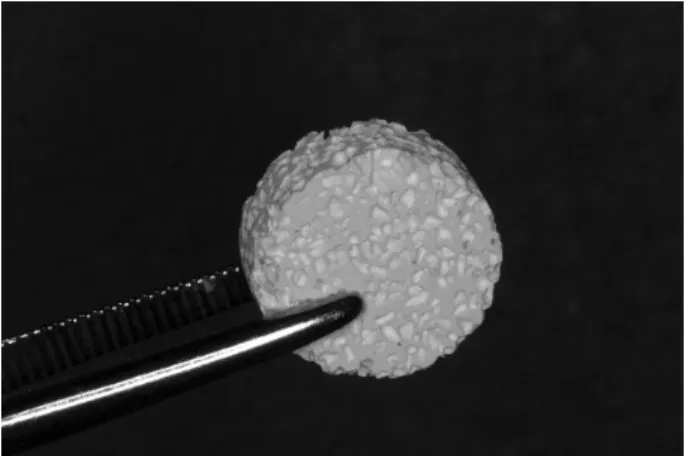
Disc-shaped MBCP implants (Biomatlante Inc., Vigneux de Bretagne, France) (3mm height and 8mm diameter) were manufactured (Fig. 1). rhBMP-2(R & D Systems Inc., Minneapolis, MN, USA) was recon-stituted and diluted in a buffer to produce a concen-tration of 0.025mg/ml. For the rhBMP-2/MBCP im-plants, MBCP implants were loaded with 0.2ml of the rhBMP-2 solutions for one hour before surgery. For the MBCP implants alone, MBCP implants were loaded with 0.2ml of buffer solutions.
Figure 1. Block type MBCP implant used in this study (3mm in height and 8mm in diameter).
3. Surgical Procedures
The animals were anaesthetized by an intramuscular injection (5mg/kg body wt.) of a 4:1 solution of ket-amine hydrochloride (Ketalar®, Yuhan Co., Seoul, Korea) and xylazine(Rompun®, Bayer Korea, Seoul, Korea). The surgical site was shaved and scrubbed with iodine. A vertical incision was made in the skin of the back. After flap reflection, a subcutaneous pocket was prepared by blunt instrument. Each animal received 1 of 2 experimental treatments: MBCP carrier control and rhBMP-2/MBCP. The skin was closed and sutured with absorbable monofilament suture (Monosyn®, Aesculap AG Co. KG, Tuttlingen, Germany).
4. Histologic and Histometric Procedures
The animals were sacrificed by CO2 asphyxiation at 2 and 8 weeks post-surgery. Block sections were re-moved and fixed in a 10% neutral buffered formalin solution for 10 days. Samples were decalcified by 5% formic acid for 14 days and embedded in paraffin. Serial sections 7um in thickness were prepared at in-tervals of 80um, stained with hematoxylin/eosin (H-E), and examined using a light microscope. The most central sections from each block were selected for histologic and histometric evaluation.
Computer-assisted histometric measurements were obtained using an automated image analysis system (Image-Pro Plus®, Media Cybernetics, Silver Spring, MD, USA) coupled with a video camera on a light mi-croscope (Olympus BX50, Olympus Optical Co., Tokyo, Japan). Sections were examined at magnifications of ×20 and ×100. Histometric parameters were defined as follows.
Total augmented area (mm2
): the area of im-planted MBCP zone.
New bone area (mm2
): the area of newly formed bone within the total augmented area.
5. Statistical Analysis
Histometric recordings from the samples were used to calculate means and standard deviations (m±SD). For comparisons between the two groups, a paired or unpaired t-test was used. The interactions between the healing times were examined using two-way anal-ysis of variance. A p-value <0.01 was considered significant.
Results
1. Clinical observation
Wound healing was generally uneventful and there were no signs and symptoms of infection or in-flammation
2. Histological Observations
1) MBCP control group
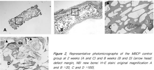
At 2 weeks, histological signs of inflammation or foreign body reactions were not generally observed. MBCP blocks were surrounded by loose connective tissue. Macropores of the block were filled with con-nective tissue. Osteogenic activity could not found histologically (Fig. 2-A, C). At 8 weeks post surgery, the implanted block was covered with dense and fi-brous connective tissue. The minimal amount of new bone formation was observed adjacent to the margins of the block. No significant resorption of the MBCP block was observed during the healing time (Fig. 2-B, D).
2) MBCP/ rhBMP-2 group
At 2 weeks, the macropores in the periphery of the block were filled with new bone. Evidence of osteo-genic activity, such as dense osteoblast-like cell lin-ing, osteoid and bone apposition along the surface of macropores was observed. Macropores in the center part were usually filled with loose fibrous connective tissue and there was a little bone-forming activity (Fig. 3-A, C).
At 8 weeks, the quantity of new bone was greater
than that observed at 2 weeks, and the specimens showed a more advanced stage of remodeling and consolidation. Some macropores with fibrous con-nective tissue could be also found in the central part of the block. The newly formed bone consisted of wo-ven and lamellar bone, and showed cement lines that were separated from the more recently deposited bone. There was no evidence of cartilage formation (Fig. 3-B, D).
Figure 2. Representative photomicrographs of the MBCP control group at 2 weeks (A and C) and 8 weeks (B and D) (arrow head: defect margin, NB: new bone; H-E stain; original magnification A and B ×20, C and D ×100).
Figure 3. Representative photomicrographs of the rhBMP-2/MBCP group at 2 weeks (A and C) and 8 weeks (B and D) (arrow head: defect margin, arrow: cement lines, NB: new bone; H-E stain; origi-nal magnification A and B ×20; C and D ×100).
3. Histometric Analysis
Table 1 and 2 show the results of histometric analysis. In the rhBMP-2/MBCP group, the quantity of the new bone was greater at 8 weeks than 2 weeks (p<0.01).
Discussion
The aim of this study was to evaluate the ectopic bone formation of rhBMP-2 using a MBCP block as a carrier in a rat subcutaneous assay model. Because the subcutaneous tissue of rats have poorer conditions than muscle or bony site or the subcutaneous tissue of other animals, the success in this region can indicate better success in other regions. The results were evaluated by histological and histometric analysis af-ter a 2- and 8-week healing inaf-terval.
In a previous study13) using a similar method to this study, it was demonstrated that rhBMP-2, when im-pregnated in an absorbable collagen sponge(ACS) and β-TCP, provoked osteoinductive activity in rat sub-cutaneous tissue at 2 weeks. However, at 8 weeks neither ACS nor the newly formed bone observed at 2 weeks were found in the rhBMP-2/ACS sites, while
rhBMP-2/β-TCP sites. This suggests that ACS does not have capacity as a carrier for rhBMP-2 in this model. The lack of space-maintaining capacity of ACS might be one of the major factors responsible for the resorption of the newly-formed bone observed in the 2-week sections after implantation. It was concluded that the carrier for the delivery of BMPs should also serve as a scaffold for bone forming cells while pro-viding sufficient space for bone formation to occur. Although favorable ectopic bone formation could be obtained using β-TCP particles, questions arise as to whether clinical applications are possible, because there is clinical difficulty in the nature of its partic-ulate form, in the manipulation and maintenance of the intended shape of the bone. Since predictable bone tissue regeneration might be one of the major goals of the clinical therapeutic results, a carrier system for rhBMPs should provide stability over time in terms of volume and shape.
MBCP consists of an intimate mixture of 40% β -TCP and 60% HA, and is obtained when a synthetic calcium deficient apatite is sintered at ≥700゚C. It is now available in blocks, particulates and as an in-jectable material in a polymer carrier18,19). MBCP has excellent biocompatibility and bioactivity, which is re-lated to its porous structure20-22). Micropores≤10μm Table 1. Total Augmented Area (group means ±SD, mm2, n=10)
Group 2 weeks 8 weeks
MBCP 21.9 ± 2.6 19.9 ± 2.8
rhBMP-2/MBCP 23.7 ± 1.9 23.5 ± 2.4
No significant difference when compared to all groups (P>0.01).
Table 2. New Bone Area (group means±SD, mm2, n=10)
Group 2 weeks 8 weeks
MBCP 0.8 ± 0.2 2.1 ± 1.1*
rhBMP-2/MBCP 3.9 ± 0.7¶ 5.8 ± 2.4*
*: Statistically significant difference compared to 2 weeks (p<0.01). ¶: Statistically significant difference compared to MBCP group (p<0.01).
degradation of the biomaterial. Macropores ≥100μm act as a scaffold for bone cells, thereby allowing cen-tripetal bone ingrowth23). The newly formed bone is in direct contact with the biomaterial surface, making this biomaterial osteoconductive. In addition to the well-documented osteoconductive effect, the os-teoinductive effect of the MBCP is recommended for use as an alternative or additive to autogenous bone for orthopedics, traumatology, odontology and dental applications24,25).
The porous structure, low resorption rate, favorable osteoconductive and bioactive properties of MBCP can be utilized by the rhBMPs carrier. The porous struc-ture of MBCP can be retained, allow the slow release of rhBMPs, and provide the appropriate scaffold to allow cells and newly formed tissues to migrate into it. In clinical aspects, the MBCP block is a very in-teresting biomaterial for use as a carrier for BMPs because it can be manipulated easily to make the in-tended shape. However, there have been few studies on its use as a carrier for BMPs29,30).
In this study, new bone formation in the macro-pores of the block was evident in the rhBMP-2/MBCP sites. The new bone area was greater at 8 weeks than at 2 weeks. The quantity of the new bone had in-creased further with the more advanced stage of remodeling. In addition, the total augmented area was stable during the healing period. In subcutaneous tis-sue of rats, this favorable results confirmed that the MBCP block is a good carrier system for tissue en-gineering using rhBMPs.
In conclusion, the use of the block type of MBCP as a carrier for rhBMP-2 is effective in inducing new bone formation. The new bone induced by the rhBMPs/MBCP block system was stable during the ob-served healing period. These results suggest that the MBCP block system can be used as a carrier system for tissue engineering using rhBMPs
References
1. Urist MR. Bone: Formation by autoinduction. Science 1965;150:893-899.
2. Ahn SH, Kim CS, Suk HJ, et al. Effect of recombinant hu-man bone morphogenetic protein- 4 with carriers in rat cal-varial defects. J Periodontol 2003;74:787-797.
3. Pang EK, Im SU, Kim CS, et al. Effect of recombinant hu-man bone morphogenetic protein-4 dose on bone formation in rat calvarial defect model. J Periodontol 2004;75:1364-1370. 4. Aldinger G, Herr G, Kusswetter W, et al. Bone
morphoge-netic protein: a review. Int Orthop 1991;15:169-177. 5. Sampath TK, Maliakal JC, Hauschka PV. Recombinant
hu-man osteogenic protein-1 (hOP-1) induces new bone for-mation in vivo with a specific activity comparable with natural bovine osteogenic protein and stimulates osteoblast proliferation and differentiation in vitro. J Biol Chem 1992;267:20352-20362.
6. Han DK, Kim CS, Jung UW, et al. Effect of a fibrin-fi-bronectin sealing system as a carrier for recombinant hu-man bone morphogenetic protein-4 on bone formation in rat calvarial defects. J Periodontol 2005;76:2216-2222. 7. Jung UW, Choi SY, Pang EK, et al. The Effect of varying
the particle size of beta tricalcium phosphate carrier of re-combinant human bone morphogenetic protein-4 on bone formation in rat calvarial defects. J Periodontol 2006;77: 765-772.
8. Yasko AW, Lane JM, Fellinger EJ, et al. The healing of segmental bone defects, induced by recombinant human bone morphogenetic protein (rhBMP-2). A radiographic, histological, and biomechanical study in rats. J Bone Joint Surg Am 1992;74:659-670.
9. Kim CS, Choi SH, Choi BK, et al. The effect of recombi-nant human bone morphogenetic protein-4 on the osteo-blastic differentiation of mouse calvarial cells affected by Porphyromonas gingivalis. J Periodontol 2002;73:1126-1132. 10. Hyun SJ, Choi SH, Chai JK, et al. Effect of recombinant
human bone morphogenetic protein-2, -4 and -7 on bone formation in rat calvarial defects. J Periodontol 2005;76: 1667-1674.
fects in dogs. J Periodontol 2002;73:63-72.
12. Lindholm TS, Gao TJ. Functional carriers for bone mor-phogenetic proteins. Ann Chir Gynaecol Suppl 1993;207:3-12. 13. Kim CS, Kim JI, Kim J, et al. Ectopic bone formation as-sociated with recombinant human bone morphogenetic pro-tein-2 using absorbable collagen sponge and beta tricalcium phosphate as carriers. Biomaterials 2005;26:2501-2507. 14. Kawamura M, Urist MR. Human fibrin is a physiologic
de-livery system for bone morphogenetic protein. Clin Orthop Relat Res 1988;235:302-310.
15. Hong SJ, Kim CS, Han DK, et al. The effect of a fibrin-fi-bronectin/ß-tricalcium phosphate/recombinant human bone morphogenetic protein-2 system on bone formation in rat calvarial defects. Biomaterials 2006;27:3810-3816.
16. Gao TJ, Kousinioris NA, Wozney JM, Winn S, Uludag H. Synthetic thermoreversible polymers are compatible with osteoinductive activity of recombinant human bone mor-phogenetic protein 2. Tissue Eng 2002;8:429-440. 17. Miki T, Imai Y. Osteoinductive potential of freeze-derived,
biodegredable, poly(glycolic acid-co-lactic acid) disks in-corporated with bone morphogenetic protein in skull de-fects of rats. Int J Oral Maxillofac Surg 1996;25:402-426. 18. Nery EB, LeGeros RZ, Lynch KL, Lee K. Tissue response to biphasic calcium phosphate ceramic with different ratios of HA/beta TCP in periodontal osseous defects. J Periodontol 1992 ;63:729-735.
19. LeGeros RZ, Lin S, Rohanizadeh R, Mijares D, LeGeros JP. Biphasic calcium phosphate bioceramics: preparation, properties and applications. J Master Sci Mater Med 2003;14:201-209.
20. Cornell CN, Lane JM. Current understanding of osteo-conduction in bone regeneration. Clin Orthop Relat Res 1998;355 Suppl:S267-273.
21. Schopper C, Ziya-Ghazvini F, Goriwoda W, et al. HA/TCP compounding of a porous CaP biomaterial improves bone formation and scaffold degradation--a long-term histological study. J Biomed Mater Res B Appl Biomater 2005;74:458-467. 22. Manjubala I, Sastry TP, Kumar RV. Bone growth in-duced by biphasic calcium phosphate ceramic in femoral defect of dogs. J Biomater Appl 2005;19:341-360.
23. Gauthier O, Bouler JM, Aguado E, Pilet P, Daculsi G. Macroporous biphasic calcium phosphate ceramics: influ-ence of macropore diameter and macroporosity percentage
24. Daculsi G, Laboux O, Malard O, Weiss P. Current state of the art of biphasic calcium phosphate bioceramics. J Mater Sci Mater Med 2003;14:195-200.
25. Habibovic P, Yuan H, van den Doel M, et al. Relevance of osteoinductive biomaterials in critical-sized orthotopic defect. J Orthop Res 2006;24:867-876.
26. Le Nihouannen D, Daculsi G, Saffarzadeh A, et al. Ectopic bone formation by microporous calcium phosphate ceramic particles in sheep muscles. Bone 2005;36:1086-1093. 27. Yuan H, van Blitterswijk CA, de Groot K, de Bruijn JD.
A comparison of bone formation in biphasic calcium phos-phate (BCP) and hydroxyapatite (HA) implanted in muscle and bone of dogs at different time periods. J Biomed Mater Res A 2006;78:139-147.
28. Le Guehennec L, Goyenvalle E, Aguado E, et al. Small-an-imal models for testing macroporous ceramic bone substitutes. J Biomed Mater Res B Appl Biomater 2005;72: 69-78.
29. Oda S, Kinoshita A, Higuchi T, Shizuya T, Ishikawa I. Ectopic bone formation by biphasic calcium phos-phate(BCP) combined with recombinant human bone mor-phogenetic protein-2 (rhBMP-2). J Med Dent Sci 1997;44: 53-62.
30. Boden SD, Kang J, Sandhu H, Heller JG. Use of recombi-nant human bone morphogentic protein-2 to achieve post-erolateral lumbar spine fusion in humans: a prospective, randomized clinical pilot trial: 2002 Volvo Award in clin-ical studies. Spine 2002;27:2662-2273.
31. Okubo Y, Bessho K, Fujimura K, et al. Comparative study of intramuscular and intraskeletal osteogenesis by recombi-nant human bone morphogenetic protein-2. Oral Surg Oral Med Oral Pathol Oral Radiol Endod 1999;87:34–38. 32. Yoshida K, Bessho K, Fujimura K, et al. Osteoinduction
capability of recombinant human bone morphogenetic pro-tein-2 in intramuscular and subcutaneous sites: an ex-perimental study. J Craniomaxillofac Surg 1998;26:112–115. 33. Daculsi G, Layrolle P. Osteoinductive properties of micro
macroporous biphasic calcium phosphate bioceramics. Key Eng Mater 2004;254–256:1005–1008.
34. Yang Z, Yuan H, Tong W, et al. Osteogenesis in extra-skeletally implanted porous calcium phosphate ceramics: variability among different kinds of animals. Biomaterials 1996;17:2131-2137.