R E S E A R C H A R T I C L E
Open Access
Factors associated with improvement or
decline in cognitive function after an
ischemic stroke in Korea: the Korean stroke
cohort for functioning and rehabilitation
(KOSCO) study
Jin A. Yoon
1, Deog Young Kim
2, Min Kyun Sohn
3, Jongmin Lee
4, Sam-Gyu Lee
5, Yang-Soo Lee
6, Eun Young Han
7,
Min Cheol Joo
8, Gyung-Jae Oh
8, Junhee Han
9, Minsu Park
1, Kyung Pil Park
10, Kyung-Ha Noh
10, Won Hyuk Chang
11,
Yong-Il Shin
1,12*and Yun-Hee Kim
11*Abstract
Background: We conducted a prospective cohort study to investigate prevalence of poststroke cognitive impairment at 3 and 12 months after stroke onset and identify clinical and demographic factors associated with improvement or decline in cognitive function between 3 months and 12 months.
Methods: We analyzed the cognitive assessments of total patients and patients older than 65 years separately. All patients with an ischemic stroke were divided into normal cognitive group (NCG) and impaired cognition group (ICG) by using a cutoff score on the Korean Mini-Mental State Examination (K-MMSE). Patients were additionally classified into 3 subgroups according to the changes in their K-MMSE scores between 3 and 12 months: Stable group with K-MMSE scores changes ranging from−2 to +2 points (−2 ≤ △MMSE ≤ +2); converter group with increase more than 3 points (3≤ △MMSE); and reverter group with decrease more than 3 points (−3 ≤ △MMSE). We also analyzed factors affecting cognitive change from 3 months to 12 months among the 3 groups including baseline medical record, stroke and treatment characteristics, and various functional assessments after 3 months. Results: This study included 2,625 patients with the first time ischemic stroke. Among these patients, 1,735 (66.1%) were classified as NCG, while 890 patients (33.9%) were belonged to the ICG at 3 month. Within the NCG, 1,460 patients (82.4%) were stable group, 93 patients (5.4%) were converter group, and 212 patients (12.2%) were reverter group at 12 months onset. Within the ICG group, 472 patients (53.0%) were stable group, 321 patients (36.1%) were converter group, and 97 patients (10.9%) were reverter group. When different factors were investigated, the three subgroups in NCG and ICG showed significant different factors affecting cognitive function from 3 to 12 month.
(Continued on next page)
* Correspondence:[email protected];[email protected]
1Department of Rehabilitation Medicine, Pusan National University School of Medicine, Research Institute of Convergence for Biomedical Science and Technology, 20, Geumo-ro, Mulgeum, Yangsan 626-770, South Korea 11
Department of Physical and Rehabilitation Medicine, Center for Prevention and Rehabilitation, Heart Vascular and Stroke Institute, Samsung Medical Center, Sungkyunkwan University School of Medicine, 50 Ilwon-dong, Gangnam-gu, Seoul 135-710, Republic of Korea
Full list of author information is available at the end of the article
© The Author(s). 2017 Open Access This article is distributed under the terms of the Creative Commons Attribution 4.0 International License (http://creativecommons.org/licenses/by/4.0/), which permits unrestricted use, distribution, and reproduction in any medium, provided you give appropriate credit to the original author(s) and the source, provide a link to the Creative Commons license, and indicate if changes were made. The Creative Commons Public Domain Dedication waiver (http://creativecommons.org/publicdomain/zero/1.0/) applies to the data made available in this article, unless otherwise stated. Yoon et al. BMC Neurology (2017) 17:9
(Continued from previous page)
Conclusions: The prevalence of cognitive impairment showed difference between 3,12 months. To analyze the cognitive change from 3 month to 12 month, the proportion stable group was dominant in NCG and converter group was higher in ICG. By investigating the influencing factors from each group, we were able to identify the predictors including the age factor.
Keywords: Ischemic stroke, Cognition, Inverter, Reverter, Risk factors
Background
Cerebrovascular stroke is considered one of the main causes of dementia [1–3]. It may decrease quality of life in addition to causing other neurological deficits [4]. Post-stroke dementia is defined as a presence of dementia identified at 3 months after an acute stroke [5]. Reasons for a stroke patient to develop dementia are still insuffi-ciently understood. It is not always a direct consequence of cerebrovascular lesions, and, in some cases, post-stroke dementia has a progressive course, suggesting a degenera-tive rather than a vascular origin [6, 7]. In previous aut-opsy series, 10 to 15% of dementias occurring after a stroke were found to be due to a combination of vascular and Alzheimer’s disease [8, 9].
Despite consensus that strokes are associated with an increased risk of post-stroke dementia, the data regard-ing prevalence at 3 months post-stroke are still conflict-ing, with reports ranging from 6% to more than 50% [10–14]. In addition, cognitive function may vary (either improve or decline) years after a stroke. Snaphaan et al. reported that post-stroke memory dysfunction varied from 23 to 55% at 3 months after a stroke, and this de-clined from 11 to 31% at 1 year after a stroke. Dede-clined cognitive function may change. A previous cohort study showed 33% of patients with mild cognitive impairments diagnosed in the first 6 months after a stroke showed improvement at 1 year. Several prospective studies have identified delayed improvement in cognitive function after strokes using various diagnostic assessment tools for dementia [15-17].
The pathophysiology of delayed cognitive change after a stroke is multifactorial, and the prevalence rate of post-stroke dementia is higher among older patients [18, 19]. Previous studies have tracked cognitive changes to identify the factors associated with delayed improvement or a decline in cognitive function after stroke. However, no large-scale study has been con-ducted to investigate the pattern of post-stroke cogni-tive changes, identify the risk factors, or compare age-related differences using repeated administration of the most commonly used screening tool.
Therefore, we conducted a prospective cohort study in conjunction with the Korean Stroke Cohort for Func-tioning and Rehabilitation (KOSCO) to identify 1) the prevalence of delayed cognitive impairment; patients
progress to either converter, stable, or reverter group after ischemic stroke and 2) clinical and demographic factors associated with improvement or decline in cogni-tive function between 3 months and 12 months after is-chemic stroke. The present study is the first to involve a large and well-characterized Korean cohort, a battery of short cognitive and functional assessments, and a 1-year follow-up.
Methods Study design
KOSCO is a large, multi-centered, prospective cohort study of all acute, first-time stroke patients admitted to participating hospitals in nine distinct areas of Korea. A written informed consent is obtained from all patients prior to inclusion in the study, and the study protocols were approved by the ethics committee of each hospital. The detailed rationale and protocols of KOSCO were de-scribed in a previous article [20].
Study subjects
All consecutive patients with an acute, first-time IS admitted to the representative hospitals were asked to participate in the study. The inclusion criteria were: 1) first-time ischemic stroke with corresponding lesion on a MRI/A scan, 2) age ≥19 years at stroke onset, and 3) onset of symptoms within 7 days prior to inclusion. Ex-clusion criteria were: 1) recurrent stroke; 2) hemorrhagic stroke; 3) traumatic intracerebral hemorrhage; 4) previ-ously diagnosed dementia or cognitive impairment; 5) persistent aphasia; and 6) history of systemic diseases known to involve the central nervous system.
Procedure
All eligible patients were recruited from August 2012 to April 2015 at the time of stroke evaluation. After provid-ing a written informed consent, patients were formally enrolled in the study. If a patient was unable to provide informed consent, the consent was obtained from the patient’s legally authorized representative.
Demographic and clinical characteristics
Baseline demographic and clinical characteristics of en-rolled patients were evaluated at 3 months. A complete enumeration survey of all patients was performed using
a review of medical records upon the first admission. Survey items included demographic data and presence of cerebrovascular risk factors using standardized, struc-tured questionnaires. The items were classified accord-ing to the current guidelines of the American Heart Association [21]. Comorbidities were assessed using the Charlson comorbidity index [22]. Initial stroke severity was recorded at the time of hospital arrival using the Korean National Institute of Health Stroke Scale (K-NIHSS) for ischemic strokes [23]. Physical examin-ation findings and laboratory measures were also re-corded. The course of the disease during admission was documented including information on medication use, treatments such as intravenous or arterial thrombolysis, and complications. Patients that re-ceived rehabilitation at 3 months were transferred to the rehabilitation center to initiate active rehabilita-tion after acute management at the neuroscience cen-ter. The remaining patients that did not receive any rehabilitation treatments were discharged or trans-ferred to other hospitals instead of being transtrans-ferred to the Rehabilitation Medicine Department.
Classification of ischemic stroke; etiology, and neuroimaging
The etiologies of ischemic strokes were classified accord-ing to the TOAST criteria [24]. Etiology was determined based on neuro-imaging, medical history, and use of medication. MRI scans were reviewed by neuroimaging specialists in each institute. Ischemic strokes were classi-fied according to arterial territory and as either lacunar or territorial.
Cognitive assessment
To identify influencing factors by age, we analyzed the Korean Mini-Mental State Examination (K-MMSE) at 3 months separately between total patients and patients older than 65 years. To analyze changes in cognitive function, all patients were divided into normal cognitive group (NCG) and impaired cognition group (ICG) by
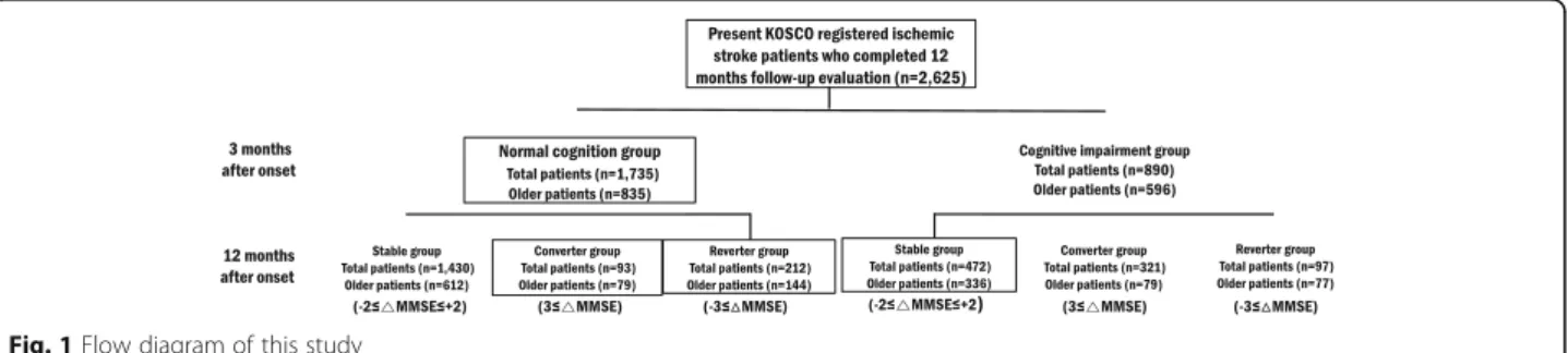
using standard deviation score after correcting raw scores by age, sex and education level of the patients [25]. Patients were again classified according to the changes in their K-MMSE scores between 3 and 12 months after stroke onset into stable groups (NCG-SG, ICG-SG) with K-MMSE changes ranging from −2 to +2 points (−2 ≤ △MMSE ≤ +2), converter groups (NCG-CG, ICG-CG) with increases of K-MMSE more than 3 points (3≤ △MMSE), and reverter groups (NCG-RG, ICG-RG) with score decreases of K-MMSE more than 3 points (−3 ≤ △MMSE). Factors affecting cognitive change from 3 months to 12 months including baseline medical record, stroke and treatment characteristics, and various func-tional assessments after 3 months were analyzed among the groups (Fig. 1).
Functional assessment battery
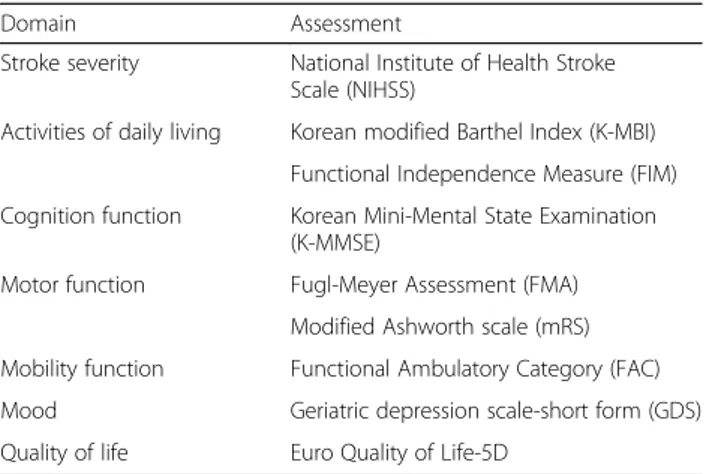
At 3 months after stroke onset, a face-to-face functional assessment was performed. Assessments included the K-NIHSS for stroke severity, Functional Independence Measure (FIM) [26], Korean modified Barthel Index (K-MBI) [27] for activities of daily living, Fugl-Meyer As-sessment (FMA) [28] for motor function, Functional Ambulatory Category (FAC) [29] for mobility and gait, mRS (modified rankin scale) [30] for general functional level, Geriatric depression scale-short form (GDS-SF) [31] for mood, and Euro Quality of Life (EQ)-5D [32] for quality of life (Table 1).
Statistical analysis
For statistical analysis, we used descriptive statistics for the demographic and clinical characteristics, initial stroke features and treatment methods. Nominal and or-dinal data obtained from a baseline review of medical re-cords and initial stroke features were compared using frequency analysis. Scale factors were analyzed using average analysis. Chi-square test and one-way ANOVA were used to compare the influencing factors among groups. Bonferroni correction was done for post-hoc analysis of ANOVA. Statistical analysis was completed
Fig. 1 Flow diagram of this study
using SPSS for Windows version 21.0 (SPSS Inc., Chicago, IL).P < 0.05 is considered statistically significant.
Results
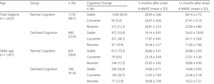
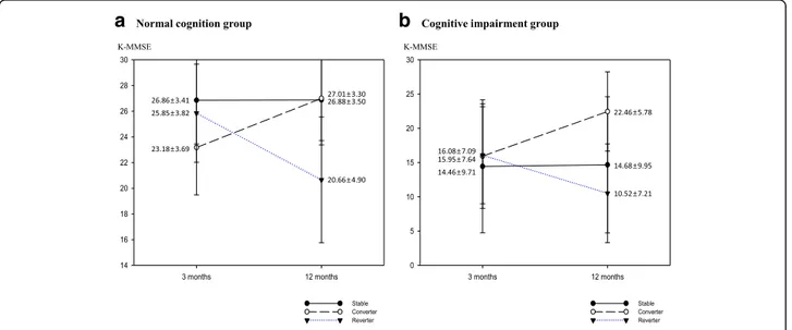
A total of 2,625 patients (older patients = 1,431) with first time ischemic stroke were included in this study. Among these patients, 1,735 (66.1%) (older patients = 835 (58.4%)) were classified as NCG, while 890 patients (33.9%) (older patients = 596 (41.6%)) were the ICG at 3 month K-MMSE assessment. Although, percentage of normal and declined cognitive function was similar for older patients at 3 months and 12 months, percentage of normal cognition group was slightly increased and per-centage of declined cognition groups was decreased in total patients (Fig. 2). Among NCG, 1,460 (82.4%) (older patients = 612 (73.3%)) were stable group, 93 patients
(5.4%) (older patients = 79 (9.5%)) were converter group, and 212 patients (12.2%) (older patients = 144(17.2%)) were re-verter group at 12 months onset. Among ICG, 472 patients (53.0%) (older patients = 336 (56.4%)) were stable group, 321 patients (36.1%) (older patients = 183(30.7%)) were converter group, and 97 patients (10.9%) (older patients = 77 (12.9%)) were reverter group (Table 2) (Figs. 3, 4). To analyze the cog-nitive change from 3 month to 12 month, the proportion stable group was dominant in NCG and converter group was higher in ICG.
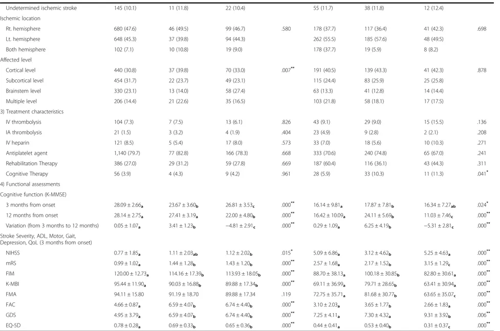
Among NCG of total patients, hypertension, and cor-tical or multiple level involvement was dominant in re-verter group, male sex, lower onset age, higher education level were dominant in stable group. In addition, func-tional assessments in stable group including NIHSS, mRS, FIM, K-MBI, FAC, GDS, and EQ-5D at 3 months were significantly better in scores compared to other groups. For the tendency of ICG of total patients, onset age, hypertension history was higher, education level was lower in reverter group. All functional assessments at 3 months showed better scores in converter groups and worse scores in reverter group (Table 3). Among separated older patients, male sex, lower onset age were dominant in stable group, educational level was lower in reverter group. Functional assessments including NIHSS, mRS, FIM, K-MBI, FAC, GDS, and EQ-5D, at 3 months showed better scores in stable group compared to others. In addition, proportion of receiving rehabilitation therapy at 3 months was lower and all functional l assessments showed better scores in converter group compared to others (Table 4).
Table 1 Functional assessments at 3 months
Domain Assessment
Stroke severity National Institute of Health Stroke Scale (NIHSS)
Activities of daily living Korean modified Barthel Index (K-MBI) Functional Independence Measure (FIM) Cognition function Korean Mini-Mental State Examination
(K-MMSE)
Motor function Fugl-Meyer Assessment (FMA)
Modified Ashworth scale (mRS) Mobility function Functional Ambulatory Category (FAC)
Mood Geriatric depression scale-short form (GDS)
Quality of life Euro Quality of Life-5D
a
b
Fig. 2 Cognitive function of patients at 3 months and 12 months after stroke onset
Discussion
In our study, total the percentage of cognitive impair-ment group did not change at 12 months compared to 3 months assessment. Otherwise, the percentage of cog-nitive impairment at 3 months, the percentage of pa-tients in the reverter group, and the percentage of patients transferring from the NCG to the ICG at 12 months were higher in older patients compared to total group analysis (Table 2) (Fig. 2). Influencing factors for delayed cognitive change were discretely determined in the NCG and ICG of total and older patients. Hyper-tension history and onset age, sex, education level were somewhat repeated influencing factors. Although pres-ence of atrial fibrillation, smoking, alcohol history showed statistical significance, large difference of
number of patients between groups made it difficult to define it as meaningful result. Another unique aspect of our study is that we included functional assessment since it could influence on patients’ delayed cognitive function. Patients with better functional assessment scores not only in cognitive field but in all other do-mains including activities of daily living, motor function, mobility and gait, general functional level, quality of life at 3 month tend to have less cognitive decline and more cognitive improvement from 3 month to 12 month. Otherwise, stroke characteristics including ischemic type, location, treatment characteristics showed no sig-nificant difference between the groups. Patients in the ICG-RG that were aged >65 years received more re-habilitation therapy. Moreover, compared to the other
Table 2 Cognitive change divided by age from 3 months to 12 months
Age Group n (%) Cognitive Change 3 months after onset 12 months after onset
n (%) K-MMSE (mean ± SD) K-MMSE (mean ± SD)
Total Subjects (n = 2,625) Normal Cognition 1,735 (66.1) Stable 1,430 (82.4) 28.09 ± 2.66 28.14 ± 2.75 Converter 93 (5.4) 23.67 ± 3.60 27.41 ± 3.19 Reverter 212 (12.2) 26.81 ± 3.53 22.00 ± 4.80 Declined Cognition 890 (33.9) Stable 472 (53.0) 16.14 ± 9.81 16.42 ± 10.09 Converter 321 (36.1) 17.87 ± 9.81 24.11 ± 5.69 Reverter 97 (10.9) 16.34 ± 7.27 11.03 ± 7.46 Older age (n = 1,431) Normal Cognition 835 (58.4) Stable 612 (73.3) 26.86 ± 3.41 26.88 ± 3.50 Converter 79 (9.5) 23.18 ± 3.69 27.01 ± 3.30 Reverter 144 (17.2) 25.85 ± 3.82 20.66 ± 4.90 Declined Cognition 596 (41.6) Stable 336 (56.4) 14.46 ± 9.71 14.68 ± 9.95 Converter 183 (30.7) 15.95 ± 7.64 22.46 ± 5.78 Reverter 77 (12.9) 16.08 ± 7.09 10.52 ± 7.21
n, Number; SD, Standard Deviation; K-MMSE, Korean Mini Mental State Examination
a
b
Fig. 3 Cognitive change of total patients from 3 months to 12 months
patient groups, the patients in the ICG-SG received less cognitive therapy, which was a component of the re-habilitation therapy. We believe that unlike the other factors, the administration of rehabilitation treatment depended on the cognitive status of the patient; however, rehabilitation was not a factor that improved cognitive function.
Stroke severity, onset age, pre-stroke cognitive func-tion, level of educafunc-tion, and bilateral lesions are well-known factors associated with development of post-stroke dementia [33–36]. In contrast, a cohort study of younger stoke patients (mean age, 60 years) showed that more than 30% of the patients with mild cognitive im-pairments between 0 and 6 months were classified as cognitively intact by 12 to 18 months [16]. For older pa-tients (mean age, 80.4 ± 3.8), about 50% of the papa-tients experienced an improvement in MMSE at 15 months [17]. As the prognosis of stroke varies according to the patient’s age at onset, identifying the factors affecting cognitive changes by age might aid in both preventing secondary cognitive decline, and enhancing post-stroke cognitive function. In our study, separating patients by age, cognitive function at 3 month, and aspect of cogni-tive change to find the propensity of each prevalence and influencing factors was meaningful. Also, compared to other cross sectional studies, our study analyzed de-layed post stroke cognitive function and focused on amount and aspect of cognitive change from 3 month to 12 month and its influencing factors.
To carry this analysis further, we examined the differ-ences in the MMSE scores among the groups by using the cutoff score of <24 points, conventionally accepted for the diagnosis of significant cognitive impairment, i.e., dementia [37]. Previous studies have established more
than 3 points of MMSE variability as a significant change for improvement or decline in cognitive function.
MMSE is the most frequently applied test for dementia screening. A systematic review and meta-analysis examin-ing cognitive tests to detect dementia found 10,263 cases of dementia identified from 36,080 participants in 108 co-hort studies. The result reported a sensitivity of 0.81 and a specificity of 0.89 for the MMSE [38]. The MMSE requires only 5–10 min to evaluate various cognitive domains (orientation, memory, language, attention, visuospatial) and is practical to use serially and routinely [39].
The overall prevalence of dementia in subjects aged greater than 65 in Korea is estimated to be 9.2%. In addition, the pooled age-specific prevalence of dementia is estimated to increase with each 5-year age band (65–69 years) [40]. This result is much higher than the estimated overall prevalence of dementia in Asian people [41]. We analyzed our data by separating patients older than 65 y/o to compare the age factors. The percentages of normal cognitive group and cognitive impaired patients and mean MMSE scores showed significant differences between total and older patient groups. In particular, the percentage of patients in the reverter group was higher and the con-verter group was lower at 12 months in older patient group, and their average MMSE showed differences by age. Otherwise, in older ICG, less factors were significant compared to other groups. This finding may be due to the lower percentage of patients when compared to the entire study population and NCG.
Limitations
First, we only used a MMSE to test cognitive function. Although there are 40 other more detailed tests for de-mentia diagnosis in healthcare settings, we required
a
b
Fig. 4 Cognitive change of the older patient group from 3 months to 12 months
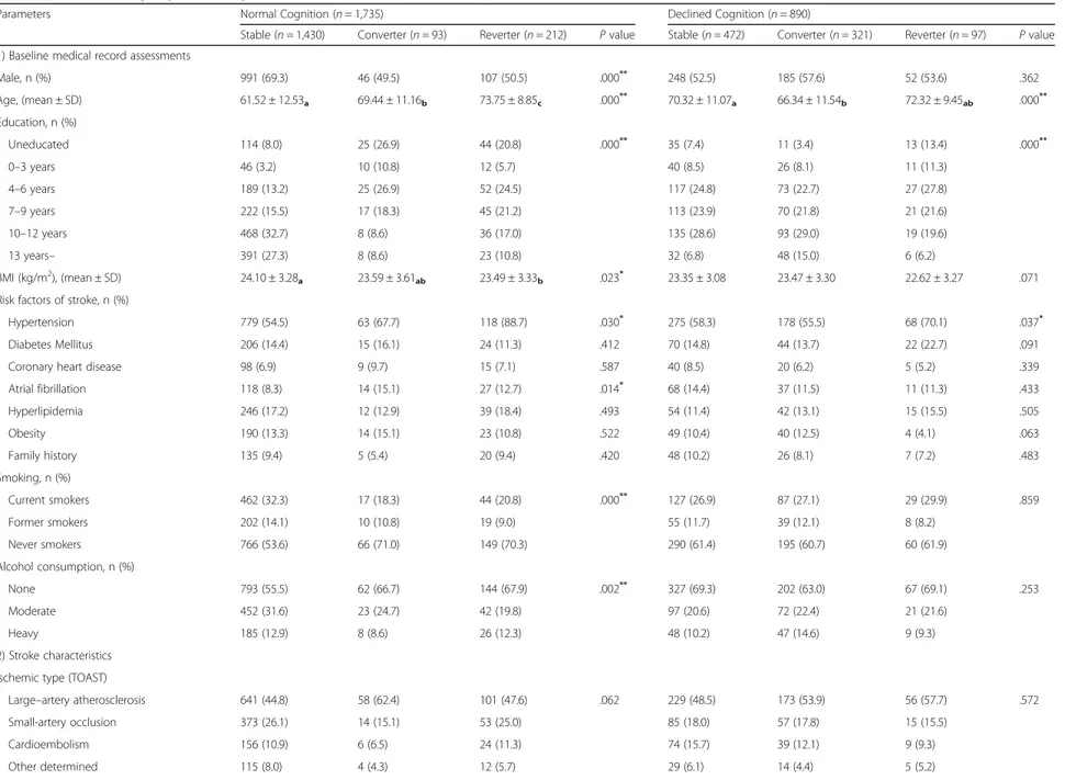
Table 3 Factors affecting cognitive change from 3 months to 12 months in total patients
Parameters Normal Cognition (n = 1,735) Declined Cognition (n = 890)
Stable (n = 1,430) Converter (n = 93) Reverter (n = 212) P value Stable (n = 472) Converter (n = 321) Reverter (n = 97) P value 1) Baseline medical record assessments
Male, n (%) 991 (69.3) 46 (49.5) 107 (50.5) .000** 248 (52.5) 185 (57.6) 52 (53.6) .362 Age, (mean ± SD) 61.52 ± 12.53a 69.44 ± 11.16b 73.75 ± 8.85c .000** 70.32 ± 11.07a 66.34 ± 11.54b 72.32 ± 9.45ab .000** Education, n (%) Uneducated 114 (8.0) 25 (26.9) 44 (20.8) .000** 35 (7.4) 11 (3.4) 13 (13.4) .000** 0–3 years 46 (3.2) 10 (10.8) 12 (5.7) 40 (8.5) 26 (8.1) 11 (11.3) 4–6 years 189 (13.2) 25 (26.9) 52 (24.5) 117 (24.8) 73 (22.7) 27 (27.8) 7–9 years 222 (15.5) 17 (18.3) 45 (21.2) 113 (23.9) 70 (21.8) 21 (21.6) 10–12 years 468 (32.7) 8 (8.6) 36 (17.0) 135 (28.6) 93 (29.0) 19 (19.6) 13 years– 391 (27.3) 8 (8.6) 23 (10.8) 32 (6.8) 48 (15.0) 6 (6.2) BMI (kg/m2), (mean ± SD) 24.10 ± 3.28a 23.59 ± 3.61ab 23.49 ± 3.33b .023* 23.35 ± 3.08 23.47 ± 3.30 22.62 ± 3.27 .071
Risk factors of stroke, n (%)
Hypertension 779 (54.5) 63 (67.7) 118 (88.7) .030* 275 (58.3) 178 (55.5) 68 (70.1) .037*
Diabetes Mellitus 206 (14.4) 15 (16.1) 24 (11.3) .412 70 (14.8) 44 (13.7) 22 (22.7) .091
Coronary heart disease 98 (6.9) 9 (9.7) 15 (7.1) .587 40 (8.5) 20 (6.2) 5 (5.2) .339
Atrial fibrillation 118 (8.3) 14 (15.1) 27 (12.7) .014* 68 (14.4) 37 (11.5) 11 (11.3) .433 Hyperlipidemia 246 (17.2) 12 (12.9) 39 (18.4) .493 54 (11.4) 42 (13.1) 15 (15.5) .505 Obesity 190 (13.3) 14 (15.1) 23 (10.8) .522 49 (10.4) 40 (12.5) 4 (4.1) .063 Family history 135 (9.4) 5 (5.4) 20 (9.4) .420 48 (10.2) 26 (8.1) 7 (7.2) .483 Smoking, n (%) Current smokers 462 (32.3) 17 (18.3) 44 (20.8) .000** 127 (26.9) 87 (27.1) 29 (29.9) .859 Former smokers 202 (14.1) 10 (10.8) 19 (9.0) 55 (11.7) 39 (12.1) 8 (8.2) Never smokers 766 (53.6) 66 (71.0) 149 (70.3) 290 (61.4) 195 (60.7) 60 (61.9) Alcohol consumption, n (%) None 793 (55.5) 62 (66.7) 144 (67.9) .002** 327 (69.3) 202 (63.0) 67 (69.1) .253 Moderate 452 (31.6) 23 (24.7) 42 (19.8) 97 (20.6) 72 (22.4) 21 (21.6) Heavy 185 (12.9) 8 (8.6) 26 (12.3) 48 (10.2) 47 (14.6) 9 (9.3) 2) Stroke characteristics Ischemic type (TOAST)
Large–artery atherosclerosis 641 (44.8) 58 (62.4) 101 (47.6) .062 229 (48.5) 173 (53.9) 56 (57.7) .572
Small-artery occlusion 373 (26.1) 14 (15.1) 53 (25.0) 85 (18.0) 57 (17.8) 15 (15.5) Cardioembolism 156 (10.9) 6 (6.5) 24 (11.3) 74 (15.7) 39 (12.1) 9 (9.3) Other determined 115 (8.0) 4 (4.3) 12 (5.7) 29 (6.1) 14 (4.4) 5 (5.2) Yoon et al. BMC Neurology (2017) 17:9 Page 7 of 12
Table 3 Factors affecting cognitive change from 3 months to 12 months in total patients (Continued)
Undetermined ischemic stroke 145 (10.1) 11 (11.8) 22 (10.4) 55 (11.7) 38 (11.8) 12 (12.4)
Ischemic location Rt. hemisphere 680 (47.6) 46 (49.5) 99 (46.7) .580 178 (37.7) 117 (36.4) 41 (42.3) .698 Lt. hemisphere 648 (45.3) 37 (39.8) 94 (44.3) 262 (55.5) 185 (57.6) 48 (49.5) Both hemisphere 102 (7.1) 10 (10.8) 19 (9.0) 178 (37.7) 19 (5.9) 8 (8.2) Affected level Cortical level 440 (30.8) 37 (39.8) 70 (33.0) .007** 191 (40.5) 139 (43.3) 41 (42.3) .878 Subcortical level 454 (31.7) 22 (23.7) 49 (23.1) 115 (24.4) 83 (25.9) 25 (25.8) Brainstem level 330 (23.1) 13 (14.0) 58 (27.4) 63 (13.3) 41 (12.8) 14 (14.4) Multiple level 206 (14.4) 21 (22.6) 35 (16.5) 103 (21.8) 58 (18.1) 17 (17.5) 3) Treatment characteristics IV thrombolysis 104 (7.3) 7 (7.5) 13 (6.1) .826 43 (9.1) 29 (9.0) 15 (15.5) .136 IA thrombolysis 21 (1.5) 3 (3.2) 4 (1.9) .404 23 (4.9) 9 (2.8) 2 (2.1) .208 IV heparin 121 (8.5) 5 (5.4) 17 (8.0) .573 33 (7.0) 18 (5.6) 10 (10.3) .271 Antiplatelet agent 1,140 (79.7) 77 (82.8) 166 (78.3) .668 333 (70.6) 240 (74.8) 65 (67.0) .241 Rehabilitation Therapy 386 (27.0) 29 (31.2) 59 (27.8) .669 187 (60.4) 116 (36.1) 43 (44.3) .311 Cognitive Therapy 56 (3.9) 4 (4.3) 9 (4.2) .961 28 (5.9) 33 (10.3) 11 (11.3) .041* 4) Functional assessments Cognitive function (K-MMSE)
3 months from onset 28.09 ± 2.66a 23.67 ± 3.60b 26.81 ± 3.53c .000** 16.14 ± 9.81a 17.87 ± 7.81b 16.34 ± 7.27ab .024*
12 months from onset 28.14 ± 2.75a 27.41 ± 3.19a 22.00 ± 4.80b .000** 16.42 ± 10.09a 24.11 ± 5.69b 11.03 ± 7.46c .000**
Variation (from 3 months to 12 months) 0.05 ± 1.07a 3.41 ± 1.23b −4.81 ± 2.91c .000** 0.29 ± 1.09a 6.25 ± 4.19b −5.31 ± 2.81c .000**
Stroke Severity, ADL, Motor, Gait, Depression, QoL (3 months from onset)
NIHSS 0.77 ± 1.85a 1.11 ± 2.03ab 1.12 ± 2.02b .015* 5.09 ± 6.86a 3.12 ± 4.62b 5.25 ± 4.63a .000** mRS 0.99 ± 1.02a 1.44 ± 1.28b 1.43 ± 1.20b .000** 2.57 ± 1.68a 2.17 ± 1.52b 3.15 ± 1.29c .000** FIM 120.00 ± 12.73a 114.16 ± 17.39b 113.93 ± 18.05b .000** 88.70 ± 38.13a 100.18 ± 30.85b 82.80 ± 30.61a .000** K-MBI 95.44 ± 11.90a 90.03 ± 16.88b 89.88 ± 17.34b .000** 69.11 ± 36.99a 79.71 ± 28.65b 63.41 ± 30.94a .000** FMA 94.11 ± 15.80 91.19 ± 18.70 89.88 ± 17.34 .119 72.75 ± 35.71a 81.68 ± 30.77b 63.65 ± 35.07c .000** FAC 4.66 ± 0.87a 6.59 ± 4.07b 6.74 ± 4.40b .000** 3.10 ± 2.03a 3.65 ± 1.77b 2.66 ± 1.83a .000** GDS 4.95 ± 3.79a 6.59 ± 4.07b 6.74 ± 4.40b .000** 7.25 ± 4.11a 7.30 ± 4.32a 9.31 ± 3.92b .006** EQ-5D 0.78 ± 0.28a 0.69 ± 0.33b 0.65 ± 0.36b .000** 0.44 ± 0.41a 0.53 ± 0.40b 0.31 ± 0.37c .000**
n, Number; SD, Standard Deviation; BMI, Body Mass Index; TOAST, Trial of Org 10172 in Acute Stroke Treatment; Rt, Right; Lt, Left; IV, Intra-Venous; IA, Intra-Artrial; K-MMSE, Korean Mini Mental State Examination; NIHSS, National Institutes of Health Stroke Scale; mRS, modified Rankin Scale; ADL, Activity of Daily Living; FIM, Functional Independence Measure; K-MBI, Korean version of Modified Barthel Index; FMA, Fugl-Meyer Assessment; FAC, Functional Ambulation Categories; GDS, Geriatric Depression Scale; QoL, Quality of Life; EQ-5D, EuroQol-5D
*p < 0.05;**p < 0.01 abcPost HOC group
Yoon et al. BMC Neurology (2017) 17:9 Page 8 of 12
Table 4 Factors affecting cognitive change from 3 months to 12 months in older patients
Parameters Normal Cognition (n = 835) Declined Cognition (n = 596)
Stable (n = 612) Converter (n = 79) Reverter (n = 144) P value Stable (n = 336) Converter (n = 183) Reverter (n = 77) P value 1) Baseline medical record assessments
Male, n (%) 375 (61.3) 37 (46.8) 66 (45.8) .000** 163 (48.5) 91 (49.7) 38 (49.4) .963 Age, (mean ± SD) 73.15 ± 5.86a 75.24 ± 6.84b 76.53 ± 6.21b .000** 76.06 ± 6.15 74.78 ± 5.55 75.95 ± 6.09 .059 Education, n (%) Uneducated 98 (16.0) 24 (30.4) 41 (28.5) .000** 33 (9.8) 10 (5.5) 12 (15.6) .307 0–3 years 34 (5.6) 9 (11.4) 10 (6.9) 36 (10.7) 20 (10.9) 9 (11.7) 4–6 years 132 (21.6) 22 (27.8) 43 (29.9) 106 (31.5) 61 (33.3) 23 (29.9) 7 - 9 years 103 (16.8) 12 (15.2) 25 (17.4) 74 (22.0) 35 (19.1) 17 (22.1) 10 - 12 years 142 (23.2) 4 (5.1) 15 (10.4) 71 (21.1) 41 (22.4) 13 (16.9) 13 years - 103 (16.8) 8 (10.1) 10 (6.9) 16 (4.8) 16 (8.7) 3 (3.9) BMI (kg/m2), (mean ± SD) 23.72 ± 3.20 23.59 ± 3.61 23.11 ± 3.43 .115 23.00 ± 2.96 23.24 ± 3.15 22.26 ± 3.23 .066
Risk factors of stroke, n (%)
Hypertension 414 (67.6) 54 (68.4) 91 (63.2) .570 212 (63.1) 116 (63.4) 55 (71.4) .371
Diabetes Mellitus 102 (16.7) 12 (15.2) 15 (10.4) .175 55 (16.4) 28 (15.3) 17 (22.1) .392
Coronary heart disease 60 (9.8) 8 (10.1) 11 (7.6) .711 27 (8.0) 16 (8.7) 5 (6.5) .831
Atrial fibrillation 78 (12.7) 11 (13.9) 22 (15.3) .712 59 (17.6) 27 (14.8) 10 (13.0) .515 Hyperlipidemia 110 (18.0) 11 (13.9) 31 (21.5) .357 39 (11.6) 19 (10.4) 12 (15.6) .489 Obesity 79 (12.9) 12 (15.2) 11 (7.6) .154 29 (8.6) 20 (10.9) 3 (3.9) .185 Family history 52 (8.5) 4 (5.1) 11 (7.6) .562 30 (8.9) 12 (6.6) 6 (7.8) .635 Smoking, n (%) Current smokers 105 (17.2) 11 (13.9) 23 (16.0) .050 67 (19.9) 27 (14.8) 19 (24.7) .285 Former smokers 107 (17.4) 10 (12.7) 12 (8.3) 39 (11.6) 28 (15.3) 8 (10.4) Never smokers 400 (65.4) 58 (73.4) 109 (75.7) 230 (68.5) 128 (69.9) 50 (64.9) Alcohol consumption, n (%) None 414 (67.6) 54 (68.4) 108 (75.0) .322 249 (74.1) 133 (72.7) 56 (72.7) .367 Moderate 141 (23.0) 20 (25.3) 23 (16.0) 63 (18.8) 28 (15.3) 15 (19.5) Heavy 57 (9.3) 5 (6.3) 13 (9.0) 24 (7.1) 22 (12.0) 6 (7.8) 2) Stroke characteristics Ischemic type (TOAST)
Large-artery atherosclerosis 278 (45.4) 51 (64.6) 67 (46.5) .052 167 (49.7) 94 (51.4) 46 (59.7) .662 Small-artery occlusion 155 (25.3) 11 (13.9) 39 (27.1) 60 (17.9) 34 (18.6) 12 (15.6) Cardioembolism 89 (14.5) 5 (6.3) 17 (11.8) 59 (17.6) 28 (15.3) 7 (9.1) Other determined 36 (5.9) 4 (5.1) 5 (3.5) 15 (4.5) 5 (2.7) 4 (5.2) Yoon et al. BMC Neurology (2017) 17:9 Page 9 of 12
Table 4 Factors affecting cognitive change from 3 months to 12 months in older patients (Continued)
Undetermined ischemic stroke 54 (8.8) 8 (10.1) 16 (11.1) 35 (10.4) 22 (12.0) 8 (10.4)
Ischemic location Rt. hemisphere 309 (50.5) 39 (49.4) 64 (44.4) .394 135 (40.2) 68 (37.2) 33 (42.9) .667 Lt. hemisphere 247 (40.4) 31 (39.2) 70 (48.6) 178 (53.0) 106 (57.9) 38 (49.4) Both hemisphere 56 (9.2) 9 (11.4) 10 (6.9) 23 (6.8) 9 (4.9) 6 (7.8) Affected level Cortical level 203 (33.2) 32 (40.5) 49 (34.0) .047* 143 (42.6) 75 (41.0) 32 (41.6) .729 Subcortical level 193 (31.5) 20 (25.3) 30 (20.8) 83 (24.7) 53 (29.0) 21 (27.3) Brainstem level 125 (20.4) 11 (13.9) 40 (27.8) 42 (12.5) 20 (10.9) 13 (16.9) Multiple level 91 (14.9) 16 (20.3) 25 (17.4) 68 (20.2) 35 (19.1) 11 (14.3) 3) Treatment characteristics IV thrombolysis 40 (6.5) 5 (6.3) 9 (6.3) .991 27 (8.0) 16 (8.7) 7 (9.1) .935 IA thrombolysis 9 (1.5) 2 (2.5) 3 (2.1) .722 20 (6.0) 4 (2.2) 1 (1.3) .049* IV heparin 52 (8.5) 5 (6.3) 9 (6.3) .575 29 (8.6) 10 (5.5) 8 (10.4) .301 Antiplatelet agent 485 (79.2) 65 (82.3) 115 (79.9) .818 239 (71.1) 131 (71.6) 53 (68.8) .901 Rehabilitation Therapy 157 (25.7) 25 (31.6) 43 (29.9) .363 133 (39.6) 54 (29.5) 36 (46.8) .015* Cognitive Therapy 26 (4.2) 4 (5.1) 5 (3.5) .844 19 (5.7) 11 (6.0) 10 (13.0) .061 4) Neuropsychological assessments Cognitive function (K-MMSE)
3 months from onset 26.86 ± 3.41a 23.18 ± 3.69b 25.85 ± 3.82c .000** 14.46 ± 9.71 15.95 ± 7.64 16.08 ± 7.09 .112
12 months from onset 26.88 ± 3.50a 27.01 ± 3.30a 20.66 ± 4.90b .000** 14.68 ± 9.95a 22.46 ± 5.78b 10.52 ± 7.21c .000**
Variation (from 3 months to 12 months) 0.03 ± 1.17a 3.84 ± 1.31b −5.19 ± 3.05c .000** 0.21 ± 1.10a 6.51 ± 4.03b −5.56 ± 2.99c .000**
Stroke Severity, ADL, Motor, Gait, Depression, QoL (3 months from onset)
NIHSS 0.78 ± 1.98a 1.15 ± 2.06a 1.32 ± 2.15b .009** 5.75 ± 7.43a 3.17 ± 4.69b 5.08 ± 4.68b .000**
mRS 1.12 ± 1.12a 1.51 ± 1.32b 1.63 ± 1.25b .000** 2.79 ± 1.68a 2.37 ± 1.54b 3.26 ± 1.29a .000**
FIM 117.40 ± 15.58a 113.08 ± 18.30ab 111.16 ± 19.83b .000** 83.68 ± 39.78a 95.74 ± 32.33b 81.34 ± 31.60a .001**
K-MBI 93.31 ± 16.11a 89.09 ± 17.82ab 87.34 ± 19.10b .000** 64.13 ± 38.83a 75.71 ± 30.51b 61.61 ± 32.50a .001**
FMA (affected side) 93.92 ± 16.11 90.53 ± 19.31 91.44 ± 17.83 .094 70.49 ± 36.80a 81.45 ± 30.54b 65.90 ± 35.97a .000**
FAC 4.51 ± 1.05a 4.15 ± 1.40b 4.00 ± 1.35b .000** 2.83 ± 2.10a 3.45 ± 1.79b 2.70 ± 1.87a .001**
GDS 5.40 ± 3.79a 6.83 ± 4.19b 7.41 ± 4.35b .000** 7.63 ± 4.09a 8.03 ± 4.27ab 9.65 ± 4.04b .022*
EQ-5D 0.76 ± 0.29a 0.67 ± 0.34b 0.63 ± 0.36b .000** 0.39 ± 0.41a 0.49 ± 0.40b 0.31 ± 0.37a .003**
n, Number; SD, Standard Deviation; BMI, Body Mass Index; TOAST, Trial of Org 10172 in Acute Stroke Treatment; Rt, Right; Lt, Left; IV, Intra-Venous; IA, Intra-Artrial; K-MMSE, Korean Mini Mental State Examination; NIHSS, National Institutes of Health Stroke Scale; mRS, modified Rankin Scale; ADL, Activity of Daily Living; FIM, Functional Independence Measure; K-MBI, Korean version of Modified Barthel Index; FMA, Fugl-Meyer Assessment; FAC, Functional Ambulation Categories; GDS, Geriatric Depression Scale; QoL, Quality of Life; EQ-5D, EuroQol-5D
*p < 0.05;**p < 0.01 abcPost HOC group
Yoon et al. BMC Neurology (2017) 17:9 Page 10 of 12
multi-domain, serial functional assessments for screen-ing and detectscreen-ing post stroke cognitive decline. Also, MMSE was optimal for our insurance benefits and med-ical policy which can be done consecutively for our large-scale cohort study [40].
Second, we excluded patients with pre-stroke cognitive decline, but we had no objective assessment data on which to base our exclusions. Instead, patients’ pre-stroke cognitive function was determined by administered ques-tionnaires and face-to-face interviews. Additional studies, such as volumetric analysis by MRI/MRA scans, may be valuable to investigate and compare the severity of stroke among the groups.
Third, we excluded patients who are not capable of 1 year follow up examination including functional as-sessments which could make selection bias. However, it could be a strength of this cohort study which differs from others and it may suggest more objective data for stroke survivors.
Conclusions
The prevalence of cognitive impairment at 3 month showed difference between total and older patient groups. To analyze the cognitive change from 3 month to 12 month, the proportion stable group was dominant in NCG and converter group was higher in ICG. By in-vestigating the influencing factors from each group, we were able to identify the early predictors including the age factor.
Abbreviations
CG:Converter group; EQ-5D: Euro quality of Life; FAC: Functional ambulatory category; FIM: Functional independence measure; FMA: Fugl-meyer assessment; GDS-SF: Geriatric depression scale-short form; ICG: Impaired cognitive group; K-MBI: Korean modified Barthel index; K-MMSE: Korean mini-mental State examination; K-NIHSS: Korean national institute of health stroke scale; KOSCO: Korean stroke cohort for functioning and rehabilitation; mRS: Modified rankin scale; NCG: Normal cognitive group; RG: Reverter group; SG: Stable group
Funding
This study was supported by the research program funded by the Korea Centers for Disease Control and Prevention (2013-2013E3301702). Availability of data and materials
The datasets analyzed during the current study are available from the corresponding author on reasonable request.
Authors’ contributions
JAY, YIS, KPP, MSP: contribution to conception and design; acquisition of data; involvement in drafting the manuscript; final approval of the version to be published. MKS, JL, DYK, SGL, YIS, GJO, KHN, WHC: contribution to conception and design; acquisition of data; final approval of the version to be published. YSL, MCJ, EYH, JHH: acquisition of data; final approval of the version to be published. YHK: contribution to conception and design; acquisition of data; revising the manuscript critically; final approval of the version to be published. All authors read and approved the final manuscript. Competing interest
The authors declare that they have no competing interest.
Consent for publication Not applicable.
Ethics approval and consent to participate
The study was approved by the Research Ethics Committee of the Pusan National University Yangsan Hospital and all patients provided written informed consent.
Author details
1Department of Rehabilitation Medicine, Pusan National University School of Medicine, Research Institute of Convergence for Biomedical Science and Technology, 20, Geumo-ro, Mulgeum, Yangsan 626-770, South Korea. 2Department and Research Institute of Rehabilitation Medicine, Yonsei University College of Medicine, 50-1 Yonsei-ro, Seodaemun-gu, Seoul 120-752, Republic of Korea.3Department of Rehabilitation Medicine, School of Medicine, Chungnam National University, 282 Munhwa-ro, Jung-gu, Daejeon 301-721, Republic of Korea.4Department of Rehabilitation Medicine, Konkuk University School of Medicine, 120-1 Neungdong-ro, Hwayang-dong, Gwangjin-gu, Seoul 143-729, Republic of Korea.5Department of Physical and Rehabilitation Medicine, Chonnam National University Medical School, 42 Jebong-ro, Donggu, Gwangju 501-757, Republic of Korea.6Department of Rehabilitation Medicine, Kyung-pook National University College of Medicine, 130 Dongdeok-ro, Jung-gu, Daegu 700-721, Republic of Korea.7Department of Rehabilitation Medicine, Jeju University Hospital, University of Jeju College of Medicine, 15 Aran 13-gil, Jeju 690-767, Republic of Korea.8Department of Rehabilitation Medicine, Wonkwang University School of Medicine, 895 Muwang-ro, Iksan, Jeonlabuk-do 570-711, Republic of Korea.9Research And Statistical Support, Research Institute of Convergence for Biomedical Science and Technology, Pusan National University Yangsan Hospital, 20, Geumo-ro, Mulgeum-eu, Yangsan 626-770, South Korea.10Department of Neurology, Pusan National University College of Medicine, Pusan National University Yangsan Hospital, 20, Geumo-ro, Mulgeum-eu, Yangsan 626-770, South Korea.11Department of Physical and Rehabilitation Medicine, Center for Prevention and Rehabilitation, Heart Vascular and Stroke Institute, Samsung Medical Center, Sungkyunkwan University School of Medicine, 50 Ilwon-dong, Gangnam-gu, Seoul 135-710, Republic of Korea.12Department of Rehabilitation Medicine, Pusan National University School of Medicine, Research Institute for Convergence of Biomedical Science and Technology, Pusan National University Yangsan Hospital, 20, Geumo-ro, Mulgeum, Yangsan 626-770, South Korea.
Received: 21 June 2016 Accepted: 7 December 2016
References
1. Hebert R, Brayne C. Epidemiology of vascular dementia. Neuroepidemiology. 1995;14:240–57.
2. Leys D, Pasquier F, Parnetti L. Epidemiology of vascular dementia. Haemostasis. 1998;28:134–50.
3. Skoog I, Nilsson L, Palmertz B, Andreasson LA, Svanborg A. A population-based study of dementia in 85-year-olds. N Engl J Med. 1993;328:153–8.
4. de Haan R, Limburg M, Van der Meulen J, Jacobs H, et al. Quality of life after stroke impact of stroke type and lesion location. Stroke. 1995;26:402–8. 5. Pohjasvaara T, Erkinjuntti T, Ylikoski R, Hietanen M, et al. Clinical
determinants of poststroke dementia. Stroke. 1998;29:75–81.
6. Tatemichi TK, Foulkes MA, Mohr JP, Hewitt JR, Hier DB, Price TR, Wolf PA. Dementia in stroke survivors in the stroke data bank cohort: prevalence, incidence, risk factors, and computed tomographic findings. Stroke. 1990;21:858–66.
7. He’non H, Pasquier F, Durieu I, Godefroy O, Lucas C, Lebert F, Leys D. Preexisting dementia in stroke patients: baseline frequency, associated factors, and outcome. Stroke. 1997;28:2429–36.
8. Tomlinson BE, Blessed G, Roth M. Observations on the brains of demented old people. J Neurol Sci. 1970;11:205–42.
9. Katzman R. Vascular disease and dementia. In: Yahr MD, editor. H. Houston Merritt memorial volume. New York: Raven; 1983. p. 153–76.
10. Madureira S, Guerreiro M, Ferro JM. Dementia and cognitive impairment three months after stroke. Eur J Neurol. 2001;8:621–27.
11. Henon H, Durieu I, Guerouaou D, Lebert F, Pasquier F, Leys D. Poststroke dementia: incidence and relationship to prestrike cognitive decline. Neurology. 2001;57:1216–22.
12. Inzitari D, Di Carlo A, Pracucci G, Lamassa M, Vanni P, Romanelli M, et al. Incidence and determinants of poststroke dementia as defined by an informant interview method in a hospital-based stroke registry. Stroke. 1998; 29:2087-93.
13. Henon H, Pasquier F, Leys D. Poststroke dementia. Cerebrovasc Dis. 2006;22:61–70.
14. Snaphaan L, de Leeuw FE. Poststroke memory function in nondemented patients: a systematic review on frequency and neuroimaging correlates. Stroke. 2007;38:198–203.
15. Desmond DW, Moroney JT, Sano M, Stern Y. Recovery of cognitive function after stroke. Stroke. 1996;27:1798–803.
16. Tham W, Auchus AP, Thong M, Goh M-L, Chang H-M, Wong M-C, Chen C. Progression of cognitive impairment after stroke: one year results from a longitudinal study of Singaporean stroke patients. J Neurol Sci. 2002; 203–204:49–52.
17. Ballard C, Rowan E, Stephens S, Kalaria R, Kenny RA. Prospective follow-up study between 3 and 15 months after stroke improvements and decline in cognitive function among dementia-free stroke survivors >75 years of Age. Stroke. 2003;34:2440–5.
18. Lowery K, Ballard C, Rodgers H, McLaren A, O’Brien J, Rowan E, Stephens S. Cognitive decline in a prospectively studied group of stroke survivors, with a particular emphasis on the 75’s. Age Ageing. 2002;31 suppl 3:24–7. 19. Pohjasvaara T, Erkinjuntti T, Vataja R, Kaste M. Clinical determinants of
post-stroke dementia in the Helsinki stroke aging memory study (SAM) cohort. Stroke. 1997;28:785–92.
20. Chang WH, Sohn MK, Lee J, et al. Korean stroke cohort for functioning and rehabilitation (KOSCO): study rationale and protocol of a multi-centre prospective cohort study. BMC Neurol. 2015;15:42.
21. Goldstein LB, Adams R, Alberts MJ, Appel LJ, Brass LM, Bushnell CD, et al. Primary prevention of ischemic stroke. Stroke. 2006;37:1583–633. 22. Bernardini J, Callen S, Fried L, Piraino B. Inter-rater reliability and annual
rescoring of the Charlson comorbidity index. Adv Perit Dial. 2004;20:125–7. 23. Oh MS, Yu KH, Lee JH, Jung S, Ko IS, Shin JH, et al. Validity and reliability of
a Korean version of the national institutes of health stroke scale. J Clin Neurol. 2012;8:177–83.
24. Amarenco P, Bogousslavsky J, Caplan LR, Donnan GA, Hennerici MG. Classification of stroke subtypes. Cerebrovasc Dis. 2009;27:493–501. 25. Kang Y, Na DL, Hahn S. A validity study on the Korean mini-mental
state examination (K-MMSE) in dementia patients. J Korean Neurol Assoc. 1997;15:300–8.
26. Dodds TA, Martin DP, Stolov WC, Deyo RA. A validation of the functional independence measurement and its performance among rehabilitation inpatients. Arch Phys Med Rehabil. 1993;74:531–6.
27. Jung HY, Park BK, Shin HS, Kang YK, Pyun SB, Paik NJ, et al. Development of the Korean version of modified barthel index (K-MBI): multi-center study for subjects with stroke. J Korean Acad Rehabil Med. 2007;31:283–97. 28. Fugl-Meyer AR, Jaasko L, Leyman I, Olsson S, Steglind S. The post-stroke
hemiplegic patient. 1. A method for evaluation of physical performance. Scand J Rehabil Med. 1975;7:13–31.
29. Holden MK, Gill KM, Magliozzi MR, Nathan J, Piehl-Baker L. Clinical gait assessment in the neurologically impaired. Reliability and meaningfulness Phys Ther. 1984;64:35–40.
30. Burn JP. Reliability of the modified Rankin scale. Stroke. 1992;23:438. 31. Lesher EL, Berryhill JS. Validation of the geriatric depression scale–short form
among inpatients. J Clin Psychol. 1994;50:256–60.
32. Greiner W, Claes C, Busschbach JJ, von der Schulenburg JM. Validating the EQ-5D with time trade off for the German population. Eur J Health Econ. 2005;6:124–30.
33. Pendlebury ST, Rothwell PM. Prevalence, incidence, and factors associatted with pre-stroke and poststroke dementia: a systematic review and meta-analysis. Lancet Neurol 2009;8(11):1006–18.
34. Inzitari D, Di Carlo A, Pracucci G, Lamassa M, Vanni P, Romanelli M, Spolveri S, Adriani P, Meucci I, Landini G, Ghetti A. Incidence and determinants of poststroke dementia as defined by an informant interview method in a hospital-based stroke registry. Stroke. 1998;29:2087–93.
35. Desmond DW, Moroney JT, Paik MC. Frequency and clinical determinants of dementia after ischemic stroke. Neurology. 2000;54:1124–31.
36. Barba R, Martinez ES, Rodriguez GE, Pondal M, Vivancos J, Del Ser T. Poststroke dementia: clinical features and risk factors. Stroke. 2000;31: 1494–501.
37. Tatemichi TK, Desmond DW, Paik M, et al. The mini-mental state examination as a screen for dementia following stroke [abstract]. J Clin Exp Neuropsychol. 1991;13:419.
38. Tsoi K, Chan J, Hirai H, Wong S, Kwok T. Cognitive tests to detect dementia a systematic review and meta-analysis. JAMA Intern Med. 2015;175:1450–8. 39. Folstein MF, Folstein SE, McHugh PR.“Mini-mental state”: a practical method
for grading the cognitive state of patients for the clinician. J Psychiatr Res. 1975;12:189–98.
40. Kim YJ, Han JW, So YS, Seo JY, Kim KY, Kim KW. Prevalence and trends of dementia in Korea: a systematic review and meta-analysis. J Korean Med Sci. 2014;29:903–12.
41. Alzheimer’s Disease International. World Alzheimer’s report 2009. London: Alzheimer’s Disease International, 2009. p. 26.
• We accept pre-submission inquiries
• Our selector tool helps you to find the most relevant journal • We provide round the clock customer support
• Convenient online submission • Thorough peer review
• Inclusion in PubMed and all major indexing services • Maximum visibility for your research
Submit your manuscript at www.biomedcentral.com/submit