저작자표시-비영리-변경금지 2.0 대한민국 이용자는 아래의 조건을 따르는 경우에 한하여 자유롭게 l 이 저작물을 복제, 배포, 전송, 전시, 공연 및 방송할 수 있습니다. 다음과 같은 조건을 따라야 합니다: l 귀하는, 이 저작물의 재이용이나 배포의 경우, 이 저작물에 적용된 이용허락조건 을 명확하게 나타내어야 합니다. l 저작권자로부터 별도의 허가를 받으면 이러한 조건들은 적용되지 않습니다. 저작권법에 따른 이용자의 권리는 위의 내용에 의하여 영향을 받지 않습니다. 이것은 이용허락규약(Legal Code)을 이해하기 쉽게 요약한 것입니다. Disclaimer 저작자표시. 귀하는 원저작자를 표시하여야 합니다. 비영리. 귀하는 이 저작물을 영리 목적으로 이용할 수 없습니다. 변경금지. 귀하는 이 저작물을 개작, 변형 또는 가공할 수 없습니다.
A Thesis
For the Degree of Masters of Veterinary Medicine
Transient upregulation of Nerve injury induced
protein 1 in the course of neuroinflammation in
rat spinal cord injury
Department of Veterinary Medicine
GRADUATE SCHOOL
JEJU NATIONAL UNIVERSITY
W.M.P.D. Ekanayake
2020.2.
II
Contents
Abstract………..…….
1
Introduction……….………
2
Materials and Methods……….………...
3
Results……….………
9
Discussion……….………..
19
III
Transient upregulation of Nerve injury
induced protein 1 in the course of
1 1. Abstract
Nerve injury-induced protein-1 (Ninjurin-1) is a cell surface protein that induces neurite growth in the rat sciatic nerve and plays an important role in the transmigration of macrophages to inflammatory lesions in the central nervous system (CNS). The present study evaluated Ninjurin-1 expression in rats following clip compression-induced spinal cord injury (SCI) to elucidate the involvement of this protein. Western blot analyses revealed that Ninjurin-1 was constitutively expressed in sham controls whereas, compared to the sham controls, Ninjurin-1 levels in SCI rats exhibited an upregulation until day 4 post-injury (PI) and then slightly declined thereafter. Immunohistochemistry analyses revealed weak Ninjurin-1 immunostaining in vascular endothelial cells, ependymal cells, and some glial cells in sham controls whereas SCI rats exhibited staining in macrophages/microglia and reactive astrocytes in the lesions of injured spinal cords. Taken together, these findings suggest that Ninjurin-1 was secreted by a variety of cells, including vascular endothelial cells, macrophages/microglia, and astrocytes that play pro-inflammatory roles immediately after injury and/or contribute to regeneration of the spinal cord following clip compression injury.
2 2. Introduction
Spinal cord injury (SCI) is caused by trauma to the spinal cord due to compression, contusion, or laceration (Mothe and Tator, 2013) and is generally accompanied by secondary degeneration in conjunction with the infiltration of inflammatory cells (Shin et al., 2013, Jung et al., 2003, Ahn et al., 2015). The neuropathological outcomes after SCI are characterized by local edema, axonal degeneration, hemorrhaging, and the infiltration of inflammatory cells. Additionally, a secondary series of events results in exudation through the damaged blood brain barrier, blood flow restriction, vasospasm, inflammation, and demyelination, which causes the formation of cystic cavitation as well as glial and connective tissue scarring (Altinova et al., 2019, Mothe and Tator, 2013, Ahn et al., 2012a).
Nerve injury-induced protein (Ninjurin)-1 is a member of the mammalian Ninjurin family that is expressed at low levels in the normal sciatic nerve but is upregulated in axons and Schwann cells following sciatic nerve injury (Araki and Milbrandt, 1996). Ninjurin-1 is also involved in the migration of T-cells to the intraluminal surface of blood vessels in the central nervous system (CNS) of rodents with experimental autoimmune encephalomyelitis (Ifergan et al., 2011b, Ahn et al., 2009). Additionally, this protein plays a key role in the migrations of monocytes, macrophages, and dendritic cells to CNS inflammatory lesions (Ifergan et al., 2011a) and contributes to development and repair in the peripheral nervous system (Araki et al., 1997). In vitro experiments have demonstrated that Ninjurin-1 promotes neurite extension in dorsal root ganglion cultures (Araki and Milbrandt, 1996). Additionally, Ninjurin-1 is dynamically expressed in different immune cells at different time points following acute brain ischemia in rats, which suggests that it performs critical roles during the modulations of acute and delayed immune responses in the post-ischemic brain (Lee et al., 2016). Although these findings indicate that Ninjurin-1 is involved in cell trafficking and axonal growth following SCI, its precise roles in these processes remain unclear. Thus, the present study evaluated Ninjurin-1 expression in a rat model of SCI to further elucidate its dynamic cellular expression and possible importance during spinal pathogeneses.
3 3. Materials and methods
3.1. Animals
The present study included 30 male and female Sprague Dawley rats (180–220 g and 6–8 weeks) that were obtained from Central Lab Inc. (Seocho-gu; Seoul, Korea). All rats were allowed to acclimate to the laboratory for 1 week prior to use. All experimental procedures were conducted in accordance with the Guidelines for the Care and Use of Laboratory Animals of Jeju National University (Permission No. 2018-0029) and all animal protocols conformed to current international laws and policies of the National Institutes of Health (NIH) Guide for the Care and Use of Laboratory Animals (NIH Publication No. 85-23, 1985, revised 1996). Additionally, every effort was made to minimize the number of animals used in this study and to reduce pain and suffering.
3.2. Antibodies and antisera
The following antibodies and antisera were used in the present study: purified mouse monoclonal anti-Ninjurin-1 antibody (Cat. 610776, Lot. 4108537, BD Transduction LaboratoriesTM; San Diego, CA, USA) as a primary antibody, rabbit polyclonal ionized calcium binding adaptor protein (Iba)-1 (019-19741, Lot. LKH4161, Wako Pure Chemical Industries, Ltd.; Osaka, Japan) to label microglia and macrophages, rabbit polyclonal anti-glial fibrillary acidic protein (GFAP) antibody (MAB360, Lot. 2041104, Sigma-Aldrich; St. Louis, MO, USA) to identify astrocytes, and mouse monoclonal anti-β-actin (Cat. A5441, Sigma-Aldrich) as a control.
4 Table 1. Antibodies characterization
Antigen Immunogen Manufacturer, species, type Dilution
GFAP Purified GFAP from cow spinal cord DAKO (Z0334, Lot. 00066092), rabbit, polyclonal
1:2,000
Iba-1 Synthetic peptide corresponding to C-terminus of Iba-1
Wako Pure chemical Industries, Ltd. (019-19741, Lot. LKH4161), rabbit, polyclonal
1:1,000
Ninjurin-1 Human Ninjurin-1 aa 1-152 BD Transduction Laboratories (610776, Lot. 4108537), mouse, monoclonal
1:500
β-actin Synthetic β–cytoplasmic actin N-terminal peptide conjugated to KLH
Sigma-Aldrich (a5441, Lot. 028K4826), mouse, monoclonal
1:10,000
Secondary antibodies for immunohistochemistry
Biotinylated goat anti-rabbit IgG Vector Laboratories (PK-6101, VECTASTAIN Elite ABC Rabbit IgG Kit)
1:100
Biotinylated horse anti-mouse IgG Vector Laboratories (PK-6102, VECTASTAIN Elite ABC Rabbit IgG Kit)
1:100
Secondary antibodies for immunofluorescence Fluorescein isothiocyanate-conjugated anti-rabbit IgG
Sigma-Aldrich (F0382, Lot.018K60572) 1:50
Secondary antibodies for Western blot analysis
Peroxidase anti-mouse IgG (H+L) Vector Laboratories (PI-2000, Lot. Z1212) 1:1,000
Abbreviations: aa, amino acid; GFAP, glial fibrillary acidic protein; Iba-1, ionized calcium binding
5
3.3. Surgical procedure to induce SCI
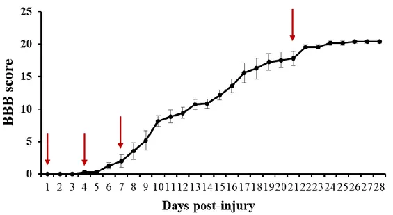
The clip compression injury was performed as previously described by our research group (Ahn et al., 2012b, Kim et al., 2017). Briefly, the rats were anesthetized with Zoletil® 50 (Virbac, France) and subjected to laminectomy at the thoracic vertebrae (T9/T10). Next, the thoracic spinal cord was compressed using a vascular clip (Stoelting; Wood Dale, IL, USA) at an occlusion pressure of 15–20 g for 1 min. Immediately thereafter, the wounds, including the muscles and skin layers, were closed. Sham-operated control rats underwent only the laminectomy procedure. Except for the sham controls, all rats who were operated lost hind limb locomotion soon after the SCI procedure; behavioral function after the SCI was evaluated using the Basso, Beattie, and Bresnahan (BBB) scoring system (Basso et al., 1996).
3.4. Tissue preparation and histological examinations
All experimental animals were sacrificed using CO2 and spinal cord tissues, including the
thoracic lesion including the core injury (T8–T11), were collected from the sham controls and SCI rats at the following time points: day (D) 1 post-injury (PI), D4PI, D7PI, and D21PI (n = 6 per time point). Figure 1 displays the behavioral scoring for locomotion after SCI at each time point. After the animals were sacrificed at the respective time points and the samples were removed, the tissues were fixed with 4% (v/v) paraformaldehyde in phosphate-buffered saline (PBS; pH 7.4) for 48 h. Then, sagittal sections (5 µm thick) were stained with hematoxylin and eosin and also used for immunohistochemistry. For Western blot analyses, the harvested spinal cords were stored at −80°C prior to use.
6
Table 2. Basso, Beattie, and Bresnahan locomotor rating scale 0 No observable hind limb (HL) movement
1 Slight movement of one or two joints, usually the hip and/or knee
2 Extensive movement of one joint or extensive movement of one joint and slight movement of one other joint
3 Extensive movement of two joints
4 Slight movement of all three joints of the HL
5 Slight movement of two joints and slight movement of the third 6 Extensive movement of two joints and slight movement of the third 7 Extensive movement of all three joints of the HL
8 Sweeping with no weight support or plantar placement of the paw with no weight support 9 Plantar placement of the paw with weight support in stance only (ie., when stationary) or
occasional, frequent, or consistent weight supported dorsal stepping and no plantar stepping
10 Occasional weight supported plantar steps: no FL-HL coordination
11 Frequent to consistent weight supported plantar steps and no FL-HL coordination
12 Frequent to consistent weight supported plantar steps and occasional FL-HL coordination 13 Frequent to consistent weight-supported plantar steps and frequent FL-HL coordination 14 Consistent weight supported plantar steps, consistent FL-HL coordination, and
predominant paw position during locomotion is rotated (internally or externally) when it makes initial contact with the surface as well as just before it is lifted off at the end of stance; or frequent plantar stepping, consistent FL-HL coordination, and occasional dorsal stepping
15 Consistent plantar stepping and consistent FL-HL coordination and no toe clearance or occasional toe clearance during forward limb advancement; predominant paw position is parallel to the body at initial contact
16 Consistent plantar stepping and consistent FL-HL coordination during gait and toe clearance occurs frequently during forward limb advancement ; predominant paw position is parallel to the body at initial contact
17 Consistent plantar stepping and consistent FL-HL coordination during gait and toe clearance, predominant paw position is parallel at initial contact lift off
18 Consistent plantar stepping and consistent FL-HL coordination during gait and toe clearance occurs consistently during forward limb advancement; predominant paw position is parallel at initial contact and rotated at lift off
19 Consistent plantar stepping and consistent FL-HL coordinated gait, consistence toe clearance, predominant paw position is parallel at initial contact and lift off, and trunk instability; tail consistently up
20 Consistent plantar stepping and consistent coordinated gait, consistent toe clearance, predominant paw position is parallel at initial contact and lift off, and trunk instability; tail consistently up
21 Consistent planter stepping and coordinated gait, consistent toe clearance, predominant paw position is parallel throughout stance, and consistent trunk stability; tail consistently up
7
3.5. Western blot analysis
Western blot analyses were performed as previously described (Ahn et al., 2015). Briefly, the spinal cord samples, including the core region (length: 1 cm), were homogenized and then electrophoresed and immunoblotted onto nitrocellulose membranes (Schleicher and Schuell; Keene, NH, USA). Then, each membrane was incubated with primary antibodies for mouse anti-Ninjurin-1 (1:1000, BD Transduction Laboratories) and mouse anti-β-actin (Sigma-Aldrich). Subsequently, the membranes were reacted with appropriate secondary antibodies and visualized with BS-ECL Plus kits (W6002, Biosesang; Gyeonggi, Korea). Immunoblot signals were detected with the Fusion Solo 6X system (Vilber Lourmat; Collegien, France) and analyzed using FUSION© software; the expression values were normalized to that of β-actin. All data were analyzed with post-hoc Student-Newman-Keuls tests for multiple comparisons and are expressed as a mean ± standard error (SE); p-values < 0.05 were considered to indicate statistical significance.
3.6. Immunohistochemistry
Immunohistochemistry was performed using a commercial avidin-biotin complex kit (Vector Laboratories; Burlingame, CA, USA) as previously described by our research group (Ahn et al., 2012b). Briefly, deparaffinized sections were treated with 0.01 M citrate buffer (pH 6.0) in a microwave for 2 min and then 0.3% hydrogen peroxide in methyl alcohol was applied for 20 min to block endogenous peroxidase activity. Next, the sections were incubated with 10% (v/v) normal horse serum in PBS prior to incubation with the primary mouse anti-Ninjurin-1 antibody for 1 h at room temperature. After three washes in PBS, a peroxidase reaction was developed using a diaminobenzidine (DAB) substrate kit (Vector Laboratories). Then, the sections were counterstained with hematoxylin prior to mounting.
8
3.7. Double-labeling immunofluorescence
For the double-labeling procedure, the sections were treated with 10% normal horse serum for 1 h, then with mouse monoclonal anti-Ninjurin-1 antibody overnight at 4°C, and then with tetra-methyl rhodamine isothiocyanate-conjugated anti-mouse IgG (1:50, T5393, Lot. 103K9165, Sigma-Aldrich) for 1 h at room temperature. Next, the sections were washed and incubated with rabbit polyclonal anti-GFAP (1:1000, EMD Millipore Corp.; Temecula, CA, USA) or anti-Iba-1 (1:1000, Wako Pure Chemical Industries, Ltd.) overnight at 4°C. After washing, the sections were incubated with fluorescein isothiocyanate-conjugated anti-rabbit IgG (1:50, F0382, Lot. 018K60572, Sigma-Aldrich) for 1 h at room temperature. Next, the immunofluorescence-stained specimens were examined under a fluorescence microscope (BX-51; Olympus, Tokyo, Japan) and, following photography, the images were merged using Adobe Photoshop 7.0 software (Adobe Systems; San Jose, CA, USA).
9 4. Results
4.1. Behavioral and histopathological examinations
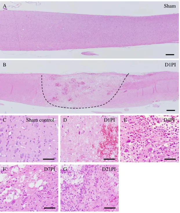
No hind limb paralysis was noted in the sham-operated rats. However, all rats with clip compression injury exhibited hind limb paralysis that improved on subsequent days (Fig. 1). Similarly, the spinal cord tissues of sham-operated rats did not show pathological lesions (Fig. 2A and C). By contrast, on D1PI, spinal cord tissues with clip compression injury exhibited a severe loss of cellularity in conjunction with edema and hemorrhaging in the core region (Fig. 2D); however, lesions were rarely observed in the penumbra region (Fig. 2D). On D4PI, although hemorrhaging and edema were less frequently observed, there was a prominent and massive infiltration of inflammatory cells (Fig. 2E) that was observable throughout D7PI and D21PI (Fig. 2F and G). Collectively, all pathological findings in the present study were comparable to those of previous reports (Ahn et al., 2012a, Shin, 2007, Moon et al., 2008, Kim et al., 2003).
10
Figure 1. Locomotor function scoring as evaluated by Basso, Beattie, and Bresnahan (BBB) scoring
(n = 30/daily).11
Figure 2. Histopathological changes in the spinal cord after clip compression injury (A–G) visualized with hematoxylin and eosin staining. Longitudinal sections of spinal cord tissues from (A) sham controls and (B) D1PI rats; the dotted line delineates the injury boundary. Higher magnification images of (C) sham controls and SCI rats on (D) D1PI, (E) D4PI, (F) D7PI, and (G) D21PI illustrating the histopathological changes on each day. Scale bars (A, B), 50 µm; (C–G), 100 µm.
Sham
control
D1PI
D1PI
D4PI
D7PI
D21PI
Sham control
A
G
F
E
D
C
B
12
4.2. Ninjurin-1 levels transiently increased after SCI
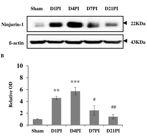
Western blot analyses were performed to evaluate variations in the expression levels of Ninjurin-1 in spinal cord tissues following clip compression injury (Fig. 3). Ninjurin-1 was weakly expressed in the sham controls; this value was arbitrarily set to “1”. On D1PI and D4PI, the relative expression levels of Ninjurin-1 significantly increased by four- to five-fold (D1PI, relative optical density [OD]: 4.58 ± 0.28-fold, n = 6, p < 0.01; D4PI, relative OD: 5.71 ± 0.63-fold, n = 6, p < 0.001 vs. sham control) whereas these levels exhibited a marked decrease on D7PI compared to D4PI (relative OD: 2.47 ± 0.78-fold, n = 6, p < 0.05). On D21PI, the Ninjurin-1 expression levels had decreased to control levels (relative OD: 1.41 ± 0.39-fold, n = 6, p < 0.01 vs. D4PI).
13
Figure 3. Western blot analysis of Ninjurin-1 levels in the spinal cord tissues of sham controls and rats exposed to clip compression injury. (A) Representative immunoblot images of Ninjurin-1 levels (approx. 22 kDa) and ß-actin (43 kDa) in the spinal cord tissues of sham controls and SCI rats on D1PI, D4PI, D7PI, and D21PI. (B) Bar graph showing the semi-quantitative analyses of the data (mean ± SE, n = 3 per group). The relative expression levels of Ninjurin-1 were calculated after normalization to the ß-actin band for each sample. Ninjurin-1 expression levels transiently increased on D1PI, peaked at D4PI, showed a tendency for a marked reduction on D7PI, and returned to control levels on D21PI.
Ninjurin-1
ß-actin
22KDa
43KDa Sham D1PI D4PI D7PI D21PI
A B
0
2
4
6
8
10
Sham D1PI D4PI D7PI D21PI
R el at ive O D
**
***
# ##14
4.3. Immunohistochemical localization of Ninjurin-1 in SCI lesions
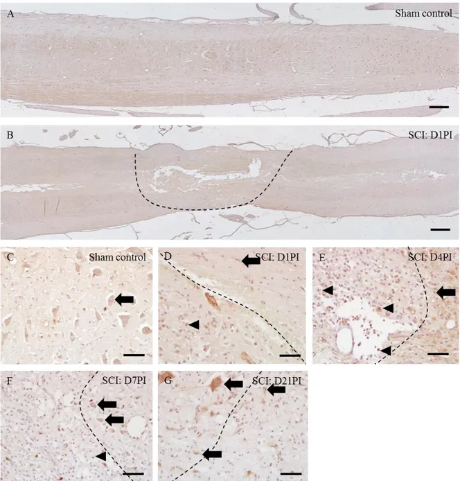
Ninjurin-1 in sham-control spinal cords (Figs. 4C) was immuno-stained in neurons (Fig. 4C arrow), vascular endothelial cells and glial cells (later identified as either astrocytes or microglia). At D1PI (Fig. 4D), Ninjurin-1 was immune-detected in neurons, vascular endothelial cells, glial cells (later identified as either astrocytes or microglia) (Fig. 4D arrow) and few round cells (later identified as Iba1-positive macrophages) (Figs. 4D arrowhead). At D4PI (Fig. 4E), some round cells infiltrated in the core region were intensely stained for Ninjurin-1 (Fig. 4E, arrowheads) while some glial cells (Fig. 4E arrow) were also immune detected. The immunostaining of Ninjurin-1 at D7PI was detected in neurons, glia (Fig. 4F arrows) as well as in round cells (Fig. 4F arrowhead) (later identified as Iba-1-positive macrophage). At D21PI, neurons and glial cells were immunostained with Ninjurin-1 (Fig. 4G arrows), with higher intensity of Ninjurin-1 in glial cells. At D21PI, Ninjurin-1 immune-positive inflammatory cells were hardly detected. Overall immunostaining pattern of Ninjurin-1 in the spinal cords was consistent with the results of the Western blot analysis.
15
Figure 4. Immunohistochemical localization of Ninjurin-1 in the spinal cords of sham control rats (A and C), D1PI (B and D), D4PI (E), D7PI (F) and D21PI (G). Immunostaining of Ninjurin-1, with hematoxylin counterstaining, was performed in longitudinal sections of spinal cords of (A) sham control, (B) injured spinal cord at D1PI. In post-SCI, both core region and penumbra region are focused in each figure (D-G). Neurons/glial cells are shown in arrows while arrowheads show inflammatory cells. Scale bars = 20μm (A & B), 100μm (C-G).
16
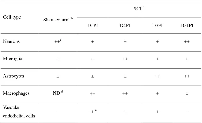
Table 3. Immunoreactivity of Ninjurin-1 in the spinal cords of controls and SCI ratsa
Cell type
Sham control b
SCI b
D1PI D4PI D7PI D21PI
Neurons ++c + + + ++ Microglia + ++ ++ + + Astrocytes ± ± ± ++ ++ Macrophages ND d ++ ++ + ± Vascular endothelial cells - ++ e + + - a
Three different sections (n = 3) per group were examined by three blind observers.
b
The spinal cords of rats were obtained from sham control, day 1 post injury (D1PI), D4PI, D7PI and D21PI.
c
The presence of Ninjurin-1-immunoreactive cells in the spinal cords of each group was scored as negative (-), < 20 positive cells (+), 20-50 positive cells (++), and > 50 positive cells (+++) under x20 magnification using an objective lens.
d
ND, macrophages were not detected in the spinal cords.
e
Intensity of Ninjurin-1 staining in the endothelial cells of vessels was classified as weak (+), moderate (++), or intense (+++) by three blind observers.
17
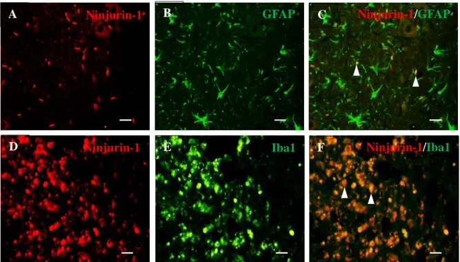
4.4. Phenotypes of Ninjurin-1-expressing cells in SCI lesions
To confirm the phenotypes of the cells that expressed Ninjurin-1, double-labeling immunofluorescence procedures for anti-Ninjurin-1 and anti-GFAP or anti-Iba-1, representative markers of astrocytes and microglia/macrophages, respectively (Velasco et al., 1980, Ahmed et al., 2007), were performed. On D7PI, Ninjurin-1 (Fig. 5A and D, shown in red) was co-localized with GFAP (Fig. 5B, shown in green) or Iba-1 (Fig. 5E, shown in green), demonstrating that astrocytes (Fig. 5C, merged image) and macrophages/microglia (Fig. 5F, merged image) expressed Ninjurin-1 post-SCI.
18
Figure 5. Double immunofluorescence staining of (A, D) Ninjurin-1, (B) GFAP, and (E) Iba-1 in spinal cord tissues following clip compression injury. Double immunofluorescence staining of the sham control sections was performed to detect (A) Ninjurin-1 (red) and (C) GFAP (green) as well as (D) Ninjurin-1 (red) and (F) Iba-1 (green). Arrowheads indicate Ninjurin-1-expressing GFAP-positive astrocytes/Ninjurin-1-expressing Iba-1-positive macrophages/microglia. Scale bars, 20 μm.
GFAP
Ninjurin-1
Iba1
Ninjurin-1
Ninjurin-1
/
GFAP
Ninjurin-1
/
Iba1
A
B
C
D
E
F
19 5. Discussion
The present study is the first to show that Ninjurin-1 levels are transiently upregulated in the lesioned areas of injured spinal cords using a clip compression rat model of SCI. Although these findings suggest that Ninjurin-1 plays a role in the pathogenesis of early neuroinflammation, its precise role during the neuroinflammation process remains to be fully elucidated.
Ninjurin-1 was initially described as a molecule involved in neuron-glia interactions that promotes axonal sprouting and nerve repair in the rat sciatic nerve; it is also upregulated in the neurons and Schwann cells of distal nerve segments following nerve transection or crush injury (Araki et al., 1997). Subsequently, it was revealed that Ninjurin-1 is also involved in cell adhesion, which is essential for the formation of early inflammatory reactions. As a cell adhesion molecule, Ninjurin-1 plays an important role in embryonic development, organogenesis, and even tissue regeneration after an injury (Lee et al., 2010, Araki et al., 1997). In cases of ischemia in the CNS, Ninjurin-1 can be found in inflammatory cells as well as some glial cells (Lee et al., 2016). Moreover, Ninjurin-1 is upregulated in myeloid cells and inflamed endothelial cells in the spinal cord in mice with EAE (Ahn et al., 2014). The transient increases in Ninjurin-1 levels and myeloid cell expression in the early stages of SCI observed in the present study are consistent with the findings of previous studies, including those investigating brain ischemia (Lee et al., 2016) and EAE (Ahn et al., 2014). Taken together, these findings indicate that Ninjurin-1 plays a role in the migration of inflammatory cells following SCI.
Although the role that Ninjurin-1 plays during CNS inflammation remains under debate, this protein has been linked to the induction of inflammatory processes in the CNS. For example, the functional repression of Ninjurin-1 reduces leukocyte infiltration and clinical disease activity in mice with EAE (Ifergan et al., 2011b). Additionally, Ninjurin-1 is involved in the interaction between leukocytes and vascular endothelial cells during vascular remodeling and CNS inflammation (Lee et al., 2010). Therefore, in conjunction with these previous studies, the present findings indicate that
20
Ninjurin-1 is related to leukocyte recruitment through cell adhesion activities and functions as a pro-inflammatory mediator.
A full understanding of the expression of Ninjurin-1 in macrophages during the early stages of SCI will also require further investigation. In ischemic brains, Ninjurin-1 expression is induced in two specific populations of macrophages, microglia-derived macrophages and bone marrow-derived macrophages (Lee et al., 2016), which are both involved in the suppression of inflammation. Furthermore, in vitro studies have shown that Ninjurin-1 promotes neurite growth during axonal regeneration (Araki and Milbrandt, 1996). In SCI rats in the present study, some axons in the lesion periphery exhibited intense Ninjurin-1 immunoreactivity, which indicates that Ninjurin-1 is involved in axonal regeneration. Similarly, Ninjurin-1 derived from inflammatory cells supports adjacent axonal regeneration, at least in a rat model of SCI. Astrogliosis is another secondary consequence of CNS injury, including in EAE (Park et al., 2018), SCI (Moon et al., 2004), and brain ischemia (Park et al., 2018). Even though Nijurin-1 is constitutively expressed in astrocytes in CNS tissues, reactive astrocytes with intense Ninjurin-1 immunoreactivity are involved in the resolution of CNS inflammation in the later stages of SCI.
Collectively, the present results suggest that transient increases in Ninjurin-1 expression levels are associated with macrophage activation and reactive astrogliosis and that these changes could activate inflammatory macrophages and/or modulate cell trafficking. The present study assessed the potential roles of Ninjurin-1 following SCI and showed that the changes in these protein levels are associated with cell migration and neuroregeneration.
21
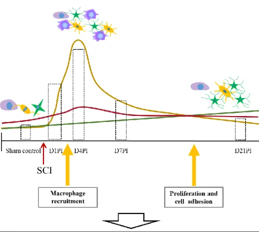
Figure 6. Schematic illustration of spatiotemporal changes in Ninjurin-1 levels in rat spinal cord tissues following clip compression injury. Ninjurin-1 was constitutively expressed in neurons and glial cells in the spinal cord tissues of sham controls. SCI-induced clip compression of the spinal cord resulted in the expression of Ninjurin-1 within infiltrating macrophages other than neurons and glial cells. Soon after SCI, Ninjurin-1 was upregulated and expressed in infiltrating macrophages, reactive glia, and some neurons that may be important for cell adhesion, proliferation, and migration. However, subsequent Ninjurin-1 expression was primarily associated with reactive glia and some neurons that may be further involved in neurite growth, glial border formation, and lesion reduction.
22 6. References
1. AHMED, Z., SHAW, G., SHARMA, V. P., YANG, C., MCGOWAN, E. & DICKSON, D. W. 2007. Actin-binding proteins coronin-1a and IBA-1 are effective microglial markers for immunohistochemistry. Journal of Histochemistry & Cytochemistry, 55, 687-700.
2. AHN, B. J., LE, H., SHIN, M. W., BAE, S. J., LEE, E. J., WEE, H. J., CHA, J. H., LEE, H. J., LEE, H. S., KIM, J. H., KIM, C. Y., SEO, J. H., LO, E. H., JEON, S., LEE, M. N., OH, G. T., YIN, G. N., RYU, J. K., SUH, J. K. & KIM, K. W. 2014. Ninjurin1 deficiency attenuates susceptibility of experimental autoimmune encephalomyelitis in mice. J Biol Chem, 289, 3328-38.
3. AHN, B. J., LEE, H.-J., SHIN, M. W., CHOI, J.-H., JEONG, J.-W. & KIM, K.-W. 2009. Ninjurin1 is expressed in myeloid cells and mediates endothelium adhesion in the brains of EAE rats. Biochemical and biophysical research communications, 387, 321-325.
4. AHN, M., LEE, C., JUNG, K., KIM, H., MOON, C., SIM, K.-B. & SHIN, T. 2012a. Immunohistochemical study of arginase-1 in the spinal cords of rats with clip compression injury. Brain research, 1445, 11-19.
5. AHN, M., MOON, C., PARK, C., KIM, J., SIM, K. B. & SHIN, T. 2015. Transient activation of an adaptor protein, disabled-2, in rat spinal cord injury. Acta Histochem, 117, 56-61. 6. AHN, M., YANG, W., KIM, H., JIN, J. K., MOON, C. & SHIN, T. 2012b.
Immunohistochemical study of arginase-1 in the spinal cords of Lewis rats with experimental autoimmune encephalomyelitis. Brain Res, 1453, 77-86.
7. ALTINOVA, H., HAMMES, S., PALM, M., GERARDO-NAVA, J., ACHENBACH, P., DEUMENS, R., HERMANS, E., FUHRMANN, T., BOECKER, A., VAN NEERVEN, S. G. A., BOZKURT, A., WEIS, J. & BROOK, G. A. 2019. Fibroadhesive scarring of grafted collagen scaffolds interferes with implant-host neural tissue integration and bridging in experimental spinal cord injury. Regen Biomater, 6, 75-87.
8. ARAKI, T. & MILBRANDT, J. 1996. Ninjurin, a novel adhesion molecule, is induced by nerve injury and promotes axonal growth. Neuron, 17, 353-361.
9. ARAKI, T., ZIMONJIC, D. B., POPESCU, N. C. & MILBRANDT, J. 1997. Mechanism of homophilic binding mediated by ninjurin, a novel widely expressed adhesion molecule.
Journal of Biological Chemistry, 272, 21373-21380.
10. BASSO, D. M., BEATTIE, M. S. & BRESNAHAN, J. C. 1996. Graded histological and locomotor outcomes after spinal cord contusion using the NYU weight-drop device versus transection. Experimental neurology, 139, 244-256.
23
S., BOURBONNIÈ RE, L., DUNAY, I. R., BOUTHILLIER, A. & MOUMDJIAN, R. 2011a. Role of ninjurin‐1 in the migration of myeloid cells to central nervous system inflammatory lesions. Annals of neurology, 70, 751-763.
12. IFERGAN, I., KEBIR, H., TEROUZ, S., ALVAREZ, J. I., LECUYER, M. A., GENDRON, S., BOURBONNIERE, L., DUNAY, I. R., BOUTHILLIER, A., MOUMDJIAN, R., FONTANA, A., HAQQANI, A., KLOPSTEIN, A., PRINZ, M., LOPEZ-VALES, R., BIRCHLER, T. & PRAT, A. 2011b. Role of Ninjurin-1 in the migration of myeloid cells to central nervous system inflammatory lesions. Ann Neurol, 70, 751-63.
13. JUNG, K., MIN, D. S., SIM, K. B., AHN, M., KIM, H., CHEONG, J. & SHIN, T. 2003. Upregulation of phospholipase D1 in the spinal cords of rats with clip compression injury.
Neurosci Lett, 336, 126-30.
14. KIM, D. H., HEO, S. D., AHN, M. J., SIM, K. B. & SHIN, T. K. 2003. Activation of embryonic intermediate filaments contributes to glial scar formation after spinal cord injury in rats. Journal of veterinary science, 4, 109-112.
15. KIM, Y., KIM, J., AHN, M. & SHIN, T. 2017. Lithium ameliorates rat spinal cord injury by suppressing glycogen synthase kinase-3beta and activating heme oxygenase-1. Anat Cell
Biol, 50, 207-213.
16. LEE, H.-K., LEE, H., LUO, L. & LEE, J.-K. 2016. Induction of nerve Injury-Induced protein 1 (Ninjurin 1) in myeloid cells in rat brain after transient focal cerebral ischemia.
Experimental neurobiology, 25, 64-71.
17. LEE, H. J., AHN, B. J., SHIN, M. W., CHOI, J. H. & KIM, K. W. 2010. Ninjurin1: a potential adhesion molecule and its role in inflammation and tissue remodeling. Mol Cells, 29, 223-7.
18. MOON, C., HEO, S., AHN, M., KIM, H., SHIN, M., SIM, K. B., KIM, H. M. & SHIN, T. 2004. Immunohistochemical study of osteopontin in the spinal cords of rats with clip compression injury. J Vet Med Sci, 66, 1307-10.
19. MOON, C., LEE, T.-K., KIM, H., AHN, M., LEE, Y., KIM, M. D., SIM, K.-B. & SHIN, T. 2008. Immunohistochemical study of cathepsin D in the spinal cords of rats with clip compression injury. Journal of Veterinary Medical Science, 70, 937-941.
20. MOTHE, A. J. & TATOR, C. H. 2013. Review of transplantation of neural stem/progenitor cells for spinal cord injury. International Journal of Developmental Neuroscience, 31, 701-713.
21. PARK, J. H., CHO, J. H., AHN, J. H., CHOI, S. Y., LEE, T. K., LEE, J. C., SHIN, B. N., HONG, S., JEON, Y. H., KIM, Y. M., HWANG, I. K., LEE, Y. J., WON, M. H. & KANG, I. J. 2018. Neuronal loss and gliosis in the rat striatum subjected to 15 and 30 minutes of
24
middle cerebral artery occlusion. Metab Brain Dis, 33, 775-784.
22. SHIN, T. 2007. Increases in the phosphorylated form of caveolin-1 in the spinal cord of rats with clip compression injury. Brain research, 1141, 228-234.
23. SHIN, T., AHN, M., MOON, C., KIM, S. & SIM, K. B. 2013. Alternatively activated macrophages in spinal cord injury and remission: another mechanism for repair? Mol
Neurobiol, 47, 1011-9.
24. VELASCO, M. E., DAHL, D., ROESSMANN, U. & GAMBETTI, P. 1980. Immunohistochemical localization of glial fibrillary acidic protein in human glial neoplasms.
25 Acknowledgement
This document contains my Master’s thesis, the final document for my master course in Veterinary Medicine at the Jeju National University. It describes the results of my research on transient upregulation of Nerve injury induced protein 1 in the course of neuroinflammation in rat spinal cord injury model. It was challenging for me to learn and adapt to the new environment at Jeju and therefore finishing this research project means lot to me and could not have been possible without the help of many people.
I would like to start by thanking my supervisor, Professor Shin Taekyun from Jeju National University. With your guidance and enthusiasm, we found a research approach that worked out. Thanks for your continuous feedback that was always valuable.
I would like to thank my fellow colleagues and supervisors at the laboratory of Veterinary Anatomy. Dr. Ahn Meejung, thank you so much for the courage and support you gave me all the time and teaching me many of the lab techniques, correcting my mistakes and validating my results. Without your kind guidance this task could not have been a reality.
I would like to convey my thanks to Dr. Kim Jeongtae, who also guided me for the research work, taught much more laboratory techniques, surgical procedure of spinal cord injury and helped to develop the ability for independent research working. I always appreciated your continuous feedback and sharp criticisms. Further, my thanks goes to Mrs.Kim, Ms.Yuna Choi and all other lab members who helped me in various terms.
Of course, this acknowledgement would not be complete without thanking my parents and friends who supported me throughout my entire study and courage my activities. Thanks to the loving support and faith of my parents, I was able to finish my study and make it a great time to look back on.