Research Article
Mitochondrial DNA Copy Number in Peripheral
Blood Is Independently Associated with Visceral Fat
Accumulation in Healthy Young Adults
Jee-Yon Lee,
1Duk-Chul Lee,
1Jee-Aee Im,
2and Ji-Won Lee
11Department of Family Medicine, Severance Hospital, Yonsei University, College of Medicine, 250 Seongsanno,
Seodaemun-gu 120-752, Republic of Korea
2Sport and Medicine Research Center, INTOTO Inc., 401 Dawoo BD, 90-6 Daeshin-Dong, Seodaemun-gu,
Seoul 120-160, Republic of Korea
Correspondence should be addressed to Ji-Won Lee; [email protected]
Received 11 July 2013; Revised 22 December 2013; Accepted 1 January 2014; Published 24 February 2014 Academic Editor: Debra Waters
Copyright © 2014 Jee-Yon Lee et al. This is an open access article distributed under the Creative Commons Attribution License, which permits unrestricted use, distribution, and reproduction in any medium, provided the original work is properly cited.
Aims. Visceral obesity is associated with an increased risk of cardiometabolic diseases and it is important to identify the underlying
mechanisms. There is growing evidence that mitochondrial dysfunction is associated with metabolic disturbances related to visceral obesity. In addition, maintaining mitochondrial DNA (mtDNA) copy number is important for preserving mitochondrial function. Therefore, we investigated the relationship between mtDNA copy number and visceral fat in healthy young adults. Methods. A total of 94 healthy young subjects were studied. Biomarkers of metabolic risk factors were assessed along with body composition by computed tomography. mtDNA copy number was measured in peripheral leukocytes using real-time polymerase chain reaction (PCR) methods. Results. The mtDNA copy number correlated with BMI (𝑟 = −0.22, 𝑃 = 0.04), waist circumference (𝑟 = −0.23, 𝑃 = 0.03), visceral fat area (𝑟 = −0.28, 𝑃 = −0.01), HDL-cholesterol levels (𝑟 = 0.25, 𝑃 = 0.02), and hs-CRP (𝑟 = 0.32, 𝑃 = 0.02) after adjusting for age and sex. Both stepwise and nonstepwise multiple regression analyses confirmed that visceral fat area was independently associated with mtDNA copy number (𝛽 = −0.33, 𝑃 < 0.01, 𝛽 = 0.32, and 𝑃 = 0.03, resp.). Conclusions. An independent association between mtDNA content and visceral adiposity was identified. These data suggest that mtDNA copy number is a potential predictive marker for metabolic disturbances. Further studies are required to understand the causality and clinical significance of our findings.
1. Introduction
The prevalence of obesity is increasing worldwide. Obesity is a well-known risk factor for numerous health problems, including cardiovascular disease (CVD), diabetes
melli-tus (DM), and cancer [1, 2]. Recent data shows that the
regional distribution of body fat, rather than overall obesity,
contributes to disease processes [3]. Visceral fat is more
metabolically active than subcutaneous fat [4] and affects
the development of metabolic disturbances, including insulin
resistance [5] and dyslipidemia [6]. The precise role of
visceral adiposity in metabolic disturbance is still unknown but proinflammatory cytokines and adipokines secreted by
visceral adipocytes are believed to be involved [7].
Mitochondria are organelles that play an important role in the energy synthesis of the cells. Mitochondria synthe-size the molecules essential for the body metabolism and
influence metabolic homeostasis of the entire body [8].
Mitochondrial function decreases with aging and mitochon-drial dysfunction is related to various age-related conditions
including type 2 DM and CVD [9]. Mitochondria are highly
vulnerable to oxidative damage [10, 11] and mitochondrial
dysfunction induced by oxidative damage is considered to contribute to the development of cardiometabolic diseases
[9].
Mitochondrial DNA copy number, which reflects the content of mtDNA, is associated with mitochondrial gene
stability and mitochondrial biogenesis [12]. Mitochondrial
Volume 2014, Article ID 586017, 7 pages http://dx.doi.org/10.1155/2014/586017
dysfunction reduces the contents of mitochondria, which is
expressed as a decreased mtDNA copy number [12].
Fur-thermore, reduced mitochondrial DNA content of peripheral blood as well as specific organs was associated with the
development of IR, type 2 DM [13], cognitive function [14],
and metabolic syndrome [15]. Adipose tissue is the main
source of cytokines and adipokines that increase systemic oxidative stress. Thus, obesity may decrease mitochondrial function. Previous results show that human obesity is asso-ciated with mitochondrial dysfunction. However, few studies have investigated the quantitative changes in mitochondria according to increased adiposity. The studies that have been performed have yielded mixed results. Furthermore, the potential differences in mitochondrial content according to the regional distribution of adiposity were not fully evaluated. Adipose tissue is the main source of cytokines and
adipokines that increase systemic oxidative stress [16].
Thus, obesity may decrease mitochondrial function. Previous results show that human obesity is associated with
mito-chondrial dysfunction [17]. However, few studies have
inves-tigated the quantitative changes in mitochondria according to increased adiposity. The studies that have been performed
have yielded mixed results [18,19]. Furthermore, the
poten-tial differences in mitochondrial content according to the regional distribution of adiposity were not fully evaluated.
Therefore, we investigated the association between peripheral blood mtDNA copy number and visceral fat accumulation among 94 healthy young-aged people.
2. Materials and Methods
2.1. Study Sample. This was a secondary data analysis from
the Yonsei Aging Cohort, which was designed to investigate
health-related markers among people of various ages [20].
Participants visited the Department of Family Medicine at Severance Hospital for routine health checkups and not for investigations or treatments of specific symptoms or diseases. All subjects participated in the study voluntarily, and written informed consent was obtained from each participant. Questionnaires about lifestyles and underlying medical conditions, overnight-fasting blood tests, and fat measurements with computed tomography were performed as baseline tests. Two additional samples of blood were collected from participants who agreed to store their blood samples for 10 years for future analysis. An additional separate written informed consent was obtained from each participant before performing the additional laboratory test with the stored blood samples.
Because the aim of our study was to investigate the asso-ciation between visceral obesity and mtDNA copy number in healthy young participants, we selected 203 people aged from 20 to 40 years. Mitochondrial DNA copy numbers were measured with the stored blood samples. Thus we excluded 75 participants who did not agree to store their blood samples. Fifteen additional participants were excluded, because data for their abdominal visceral fat areas were missing. To select a healthy population, participants with histories of hypertension, diabetes mellitus, coronary artery
occlusive disease, chronic liver disease, chronic renal disease, or cancer were not included. Subjects who participated in regular exercise were also excluded from the data analysis. Regular exercise was defined as physical exercise or physical work that was performed for more than 30 minutes, three times per week. We also excluded participants who used med-ications, including antihypertensive agents, lipid-reducing drugs, oral hypoglycemic agents, and nutrient supplements, which could affect cardiometabolic functions. Ninety-four patients were included in our analyses. The study complied with the Declaration of Helsinki, and the institutional review board of Yonsei University College of Medicine approved this study.
2.2. Measurements. All participants were questioned about
lifestyle factors, including alcohol consumption and smoking. Alcohol consumption was defined as drinking alcohol more frequently than once per week. Smoking was defined as current cigarette smoking.
Anthropometric measurements were made by a single examiner. After a 10-minute resting period, blood pressure was measured in the sitting position. Body mass index was
calculated as weight (kg) divided by height squared (cm2).
Abdominal fat tissue area was calculated using computed tomography (Tomoscan 350; Philips, Mahwah, NJ, USA) as
described previously [21].
Blood samples were collected after at least an 8-hour overnight fasting period. Fasting glucose, high sensitive C-reactive protein (hs-CRP), total cholesterol, triglyceride, and high-density lipoprotein (HDL) cholesterol levels were measured using an ADVIA 1650 chemistry system (Siemens Medical Solution, Tarrytown, NY, USA). Fasting insulin levels were determined using electrochemiluminescence immunoassays with an Elecsys 2010 (Roche, Indianapolis, IN, USA). Insulin resistance was calculated using the homeosta-sis model assessment of insulin rehomeosta-sistance (HOMA-IR) index: (insulin [𝜇IU/mL] × fasting blood glucose [mg/dL]/18)/22.5.
2.3. Measurement of Mitochondrial DNA Copy Numbers in Peripheral Blood. To reduce variations in measurements, one
examiner measured all parameters throughout the study. mtDNA in peripheral leukocytes was extracted from 1 mL of whole blood using the QIAamp Tissue Kit 250 (Qiagen Inc., Valencia, CA, USA). The relative mtDNA copy number was measured by a real-time polymerase chain reaction (QPCR) and corrected by simultaneous measurement of the nuclear
DNA according to the method of Wong and Cortopassi [22]
and Liu et al. [11]. Reactions were performed using a Light
Cycler-Fast Start DNA Master SYBR Green I kit, purchased from Roche Applied Science (Pleasanton, CA, USA). The
forward and reverse primers of𝛽-globin (used to amplify a
268 bp product) were 5
-GAAGAGCCAAGGACAGGTAC-3 and 5-CAACTTCATCCACGTTCACC-3, respectively.
The forward and reverse primers of the mitochondrial gene
(ND1 gene) used to amplify a 153 bp product were 5
-AACATACCCATGGCCAACCT-3 and 5
-AGCGAAGGG-TTGTAGTAGCCC-3, respectively. After denaturation at
0.1 seconds, 58∘C for 6 seconds, and 72∘C for 18 seconds for 40 cycles. A total of 20 ng of DNA was used and the number of PCR cycles to reach this amount of DNA was defined as the threshold cycle (Ct). The following equation was used
to quantify the mtDNA copy number relative to 𝛽-globin:
relative copy number =2ΔCt(ΔCt = Ct𝛽-globin− CtND1) [23].
The intra-assay and interassay coefficients of variation of Ct values for the ND1 gene were 4.5% and 5.8%, respectively.
2.4. Statistical Analyses. Normally distributed data are
ex-pressed as the mean ± standard deviation (SD).
Nonnor-mally distributed data are expressed as median and interquar-tile range. mtDNA, fasting insulin, HOMA-IR, total choles-terol, triglyceride, and hs-CRP were log transformed to improve the skewness of the distribution. Pearson correlation analyses were performed to evaluate relationships between mtDNA and other metabolic variables. Stepwise multiple linear regression analysis was performed to identify factors that contributed to mtDNA copy number. If there was a significant correlation (𝑟 > 0.7) between two variables, only one variable was selected and entered into the model to avoid multicollinearity. In addition, nonstepwise multiple linear
regression analysis was performed. Variables with𝑃 < 0.05
in the univariate analysis and clinically important variables, including age, BMI, and HOMA-IR, were entered into the nonstepwise analysis.
We performed all statistical analyses using the Statistical Package for the Social Sciences, version 18.0 (SPSS Inc.,
Chicago, IL, USA). Statistical significance was defined as𝑃 <
0.05.
3. Results
The clinical characteristics of the study subjects are shown inTable 1. The mean age of the study subjects was 32.26± 9.14 years, and the median (25–75th percentile) mtDNA copy number was 302.08 (48.98–891.25). After adjusting for age and sex, mtDNA copy numbers positively correlated with HDL-cholesterol levels (𝑟 = 0.25, 𝑃 = 0.02) and negatively correlated with BMI (𝑟 = −0.22, 𝑃 = 0.04), waist circum-ference (𝑟 = −0.23, 𝑃 = 0.03), visceral fat area (𝑟 = −0.28,
𝑃 = −0.01), and hs-CRP (𝑟 = 0.32, 𝑃 = 0.02) (Table 2).
The mean mtDNA level in the nonsmoking group (2.50±
0.81) was significantly higher than that of the smoking group
(1.80± 0.74, 𝑃 < 0.001). In addition, the mean mtDNA level
of female subjects (2.57± 0.80) was significantly higher than
that of male subjects (2.15± 0.83, 𝑃 = 0.02). There were no
significant differences in mean mtDNA levels between
sub-jects that consumed alcohol (2.17± 0.76) and subjects that
did not (2.42± 0.88, 𝑃 = 0.17).Figure 1shows the different
relationships between mtDNA copy number and abdominal adiposity according to the regional fat distribution. The mtDNA copy numbers negatively correlated with visceral fat area. However, there was no significant correlation with subcutaneous fat.
In stepwise multiple linear regression analyses, visceral fat area, hs-CRP, HDL-cholesterol, and smoking accounted for 35% of the variance in mtDNA copy number. Thus,
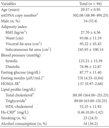
Table 1: Clinical characteristics of study subjects (𝑛 = 94).
Variables Total (𝑛 = 94)
Age (years) 29.57 ± 0.95
mtDNA copy number# 302.08 (48.98–891.25)
Male (𝑛, %) 54 (57.4)
Adiposity index
BMI (kg/m2) 27.70 ± 4.36
Waist (cm) 93.06 ± 11.19
Visceral fat area (cm2) 95.22 ± 45.45
Subcutaneous fat area (cm2) 245.93 ± 100.14
Blood pressure (mmHg)
Systolic 125.21 ± 15.59
Diastolic 76.96 ± 12.87
Fasting glucose (mg/dL) 87.77 ± 11.40
Fasting insulin (𝜇IU/mL)# 7.51 (4.53–11.84)
HOMA-IR# 1.57 (0.97–2.68) Lipid profile (mg/dL) Total cholesterol# 181.00 (164.00–215.25) Triglyceride# 89.00 (63.00–131.25) HDL-cholesterol 52.25 ± 11.92 Hs-CRP#(mg/L) 0.46 (0.10–1.42) Smoking (𝑛, %) 23 (24.5) Alcohol consumption (𝑛, %) 34 (36.2)
Note: BMI: body mass index; HOMA-IR: Homeostasis Model of Assessment of Insulin Resistance; HDL: high-density lipoprotein; LDL: low-density lipoprotein; hsCRP: high sensitive C reactive protein.
Alcohol consumption was defined as drinking alcohol more frequently than once per week.
Normally distributed data are shown as the mean (±SD).
#Non-normally distributed data are presented as medians (25–75 percentiles) and analyzed after log-transformation to correct for skewed distribution.
these variables were considered to be explanatory variables for mtDNA copy number. In addition, nonstepwise multiple regression analyses indicated that visceral fat, smoking, and hs-CRP levels were independently associated with mtDNA copy numbers, as these variables accounted for 58% of the
variance (Table 3).
4. Discussion
Our cross-sectional study revealed a relationship between peripheral blood mtDNA copy number and visceral obesity in a healthy Korean young-aged population. This associa-tion remained significant after adjusting for BMI and other confounding factors. In addition, our study showed a sig-nificant relationship between mtDNA copy number with smoking, the components of metabolic syndrome (waist circumference, blood pressure, and HDL-cholesterol), and cardiovascular risk factors (systolic and diastolic BP), which
is consistent with the findings of previous studies [15,24].
The mitochondrion is an organelle with diverse functions, including energy synthesis, cellular remodeling, and regula-tion of cell metabolism. Mitochondrial dysfuncregula-tion induces various metabolic diseases, including insulin resistance,
0.00 50.00 100.00 150.00 200.00 250.00
Visceral fat area (cm2)
−2.00 0.00 2.00 4.00 6.00 log tra n sf o rmed m tD N A −2.00 0.00 2.00 4.00 6.00 log tra n sf o rmed m tD N A R2linear= 0.161 0.00 200.00 400.00 600.00
Subcutaneous fat area
Figure 1: The relationship between abdominal visceral fat area, abdominal subcutaneous fat area, and mtDNA copy numbers. Coefficients (𝑟) and 𝑃 values were calculated using the Pearson correlation model.
Table 2: The correlation between mtDNA copy numbers# and
various parameters.
Variables Unadjusted Age, sex adjusted
𝑟 𝑃-value 𝑟 𝑃-value
Age (years) −0.28 0.01
Adiposity index −0.25 0.01 −0.22 0.04
BMI (kg/m2) −0.33 <0.01 −0.23 0.03
Waist (cm) −0.40 <0.01 −0.28 0.01
Visceral fat area (cm2)# −0.16 0.14 −0.21 0.09
Subcutaneous fat area (cm2)
Blood pressure (mmHg)
Systolic −0.22 0.04 −0.11 0.32
Diastolic −0.29 0.01 −0.15 0.17
Fasting glucose (mg/dL) 0.19 0.09 0.14 0.21
Fasting insulin (𝜇IU/mL)# −0.12 0.23 −0.17 0.12
HOMA-IR# −0.07 0.15 −0.12 0.27 Lipid profile (mg/dL) Total cholesterol# −0.09 0.42 −0.03 0.80 Triglyceride# −0.37 <0.01 −0.18 0.09 HDL-cholesterol# 0.37 <0.01 0.25 0.02 Hs-CRP (mg/L) −0.42 <0.01 0.32 0.02
#Values analyzed after log-transformation to correct for skewed distribution. Coefficients (𝑟) and 𝑃 values were calculated using the Pearson correlation model.
type-2 diabetes mellitus, and CVD [9]. Multiple
biochem-ical mechanisms, including impaired fatty-acid oxidation, and mitochondrial reactive oxygen stress, explain the link between mitochondrial dysfunction and pathologic
condi-tions [25]. Although mitochondria are present in all types
of cells, increasing evidence indicates that mitochondrial function in adipocytes is important for metabolic regulation. In an experimental animal model, rats with visceral obesity showed defective oxidative metabolism and reduced
mito-chondrial gene expression [26]. Kraunsøe et al. reported that
Table 3: Multiple regression analyses for mtDNA copy number#.
(a) Stepwise model
𝛽 coefficient SE 𝑃-value
Visceral fat area −0.33 0.00 <0.01
Hs-CRP (mg/L) −0.32 0.05 <0.01
Smoking#(%) −0.21 0.18 0.01
HDL-cholesterol (mg/dL) 0.21 0.39 0.04
𝑟2 = 0.35. Variables included in the stepwise model for mtDNA were age, sex, BMI, alcohol consumption, smoking, systolic BP, total cholesterol, HDL-cholesterol, fasting glucose, HOMA-IR and visceral fat area. To avoid multi-collinearlity, diastolic BP, triglycerides and subcutaneous fat area were not included in the stepwise model.
#Values analyzed after log-transformation to correct for skewed distribu-tion.
(b) Non-stepwise model
Variables 𝛽 coefficient SE 𝑃-value
Age (years) −0.05 0.02 0.30
Male (%) 0.03 0.20 0.89
BMI (kg/m2) −0.12 0.03 0.43
Smoking (%) −0.42 0.20 0.03
Systolic BP (mm Hg) 0.00 0.01 0.69
Visceral fat area (cm2) −0.32 0.00 0.03
HDL-cholesterol (mg/dL) 0.21 0.42 0.07
Fasting glucose (mg/dL) −0.04 0.00 0.66
HOMA-IR −0.04 0.16 0.41
hs-CRP (mg/L) −0.19 0.06 0.02
𝑟2 = 0.58. Variables included in the non-stepwise model for mtDNA were age, sex, BMI, smoking, systolic BP, HDL-cholesterol, fasting glucose, HOMA-IR and hs-CRP.
mitochondrial respiration was reduced in the visceral adipose tissues of obese humans compared to that in subcutaneous
fat tissues [27]. Furthermore, obese people have been shown
to have defective mitochondrial ATP formation compared
results from human clinical studies regarding the quantitative aspects of mtDNA and regional distribution of adiposity. Yin et al. reported a modest decrease in mtDNA content of omental adipocytes from obese men compared with that of
nonobese men [29]. In a Korean study, an inverse relationship
was reported between peripheral mitochondrial DNA copy
number and visceral fat mass [18]. However, some studies
showed no correlation between mtDNA copy number and regional distribution of adiposity. One study showed no correlation between mtDNA copy number and waist-hip
ratio which reflects visceral obesity [30]. Furthermore, results
that are opposite to those of our study have also been reported
[19,31,32]. The investigator of those studies suggested that
mtDNA content may increase secondary to mitochondrial dysfunction. Therefore, the association between visceral obesity and mtDNA copy number remains unclear. Our study participants were apparently healthy, young subjects from 20–40 years of age without chronic metabolic diseases. Therefore, although we could not determine causality, our results suggest that visceral fat accumulation may affect mito-chondrial DNA content in apparently healthy population without metabolic disturbances.
The precise mechanism that explains the association between mitochondrial DNA copy number and visceral fat mass remains unknown. We could not find the causal factor through our cross-sectional study. However, the results suggest possible mechanisms.
First, increased chronic systemic inflammation according to the secretion of proinflammatory cytokines and adipokines may play important roles in the relationship. Visceral adi-pose tissue is the main site of secretion of proinflamma-tory cytokines, which induce mitochondrial dysfunction by affecting signaling pathways associated with mitochondrial biogenesis. Visceral adipose tissue is the main site of secretion of proinflammatory cytokines, which induce mitochondrial dysfunction by affecting signaling pathways associated with mitochondrial biogenesis. For example, in cultured fat and muscle tissue, TNF-𝛼 depleted endothelial nitric oxide syn-thase expression along with mitochondrial biogenesis defects and adipocytes with defective TNF-𝛼 signaling showed
par-tial recovery of mitochondrial function in obese mice [33]. In
our study, increased mtDNA copy number was significantly associated with decreased hs-CRP level which reflects the total inflammatory status of human body. Furthermore hs-CRP level was also positively associated with visceral fat accumulation after it was adjusted for age and sex (𝑟 = 0.5, 𝑃 < 0.05) (data not shown). Because both visceral adiposity and mitochondrial DNA copy number were associated with systemic inflammatory status, increased inflammation level may explain the observed link between visceral obesity and decreased mitochondrial contents. However, the association between visceral fat accumulation and mitochondrial copy number remained statistically significant after adjustment for hs-CRP, suggesting that the association was, at least in part, independent of systemic inflammation. Furthermore, it is impossible to find out the specific roles of proinflammatory cytokines and adipokines in the observed association in our study. Therefore, measurement of proinflammatory cytokines and adipokines should be performed in the future.
Second, free fatty acids that accumulated in the visceral adipose tissue may affect the decreased mtDNA copy num-ber. Increased levels of free fatty acids promote increased synthesis of toxic fatty-acid-delivered metabolites. These metabolites elevate the level of oxidative stress, driving
mitochondrial dysfunction [34]. Therefore increased free
fatty acids according to the visceral fat accumulation may induce the decreased mitochondrial contents.
This study has several limitations. First, the cross-sectional design of our study cannot determine a causal rela-tionship between mtDNA copy number and visceral obesity and the small sample size is another limitation of the current study. Although the correlation was statistically significant, the low power was another limitation. Therefore, we cannot generalize the results to the population at large. In addition, we did not perform fat biopsy, which is the gold standard for investigation of mitochondrial function. However, it is easier to obtain peripheral blood leukocytes than muscle tissue. And decreased mtDNA copy number in peripheral blood leukocytes correlated well with mitochondrial dysfunction in
skeletal muscle [35,36]. Finally, this study did not measure
levels of proinflammatory cytokines and adipokines. There-fore our study cannot directly investigate the role of cytokines and adipokines as mediators between visceral adiposity and reduced mitochondrial DNA copy number. We agree that assessing inflammatory cytokine and adipokine levels will provide additional important information in future studies.
In conclusion, our study shows that peripheral blood mtDNA copy number is associated with abdominal visceral fat area in 94 healthy young-aged subjects. Although the causal direction of the relationship between mtDNA copy number and visceral obesity cannot be determined, our study collectively suggests that decreased mitochondrial contents may be a mediator that links visceral obesity and metabolic disturbances. Further studies are required to better under-stand the pathophysiological and clinical significance of our findings.
Conflict of Interests
The authors declare there is no conflict of interests.
Acknowledgments
This study was supported by a faculty research Grant from Yonsei University College of Medicine for 2013 (6-2013-0021) and the Bio & Medical Technology Development Program through the National Research Foundation of Korea funded by the Ministry of Science, ICT and Future Planning (NRF-2013M3A9B6046413). The authors also greatly appreciate the participants and hospital staff for all of their efforts during this study.
References
[1] M. Feinleib, “Epidemiology of obesity in relation to health hazards,” Annals of Internal Medicine, vol. 103, no. 6, part 2, pp. 1019–1024, 1985.
[2] G. A. Bray, “Medical consequences of obesity,” Journal of Clinical
Endocrinology and Metabolism, vol. 89, no. 6, pp. 2583–2589,
2004.
[3] A. Tchernof and J. P. Despr´es, “Pathophysiology of human visceral obesity: an update,” Physiological Reviews, vol. 93, no. 1, pp. 359–404, 2013.
[4] M. M. Ibrahim, “Subcutaneous and visceral adipose tissue: structural and functional differences,” Obesity Reviews, vol. 11, no. 1, pp. 11–18, 2010.
[5] N. Abate, A. Garg, R. M. Peshock, J. Stray-Gundersen, and S. M. Grundy, “Relationships of generalized and regional adiposity to insulin sensitivity in men,” The Journal of Clinical Investigation, vol. 96, no. 1, pp. 88–98, 1995.
[6] J.-P. Despres, “Abdominal obesity as important component of insulin-resistance syndrome,” Nutrition, vol. 9, no. 5, pp. 452– 459, 1993.
[7] B. L. Wajchenberg, “Subcutaneous and visceral adipose tissue: their relation to the metabolic syndrome,” Endocrine Reviews, vol. 21, no. 6, pp. 697–738, 2000.
[8] A. Brehm, M. Krssak, A. I. Schmid, P. Nowotny, W. Waldh¨ausl, and M. Roden, “Increased lipid availability impairs insulin-stimulated ATP synthesis in human skeletal muscle,” Diabetes, vol. 55, no. 1, pp. 136–140, 2006.
[9] D. L. Johannsen and E. Ravussin, “The role of mitochondria in health and disease,” Current Opinion in Pharmacology, vol. 9, no. 6, pp. 780–786, 2009.
[10] N. Larsson and D. A. Clayton, “Molecular genetic aspects of human mitochondrial disorders,” Annual Review of Genetics, vol. 29, pp. 151–178, 1995.
[11] C. Liu, C. Tsai, C. Kuo et al., “Oxidative stress-related alteration of the copy number of mitochondrial DNA in human leuko-cytes,” Free Radical Research, vol. 37, no. 12, pp. 1307–1317, 2003. [12] L. L. Clay Montier, J. J. Deng, and Y. Bai, “Number matters: control of mammalian mitochondrial DNA copy number,”
Journal of Genetics and Genomics, vol. 36, no. 3, pp. 125–131,
2009.
[13] H. K. Lee, J. H. Song, C. S. Shin et al., “Decreased mitochondrial DNA content in peripheral blood precedes the development of non-insulin-dependent diabetes mellitus,” Diabetes Research
and Clinical Practice, vol. 42, no. 3, pp. 161–167, 1998.
[14] J. Lee, K. D. Park, J. Im, M. Y. Kim, and D. Lee, “Mitochondrial DNA copy number in peripheral blood is associated with cog-nitive function in apparently healthy elderly women,” Clinica
Chimica Acta, vol. 411, no. 7-8, pp. 592–596, 2010.
[15] J. H. Kim, J. A. Im, and D. C. Lee, “The relationship between leukocyte mitochondrial DNA contents and metabolic syn-drome in postmenopausal women,” Menopause, vol. 19, no. 5, pp. 582–587, 2012.
[16] L. Fontana, J. C. Eagon, M. E. Trujillo, P. E. Scherer, and S. Klein, “Visceral fat adipokine secretion is associated with systemic inflammation in obese humans,” Diabetes, vol. 56, no. 4, pp. 1010–1013, 2007.
[17] L. K. Heilbronn, K. G. Seng, N. Turner, L. V. Campbell, and D. J. Chisholm, “Markers of mitochondrial biogenesis and metabolism are lower in overweight and obese insulin-resistant subjects,” Journal of Clinical Endocrinology and Metabolism, vol. 92, no. 4, pp. 1467–1473, 2007.
[18] J. Song, J. Y. Oh, Y. H. Sung, Y. K. Pak, K. S. Park, and H. K. Lee, “Peripheral blood mitochondrial DNA content is related to insulin sensitivity in offspring of type 2 diabetic patients,”
Diabetes Care, vol. 24, no. 5, pp. 865–869, 2001.
[19] A. Lindinger, R. Peterli, T. Peters et al., “Mitochondrial DNA content in human omental adipose tissue,” Obesity Surgery, vol. 20, no. 1, pp. 84–92, 2010.
[20] J. Y. Lee, H. K. Lee, D. C. Lee, and J. W. Lee, “Serum carci-noembryonic antigen is associated with abdominal visceral fat accumulation in female Korean nonsmokers,” PloS ONE, vol. 7, no. 8, Article ID e43518, 2012.
[21] J. Lee, H. Lee, J. Shim et al., “Viscerally obese women with normal body weight have greater brachial-ankle pulse wave velocity than nonviscerally obese women with excessive body weight,” Clinical Endocrinology, vol. 66, no. 4, pp. 572–578, 2007. [22] A. Wong and G. Cortopassi, “Reproducible quantitative PCR of mitochondrial and nuclear DNA copy number using the LightCycler,” Methods in Molecular Biology, vol. 197, pp. 129–138, 2002.
[23] R. Higuchi, C. Fockler, G. Dollinger, and R. Watson, “Kinetic PCR analysis: real-time monitoring of DNA amplification reactions,” Bio/Technology, vol. 11, no. 9, pp. 1026–1030, 1993. [24] H. Lee, C. Lu, H. Fahn, and Y. Wei, “Aging- and
smoking-associ-ated alteration in the relative content of mitochondrial DNA in human lung,” FEBS Letters, vol. 441, no. 2, pp. 292–296, 1998. [25] M. Patti and S. Corvera, “The role of mitochondria in the
pathogenesis of type 2 diabetes,” Endocrine Reviews, vol. 31, no. 3, pp. 364–395, 2010.
[26] H. K. Eun, J. Park, H. Park et al., “Essential role of mitochondrial function in adiponectin synthesis in adipocytes,” Diabetes, vol. 56, no. 12, pp. 2973–2981, 2007.
[27] R. Kraunsøe, R. Boushel, C. N. Hansen et al., “Mitochondrial respiration in subcutaneous and visceral adipose tissue from patients with morbid obesity,” The Journal of Physiology, vol. 588, no. 12, pp. 2023–2032, 2010.
[28] D. Wlodek and M. Gonzales, “Decreased energy levels can cause and sustain obesity,” Journal of Theoretical Biology, vol. 225, no. 1, pp. 33–44, 2003.
[29] X. Yin, I. R. Lanza, J. M. Swain, M. G. Sarr, K. S. Nair, and M. D. Jensen, “Adipocyte mitochondrial function is reduced in human obesity independent of fat cell size,” Journal of Clinical
Endocrinology & Metabolism, 2013.
[30] K. S. Park, K. Lee, J. H. Song et al., “Peripheral blood mitochon-drial DNA content is inversely correlated with insulin secretion during hyperglycemic clamp studies in healthy young men,”
Diabetes Research and Clinical Practice, vol. 52, no. 2, pp. 97–
102, 2001.
[31] J. A. Maassen, “Mitochondrial diabetes: pathophysiology, clin-ical presentation, and genetic analysis,” American Journal of
Medical Genetics, vol. 115, no. 1, pp. 66–70, 2002.
[32] H. de Naeyer, D. M. Ouwens, Y. van Nieuwenhove et al., “Combined gene and protein expression of hormone-sensitive lipase and adipose triglyceride lipase, mitochondrial content, and adipocyte size in subcutaneous and visceral adipose tissue of morbidly obese men,” Obesity Facts, vol. 4, no. 5, pp. 407–416, 2011.
[33] A. Valerio, A. Cardile, V. Cozzi et al., “TNF-𝛼 downregulates eNOS expression and mitochondrial biogenesis in fat and muscle of obese rodents,” The Journal of Clinical Investigation, vol. 116, no. 10, pp. 2791–2798, 2006.
[34] P. Newsholme, C. Gaudel, and M. Krause, “Mitochondria and diabetes. An intriguing pathogenetic role,” Advances in
Experimental Medicine and Biology, vol. 942, pp. 235–247, 2012.
[35] R. Bai, C. Perng, C. Hsu, and L. C. Wong, “Quantitative PCR analysis of mitochondrial DNA content in patients with
mitochondrial disease,” Annals of the New York Academy of
Sciences, vol. 1011, pp. 304–309, 2004.
[36] A. L. Andreu, R. Martinez, R. Marti, and E. Garc´ıa-Arum´ı, “Quantification of mitochondrial DNA copy number: pre-ana-lytical factors,” Mitochondrion, vol. 9, no. 4, pp. 242–246, 2009.
Submit your manuscripts at
http://www.hindawi.com
Stem Cells
International
Hindawi Publishing Corporationhttp://www.hindawi.com Volume 2014
Hindawi Publishing Corporation
http://www.hindawi.com Volume 2014
INFLAMMATION
Hindawi Publishing Corporation
http://www.hindawi.com Volume 2014
Behavioural
Neurology
Endocrinology
International Journal of Hindawi Publishing Corporationhttp://www.hindawi.com Volume 2014
Hindawi Publishing Corporation
http://www.hindawi.com Volume 2014
Disease Markers
Hindawi Publishing Corporation
http://www.hindawi.com Volume 2014
BioMed
Research International
Oncology
Journal of Hindawi Publishing Corporationhttp://www.hindawi.com Volume 2014
Hindawi Publishing Corporation
http://www.hindawi.com Volume 2014 Oxidative Medicine and Cellular Longevity Hindawi Publishing Corporation
http://www.hindawi.com Volume 2014
PPAR Research
The Scientific
World Journal
Hindawi Publishing Corporation
http://www.hindawi.com Volume 2014
Immunology Research
Hindawi Publishing Corporation
http://www.hindawi.com Volume 2014
Journal of
Obesity
Journal ofHindawi Publishing Corporation
http://www.hindawi.com Volume 2014
Hindawi Publishing Corporation
http://www.hindawi.com Volume 2014 Computational and Mathematical Methods in Medicine
Ophthalmology
Journal ofHindawi Publishing Corporation
http://www.hindawi.com Volume 2014
Diabetes Research
Journal of Hindawi Publishing Corporationhttp://www.hindawi.com Volume 2014
Hindawi Publishing Corporation
http://www.hindawi.com Volume 2014
Research and Treatment
AIDS
Hindawi Publishing Corporation
http://www.hindawi.com Volume 2014
Gastroenterology Research and Practice
Hindawi Publishing Corporation
http://www.hindawi.com Volume 2014