Jae-R yong K im
1,4, S eung-R ock Lee
2H yun Jin C hung
1, S eongyong K im
1S uk-H w an B aek
1, Jung H ye K im
1and Y ong-S un K im
31Department of Biochemistry and Molecular Biology College of Medicine
Yeungnam University, Daegu 705-717, Korea 2Center for Cell Signaling Research Division of Molecular Life Sciences and Department of Biological Sciences
Ewha Womans University, Seoul 120-750, Korea 3Institute of Environment & Life Science Hallym Academy of Sciences
Hallym University, Anyang 431-060, Korea 4Corresponding Author: Tel, 82-53-620-4342; Fax, 82-53-654-6651; Email, [email protected] Accepted 26 September 2003
Abbreviations: Aβ, Amyloid β-peptide; AD, Alzheimer's disease; CDK, cyclin-dependent kinase; DMEM, Dulbecco's modified Ea-gle's medium; DSCR1, Down syndrome candidate region 1; MTT, 3- (4, 5-dimethylthiazol-2-yl)-2, 5-diphenyltetrazolium bromide; RT- PCR, reverse transcription polymerase chain reaction; SDS, so-dium dodecylsulfate; SEST2, Hi95/sestrin 2; STC, stanniocalcin; ZFP36L2, zinc finger protein 36.
Abstract
Am yloid
β
-peptide (A
β
), a causative m olecule in
the pathogenesis of Alzheim er's disease and the
main com ponent of senile plaques, is known to be
neurotoxic in vitro and in vivo. The m echanism s
involved in this A
β
-m ediated neurotoxicity are not
fully understood, although there is evidence to
suggest the involvem ent of oxidative stress,
alter-ations in calcium hom eostasis, and/or of CDK
ac-tivators. M any studies have suggested that A
β
may exert its toxic effect via the activation of
trans-cription factors. Therefore, w e investigated A
β
-
responsive genes in human neuroblastoma CHP134
cells using 3.1K hum an DNA m icroarrays. Am ong
the several genes overexpressed or repressed by
A
β
, RTP801, Hi95/sestrin 2, and stanniocalcin 2
were confirm ed to be A
β
-m ediated overexpression
in the cells by sem iquantitative RT-PCR. Transient
expression of the sense RTP801 gene in CHP134
cells increased sensitivity to A
β
cytotoxicity and
the expression of the antisense RTP801 gene
pro-tected the cells from the A
β
toxicity. These results
suggest that RTP801 m ight play im portant roles in
A
β
toxicity and the pathogenesis of Alzheim er's
disease.
Keywords:
amyloid β-peptide, cDNA microarray, cyto-toxicity, RTP801Introduction
Alzheimer's disease (AD) is one of the major causes of senile dementia. It is a neurodegenerative disorder, which results in the disturbance of learning and me-mory. Pathologically, insoluble aggregates (amyloid plaques) consisting of amyloid β peptide (Aβ) and cytoskeletal proteins are characteristics of the disease (Mark et al., 1996). The mechanisms involved in the Aβ-mediated neurotoxicity are not fully understood, al-though a variety of evidence suggests the involve-ment of oxidative stress (Markesbery, 1997; Miranda et al., 2000; Smith et al., 2000), alterations in calcium homeostasis (Mattson et al., 1998), and/or of CDK ac-tivators (Maccioni et al., 2001). Substantial evidence is available to show the involvements of oxidative stress in AD pathogenesis; i) increments of metal ions which accelerate the formation of free radicals (Thom-pson et al., 1988; Suh et al., 2000), ii) an increase in the oxidation of lipid (Butterfield et al., 1994; Schip-pling et al., 2000), protein (Smith et al., 1991; Aksenov et al., 1997) and DNA (Lovell et al., 1999), iii) the presence of advanced glycation end products (AGE) (Vitek et al., 1994), malondialdehyde, peroxy-nitrite, heme oxygenase-1, superoxide dismutase-1 in neurofilament tangles or senile plaques (Pappolla et al., 1992), and iv) production of hydrogen peroxide by β-amyloid peptide (Behl et al., 1994; Huang et al., 1999).
Increments of oxidative stress by Aβ result in the activation of various transcription factors, including NF-κB (Behl et al., 1994; Kaltschmidt et al., 1996) and AP-1 (Marcus et al., 1998; Jang and Surh, 2002). Thus, several studies have been applied to explore Aβ-responsive genes to gain an insight into the
mole-Identification of amyloid
β
-peptide responsive genes by cDNA
microarray technology: Involvement of RTP801 in amyloid
cular mechanisms underlying Aβ toxicity. Bcl-2, Bax (Paradis et al., 1996), and superoxide dismutase (Ak-senov et al., 1998), participated in apoptosis and oxi-dative stress, were found to be differentially expres-sed by Aβ. Other studies have been undertaken with-out the limitations imposed by an a priori hypothesis. Gadd45 (Santiard-Baron et al., 1999) and Seladin-1 (Greeve, et al., 2000) were identified as Aβ -respon-sive genes by RNA differential displays. Recent ad-vances in cDNA array technology have made it pos-sible to analyze global gene expressions (Park et al., 2002). Calcineurin Aβ was also identified to be up- regulated in AD brains by cDNA microarray tech-nology (Hata et al., 2001) and interleukin-8 was found to be overexpressed in Aβ-stimulated postmortem brain microglia (Walker et al., 2001).
In this study, we set out to identify genes differ-entially expressed in human neuroblastoma CHP134 cells treated with Aβ by using cDNA microarray tech-nology, and we found that RTP801, Hi95/sestrin 2 and stanniocalcin 2 (STC2) were overexpressed in Aβ-treated CHP134 cells. Furthermore, the transient expression of RTP801 in CHP134 cells increased Aβ- or hydrogen peroxide-mediated cytotoxicity.
M aterials and M ethods
M aterials
KNU 3.1K DNA chips were from Tricogene (Daegu, Korea). β-amyloid peptide (1-42) was from the Ameri-can Peptide Company (Sunnyvale, CA). Dulbecco's modified Eagle's medium (DMEM), Pfx Taq polyme-rase, Superscript II reverse transcriptase, and a peni-cillin-streptomycin-fungizone antibiotic solution were from Life technologies, Inc. (Gaithersburg, MD). A pCR3.1 TA cloning kit was from Invitrogen Corp. (Car-lsbad, CA), a Ready-to-go T4 DNA ligase mix from Amersham Pharmacia Biotech. (Piscataway, NJ), and gel extraction kits from Qiagen Inc. (Valencia, CA). Primers for Aβ-responsive genes and cloning of RTP801 and STC2 were from Biobasic Inc. (Canada) and are listed in Table 1.
Cell culture and A
β
treatm ent
CHP134 cells were maintained in DMEM containing 10% FBS and antibiotics. Aβ (1-42) was dissolved in sterile DW. For all subsequent experiments, cells were treated with 10 µM Aβ in DMEM +2% FBS for the indicated times.
3-(4,5-dim ethylthiazol-2-yl)-2,5-diphenyltetrazolium
brom ide (M TT) assay
CHP134 cells were seeded in 96 well plates (2×10⁴
cells/well) in DMEM containing 2% FBS and anti-biotics and incubated for 1 day. After discarding the media, cells were treated with Aβ at the indicated concentrations for 48 h. Twenty five µl of MTT solu-tion (5 mg/ml in PBS) was then added into the wells and incubated for 4 h at 37oC. The MTT solution was
discarded by aspiration, and the resulting formazan products converted by the viable cells were dissolved in 100 µl of dimethyl sulfoxide. Absorbance at 570 nm was measured using a BioRad M450 microplate reader. Cell survival was expressed as a percentage of Aβ-untreated control cells.
Total RNA and m RNA preparation
Total RNAs were extracted from CHP134 cells treated with or without 10 µM Aβ for 1 h and for 6 h by acid-phenol-guanidinium thiocyanate-chloroform extra-ction (Chomczynski and Sacchi, 1987). mRNAs were purified using an Oligotex mRNA purification kit (Qia-gen Inc.).
Fluorescence-labeled cDNA probe preparation
and hybridization
Fluorescence-labeled cDNAs were prepared from mRNAs by RT reaction using the aminoallyl labeling method (http://www.tigr.org/tdb/microarray/protocolsTIGR. shtml). The slides were prehybridized in 0.1% SDS, 5X SSC and 1% bovine serum albumin for 45 min and then hybridized with fluorescence-labeled cDNA probes in 50% formamide, 5X SSC, and 0.1% SDS, 1 µg/µl cot1 DNA, 1 µg/µl poly (A)-DNA for 16-20 h. The slides were then washed in 1X SSC and 0.2% SDS at 42oC for 4 min and in 0.1X SSC and 0.2%
SDS at room temperature for 4 min.
Scanning and im age analysis
Fluorescence intensities at immobilized targets were measured using Scanarray 4,000 with a laser con-focal microscope (GSI Lumonics). The two fluorescent images (Cy3 and Cy5) were scanned separately from a confocal microscope and analyzed using Quantarray software (version 2.0.1, GSI Lumonics). Results were also analyzed by normalizing images to adjust for the different labeling and detection efficiencies at the two different fluorescent wavelengths. We used a filter that included all genes exhibiting a minimum level of expression intensity of more than 1,000 fluorescent units (on a scale of 0-65,535 fluorescent units) for both red and green channels for each experiment.
Reverse transcription-polym erase chain
reaction (RT-PCR)
screen-ed by DNA chip analysis, RT-PCR was performscreen-ed (Noh et al., 2001). To synthesize first strand cDNA, one microgram of RNA and 1 µl of 10 µM oligodT (T25NN) were mixed to a final reaction volume of 5
µl and heated for 2 min at 72oC. After cooling on ice
for 2 min, RT was performed for 2 h at 42oC in a
10 µl reaction mixture containing 50 mM Tris-HCl, pH 8.3, 75 mM KCl, 6 mM MgCl2, 2 mM DTT, 1 mM
dNTP mix and 200 units MMLV reverse transcriptase. Following incubation for 10 min at 72oC, the reaction
mixture was stored at -70oC. Genes showing
differ-ential expression after Aβ treatment were amplified by PCR from the first strand cDNA. All primers used are summarized in Table 1. PCR was performed in a 20
µl reaction volume containing 10 mM Tris-HCl, pH 8.5, 50 mM KCl, 1.5 mM MgCl2, 200 µM dNTPs, 1
U Taq polymerase, 1 µl first-strand cDNA, and 200 nM primers. The reactions were initial denaturation for 4 min at 95oC, then 30 cycles of; 94oC 15 s, 60oC
15 s, 72oC 1 min; and final extension at 72oC for 10
min. The amplified PCR products were separated in a 1.5% agarose gel containing ethidium bromide and visualized on a UV transilluminator. The levels of amplified DNAs by RT-PCR were quantified using the UTHSCSA ImageTool program (developed at the University of Texas Health Science Center at San Antonio, Texas and available from http://ddsdx.uth-scsa.edu/dig/itdesc.html) by averaging three separate Table 1. Primers of Aβ-responsive genes for RT-PCR.
ꠚꠚꠚꠚꠚꠚꠚꠚꠚꠚꠚꠚꠚꠚꠚꠚꠚꠚꠚꠚꠚꠚꠚꠚꠚꠚꠚꠚꠚꠚꠚꠚꠚꠚꠚꠚꠚꠚꠚꠚꠚꠚꠚꠚꠚꠚꠚꠚꠚꠚꠚꠚꠚꠚꠚꠚꠚꠚꠚꠚꠚꠚꠚꠚꠚꠚꠚꠚꠚꠚꠚꠚꠚꠚꠚꠚꠚꠚꠚꠚꠚꠚꠚꠚꠚꠚꠚꠚꠚꠚꠚꠚꠚꠚꠚꠚꠚꠚꠚꠚꠚꠚꠚꠚꠚꠚꠚꠚꠚꠚꠚ
Genes Sequences Size (bp)
ꠏꠏꠏꠏꠏꠏꠏꠏꠏꠏꠏꠏꠏꠏꠏꠏꠏꠏꠏꠏꠏꠏꠏꠏꠏꠏꠏꠏꠏꠏꠏꠏꠏꠏꠏꠏꠏꠏꠏꠏꠏꠏꠏꠏꠏꠏꠏꠏꠏꠏꠏꠏꠏꠏꠏꠏꠏꠏꠏꠏꠏꠏꠏꠏꠏꠏꠏꠏꠏꠏꠏꠏꠏꠏꠏꠏꠏꠏꠏꠏꠏꠏꠏꠏꠏꠏꠏꠏꠏꠏꠏꠏꠏꠏꠏꠏꠏꠏꠏꠏꠏꠏꠏꠏꠏꠏꠏꠏꠏꠏꠏ
RTP801 RTP-1056F: CATTGAGTTGTGTGCGGG 466
RTP-1521R: AGGCTTAAACGCAGCTGC
RTP-193F TCACCATGCCTAGCCTTTG 721
RTP-913R CCCCCTCAGGTTGAAGTTC
Hi95/Sestrin 2 (SEST2) SES-744F: CTTAGGTGGCACCATGGC 339
SES-1082R: TTCTGCCTGGAAGCAACC
Down syndrome candidate region 1 (DSCR1) DSCR-1401F: TTTGGGATCGGACCTCAG 644
DSCR-2044R: GTCTCTCCCAAACCGGCT
Hypothetical protein FLJ20360 FLJ-1604F: CACAGCCCAGGCTGTTCT 397
FLJ-2000R: CCCCACAGGCATACCAAC
Stanniocalcin 2 (STC2) STC2-1707F: AAGGGAGTGGCCCCTATG 399
STC2-2105R: GCCAGGACGCAGCTTTAC
STC2-128F: AAGAACCATGTGTGCCGAG 945
STC2-1072R: GGAAAGATTTCGTGGCCA
Hypothetical protein MGC4504 MGC-645F: TGGCAGACTTCATGCAGC 531
MGC-1175R: TTCCCAGGGCTATGGATG
Zinc finger protein 36, C3H type-like 2 (ZFP36L2) ZFP36-1922F: ACTCGAACTCTGTGCCGG 446 ZFP36-2367R: ACCTATGGGCTGAGGGCT
Vaccinia related kinase 1 (VRK1) VRK1-830F: TCCAATGGCTTACTGGCC 417
VRK-1246R: TGGTTCTTGAACGGGTCTG
Neuronal PAS domain protein 2 (NPAS2) NPAS2-2280F: ACTTCAGCCATGATCGGC 467
NPAS2-2746R: CTGGAGGCCTGACGACTC
Myeloid cell differentiation protein (MCL1) MCL1-1109F: ATATTTTGGGCTTGGGGC 325
MCL1-1433R: CCCTTCCTGGCACAGCTA
Coatomer protein complex, epsilon (COPE) COPE-308F: ACTACCTCGCCCACGAGA 520
COPE-828R: GTGCTGGGACAGGACGAT
Nucleotide binding protein (MinD homolog) NUBP1-643F: CAACTTCTGCCGCAAGGT 464
NUPB1-1106R: GAAAGTGGCTTCGGACCA
Ornithine decarboxylase antizyme 2 (OAZ2) OAZ2-1304F: GTGTGCATTTGCGTCTGG 472
OAZ2-1775R: GGGCAGGCCACTTCTACA
measurements of each band as well as control.
Cloning of hum an RTP801 and STC2
Human RTP801 and STC2 cDNAs were amplified from total RNAs extracted from CHP134 cells by RT-PCR. The amplified DNA was eluted from the gel using a Qiagen gel extraction kit. The eluted RTP801 and STC2 DNAs were then ligated into pCR3.1 vec-tors using a pCR3.1 TA cloning kit and a Ready-to-go T4 DNA ligase mix. The resulting construct was veri-fied by dideoxy sequencing.
Transient expression of RTP801 and STC2
pCR3.1/RTP801 and pCR3.1/STC2 plasmids were trans-fected into CHP134 cells using the jetPEI transfection reagent (Qbiogene, Carlsbad, CA) according to the manufacturer's protocols. The transient expressions of RTP801 and STC2 were confirmed by semiquanti-tative RT-PCR.
Statistical analysis
Values are expressed as means±SD. The Student's t-test was employed for the analyses. A P-value of less than 0.05 was considered statistically significant.
R esults
A
β
cytotoxicity in CHP134 cells
Cells were treated with 1, 3 and 10 µM Aβ in media containing 2% FBS for 4 days and cell survival was determined by MTT assay. Cell survival decreased in a dose dependent manner and treatment with 10 µM Aβ resulted in significant cell death (P < 0.05) com-pared to the untreated cells (Figure 1).
cDNA chip analysis
To identify differentially expressed genes in Aβ-treated cells, we used cDNA chips spotted 3,100 human cDNAs derived from the papilla cells of hair follicles. RNA was prepared from CHP134 cells treated with or without 10 µM Aβ for 1 and for 6 h. The initial analysis of the expression data from the cDNA micro-arrays indicated that the abundance of 13 genes changed 1.5-fold or more during the course of Aβ
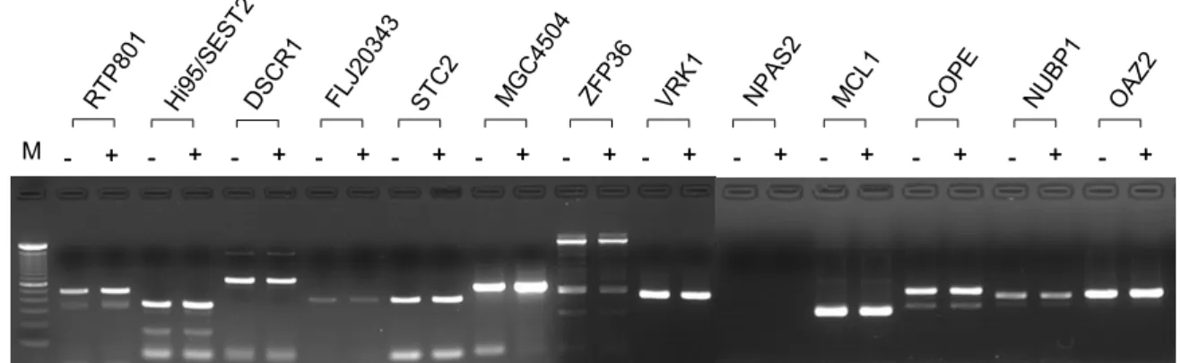
treatment (Table 2). In order to confirm the induction or repression of the 13 genes, semiquantitative RT- PCR was performed using cDNAs prepared from the RNAs of Aβ-treated or -untreated cells. Six of 13 ge-nes, RTP801, stanniocalcin 2 (STC2), hypothetical pro-tein MGC4504, Hi95/sestrin 2 (SEST2), hypothetical protein FLJ20360, and zinc finger protein 36 (ZFP36L2) were confirmed to be Aβ-responsive (Figure 2).
Induction of RTP801, STC2 and Hi95/SEST2
during A
β
treatm ent
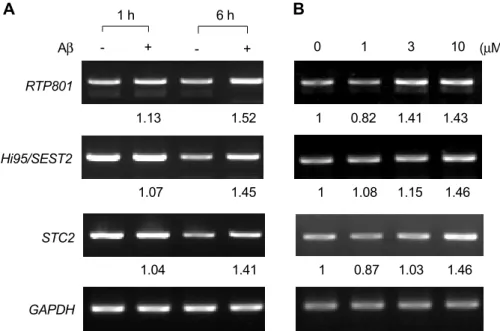
We further analyzed the expressions of 3 genes, RTP801, Hi95/SEST2, and STC2, with functions known to be associated with the oxidative stress induced by hypoxia (Budanov et al., 2002; Shoshani et al., 2002), and calcium and phosphate homeostasis (Ishibashi et al., 1998), respectively. The levels of these genes were also found to be increased in dose- and time- dependent manners in Aβ-treated cells by RT-PCR (Figure 3).
Effects of RTP801 and STC2 on
A
β
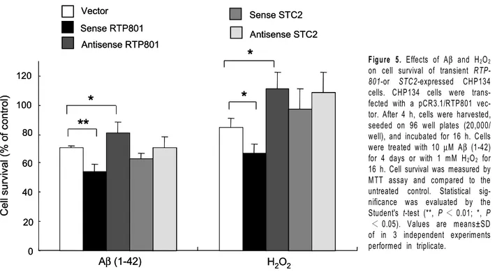
-m ediated cytotoxicity
In an attempt to investigate the effects of RTP801 and STC2 on Aβ toxicity, the protein encoding regions of RTP801 and STC2 were amplified by RT-PCR and subcloned into a pCR3.1 vector under the control of a cytomegalovirus promoter. The expressions of RTP-801 and STC2 were increased in the RTPRTP-801- or STC2- transfected cells compared to the vector transfected cells (Figure 4). Transient expression of the sense RTP801 gene in the cells showed an increase in the Aβ cytotoxicity (P < 0.01), and the expression of the antisense RTP801 gene had a protective effect again-st Aβ toxicity (P < 0.05) compared to the vector-transfected cells (Figure 5). Transient expression of the sense or the antisense STC2 gene had little ef-fects on Aβ cytotoxicity (Figure 5). These results sug-gest that RTP801 might be involved in Aβ cytotoxicity Figure 1. Aβ-induced cell death in CHP134 cells. Cells were treated with the indicated concentrations of Aβ (1-42) and then incubated for 4 days. Cell survival was measured by MTT assay. Values are means ±SD of triplicates of 3 independent experiments. *, P < 0.05 (Student's t-test). Surv iv al (% of control) 0 1 3 10 Concentration (µM)
*
50 100 0Table 2. Aβ-induced or repressed genes in CHP134 cells.
ꠚꠚꠚꠚꠚꠚꠚꠚꠚꠚꠚꠚꠚꠚꠚꠚꠚꠚꠚꠚꠚꠚꠚꠚꠚꠚꠚꠚꠚꠚꠚꠚꠚꠚꠚꠚꠚꠚꠚꠚꠚꠚꠚꠚꠚꠚꠚꠚꠚꠚꠚꠚꠚꠚꠚꠚꠚꠚꠚꠚꠚꠚꠚꠚꠚꠚꠚꠚꠚꠚꠚꠚꠚꠚꠚꠚꠚꠚꠚꠚꠚꠚꠚꠚꠚꠚꠚꠚꠚꠚꠚꠚꠚꠚꠚꠚꠚꠚꠚꠚꠚꠚꠚꠚꠚꠚꠚꠚꠚꠚꠚ
Folds Genbank
Gene name ꠏꠏꠏꠏꠏꠏꠏꠏꠏꠏꠏꠏꠏꠏꠏ Acc. No. Function RT-PCR*
1 h 6 h
ꠏꠏꠏꠏꠏꠏꠏꠏꠏꠏꠏꠏꠏꠏꠏꠏꠏꠏꠏꠏꠏꠏꠏꠏꠏꠏꠏꠏꠏꠏꠏꠏꠏꠏꠏꠏꠏꠏꠏꠏꠏꠏꠏꠏꠏꠏꠏꠏꠏꠏꠏꠏꠏꠏꠏꠏꠏꠏꠏꠏꠏꠏꠏꠏꠏꠏꠏꠏꠏꠏꠏꠏꠏꠏꠏꠏꠏꠏꠏꠏꠏꠏꠏꠏꠏꠏꠏꠏꠏꠏꠏꠏꠏꠏꠏꠏꠏꠏꠏꠏꠏꠏꠏꠏꠏꠏꠏꠏꠏꠏꠏ Calcium and phosphate
Stanniocalcin 2 (STC2) 0.77 1.85 M_003714 homeostasis 1.41
ypothetical protein MGC4504 0.88 1.79 NM_024111 Unknown 1.36
TP801 0.92 1.71 NM_019058 Cell viability 1.53
Hi95/Sestrin 2 (SEST2) 0.79 1.63 NM_031459 Cell viability 1.45
Myeloid cell differentiation
0.95 0.60 L08246 Apoptosis 1.08
protein (MCL1)
CNS development &
Down syndrome candidate 0.83 0.62 NM_004414 transcriptional function 0.90
Ornithine decarboxylase 0.91 0.66 NM_002537 Regulation of polyamine 1.07
antizyme 2 (OAZ2) synthesis
Coatomer protein complex, 1.06 0.60 NM_007263 Vesicle trafficking 0.90
epsilon (COPE)
Hypothetical protein FLJ20360 0.72 0.65 NM_017782 Unknown 0.77
Nucleotide binding protein 0.82 0.61 NM_002484 Nucleotide binding 0.96
(MinD homolog)
Zinc finger protein 36, 0.92 0.65 NM_006887 Transcription factor 0.47
C3H type-like 2 (ZFP36L2)
Vaccinia related kinase 1 0.95 0.64 NM_003384 Circardian rhythms 0.90
(VRK1)
Neuronal PAS domain
0.87 0.59 NM_002518 DNA binding No band
protein 2 (NPAS2)
ꠏꠏꠏꠏꠏꠏꠏꠏꠏꠏꠏꠏꠏꠏꠏꠏꠏꠏꠏꠏꠏꠏꠏꠏꠏꠏꠏꠏꠏꠏꠏꠏꠏꠏꠏꠏꠏꠏꠏꠏꠏꠏꠏꠏꠏꠏꠏꠏꠏꠏꠏꠏꠏꠏꠏꠏꠏꠏꠏꠏꠏꠏꠏꠏꠏꠏꠏꠏꠏꠏꠏꠏꠏꠏꠏꠏꠏꠏꠏꠏꠏꠏꠏꠏꠏꠏꠏꠏꠏꠏꠏꠏꠏꠏꠏꠏꠏꠏꠏꠏꠏꠏꠏꠏꠏꠏꠏꠏꠏꠏꠏ *Values are mean ratios of DNA levels amplified from Aβ (1-42)-treated cells to those from the untreated control.
Figure 2. RT-PCR analysis of Aβ-responsive genes. Cells were incubated in the absence (-) or in the presence (+) of 10 µM of Aβ (1-42) for 6 h and harvested. RNAs were purified from the cells and the 1st-strand cDNAs were synthesized with reverse transcriptase. Target sequences for the specific genes were amplified by PCR and the amplified DNAs were analyzed by agarose gel electrophoresis. M, 100 bp ladder. The figure shows representative data from 3 independent experiments.
- + - + -- ++ -- ++ -- + -+ - ++ -- ++ -- + -+ - ++ -- ++ -- ++ -- ++ -- ++ -- ++ M RTP 801 Hi9 5/SE ST2 DSC R1 FLJ2 0343 STC 2 MG C45 04 ZFP3 6 VR K1 NPA S2 MC L1 CO PE NU BP1 OAZ 2
and in the pathogenesis of Alzheimer's disease.
D iscussion
In the present study we identified six genes as being
Aβ-responsive in CHP134 cells by cDNA chip analysis and RT-PCR. RTP801, Hi95/SEST2 and STC2 were overexpressed in CHP134 cells treated with Aβ and the transient expression of RTP801 increased their sensitivity to Aβ cytotoxicity. RTP801 was induced by hypoxia in rat C6 glioma cells regulated by hypoxia- inducible factor-1 (HIF-1) and identified to be involved in apoptosis (Shoshani et al., 2002). Although expres-sion of the RTP801 gene in MCF7 and PC12 cells inhibited hypoxia- and H2O2-mediated apoptosis, its
function in cells is not fully understood. RTP801 is ubiquitously expressed in multiple human tissues at low levels. However, in response to hypoxia its trans-cription increases rapidly and sharply. The inducible expression of RTP801 in cells has different biological effects depending on the cell context. Shoshani et al. (2002) showed that expression of RTP801 has pro-tected MCF7 and PC12 cells from hypoxia and from H2O2-triggered apoptosis, but detrimentally affected
nondividing neuron-like PC12 cells under hypoxia and oxidative stress.
We identified that Hi95/SEST2 expression was also increased in Aβ-treated CHP134 cells. Hi95/SEST2 has been recently identified as a novel stress-responsive gene involved in the regulation of cell viability (Buda-nov et al., 2002). Hi95/SEST2 shares significant ho-mology with a p53-regulated GADD family member PA26 (Peeters et al., 2003). Increased expression of Hi95/SEST2 was induced by various cellular stresses Figure 4. semiquantitative RT-PCR of RTP801 and STC2 in CHP134
cells transfected with pCR3.1/RTP801 or pCR3.1/STC2 vectors. CHP134 cells were transfected with pCR3.1/RTP801 or pCR3.1/STC2 using a jetPEI transfection reagent. After 3 days, cells were harvested and RNAs were extracted. Transient expression of RTP801 and STC2 were confirmed by RT-PCR. RTP-183F and RTP-913R were used in Lane 1 to 5 and STC2-128F and STC2-1072R in Lane 6 to 8. Lanes 1 and 6, vector-transfected cells; Lanes 2 and 3, sense RTP801; Lanes 4 and 5, antisense RTP801; Lanes 7, sense STC2; Lane 8, antisense STC2. M, 100 bp ladder. 1 2 3 4 5 6 7 8 RTP801STC2 M GAPDH RTP801 STC2
Figure 3. Expressions of RTP801, STC2, and Hi95/SEST2 in CHP134 cells treated with Aβ. A. Cells were incubated in the absence (-) or in the presence (+) of 10 µM of Aβ (1-42) for 1 and for 6 h, and harvested. RTP801 and STC2 were amplified by RT-PCR. B. Cells were treated with the indicating concentrations of Aβ (1-42) for 6 h and harvested. RTP801 and STC2 were amplified by RT-PCR. The amplified DNAs were analyzed by agarose gel electrophoresis and their levels were quantified using a UTHSCSA ImageTool program by averaging three separate measurements of each band as well as control. This shows representative data from 3 independent experiments.
- + - + 1 h 6 h RTP801 Hi95/SEST2 STC2 GAPDH 0 1 3 10 (µM) Aβ
A
B
1.13 1.52 1 0.82 1.41 1.43 1.07 1.45 1 1.08 1.15 1.46 1.04 1.41 1 0.87 1.03 1.46including prolonged hypoxia, oxidative stress, UV-or
γ-irradiation, and doxorubicin, and the overexpression of Hi95/SEST2 full-length cDNA was found to be toxic in many types of cultured cells (Budanov et al., 2002). Hypoxia is known to induce oxidative stress in PC12 cells via Aβ and reactive oxygen species formation (Green et al., 2002). Aβ also induces oxidative stress itself by producing H2O2 (Behl et al., 1994; Huang et
al., 1999). Therefore, the Aβ-mediated overexpres-sions of RTP801 and Hi95/SEST2 genes in CHP134 cells might be associated with cellular oxidative stress. Our finding that the transient expression of the sense RTP801 gene, not the antisense RTP801 gene, ex-acerbates Aβ-or H2O2-mediated cytotoxicity in CHP134
cells suggests that the overexpression of RTP801 in Aβ-treated CHP134 cells might play an important role in cell death during Aβ-mediated oxidative stress. Stanniocalcin (STC) is a hormone that was initially identified in fish, which inhibits calcium absorption in the gills and intestines and stimulates the absorption of phosphates (Sundell et al., 1992). Mammalians have two types of STC; STC1 and STC2. STC1 has a 61% homology with fish STC and presents in kid-ney, thyroid glands, ovary, and prostates. STC2 has a 38% homology with fish STC2 and presents mainly in the pancreas (Ishibashi et al., 1998). Mammalian STC1 was suggested to play an important role in calcium homeostasis, including the absorption and secretion of calcium and phosphates. However, the function of STC2 is unknown (Jellinek et al., 2000). STC2 might also play an important role in glucose
homeostasis (Moore et al., 1999), and was identified as an estrogen-responsive gene, which was induced with estrogen receptor in human breast cancers (Bouras et al., 2002). Our data shows that STC2 is up-regul-ated by Aβ treatment. However, the transient expres-sion of sense or antisense STC2 genes was found to have little effects on Aβ cytotoxicity, suggesting that the Aβ-mediated overexpression of STC2 may not be directly associated with Aβ toxicity.
In conclusion, our results suggest that some of the novel Aβ-responsive genes play key roles in the response of neuronal cells to Aβ exposure. Further functional analysis of the novel Aβ-responsive genes is required to open up new research routes of enquiry into the pathogenesis of AD.
Acknow ledgem ent
This work was supported by grant No. R01-1999-000- 00087 from the Korea Science & Engineering Foun-dation.
R eferences
Aksenov MY, Aksenova MV, Carney JM, Butterfield DA. Oxi-dative modification of glutamine synthetase by amyloid β peptide. Free Radic Res 1997;27:267-81
Aksenov MY, Aksenova MV, Markesbery WR, Butterfield DA. Amyloid β-peptide (1-40)-mediated oxidative stress in cultured hippocampal neurons. Protein carbonyl formation, CK BB ex-pression, and the level of Cu, Zn, and Mn SOD mRNA. J
**
*
*
*
Aβ(1-42) H2O2 0 20 40 60 80 100 120 Ce ll su rv iv al (% o f co nt ro l) Vector Sense RTP801 Antisense RTP801 Sense STC2 Antisense STC2**
*
*
*
Aβ(1-42) H2O2 0 20 40 60 80 100 120 Ce ll su rv iv al (% o f co nt ro l) Vector Sense RTP801 Antisense RTP801 Sense STC2 Antisense STC2Figure 5. Effects of Aβ and H2O2 on cell survival of transient RTP-801-or STC2-expressed CHP134 cells. CHP134 cells were trans-fected with a pCR3.1/RTP801 vec-tor. After 4 h, cells were harvested, seeded on 96 well plates (20,000/ well), and incubated for 16 h. Cells were treated with 10 µM Aβ (1-42) for 4 days or with 1 mM H2O2 for 16 h. Cell survival was measured by MTT assay and compared to the untreated control. Statistical sig-nificance was evaluated by the Student's t-test (**, P < 0.01; *, P
< 0.05). Values are means±SD of in 3 independent experiments performed in triplicate.
Mol Neurosci 1998;10:181-92
Behl C, Davis JB, Lesley R, Schubert D. Hydrogen peroxide mediates amyloid β protein toxicity. Cell 1994;77:817-27 Bouras T, Southey MC, Chang AC, Reddel RR, Willhite D, Glynne R, Henderson MA, Armes JE, Venter DJ. Stannio-calcin 2 is an estrogen-responsive gene coexpressed with the estrogen receptor in human breast cancer. Cancer Res 2002;62:1289-95
Budanov AV, Shoshani T, Faerman A, Zelin E, Kamer I, Kalinski H, Gorodin S, Fishman A, Chajut A, Einat P, Skaliter R, Gudkov AV, Chumakov PM, Feinstein E. Identification of a novel stress-responsive gene Hi95 involved in regulation of cell viability. Oncogene 2002;21:6017-31
Butterfield DA, Hensley K, Harris M, Mattson M, Carney J. β-Amyloid peptide free radical fragments initiate synapto-somal lipoperoxidation in a sequence-specific fashion: impli-cations to Alzheimer's disease. Biochem Biophys Res Com-mun 1994;200:710-5
Chomczynski P, Sacchi N. Single-step method of RNA isola-tion by acid guanidinium thiocyanate-phenol-chloroform extra-ction. Anal Biochem 1987;162:156-9
Green KN, Boyle JP, Peers C. Hypoxia potentiates exocyto-sis and Ca2+ channels in PC12 cells via increased amyloid β peptide formation and reactive oxygen species generation. J Physiol 2002;541:1013-23
Greeve I, Hermans-Borgmeyer I, Brellinger C, Kasper D, Gomez-Isla T, Behl C, Levkau B, Nitsch RM. The human DIMINUTO/DWARF1 homolog seladin-1 confers resistance to Alzheimer's disease-associated neurodegeneration and oxi-dative stress. J Neurosci 2000;20:7345-52
Hata R, Masumura M, Akatsu H, Li F, Fujita H, Nagai Y, Yamamoto T, Okada H, Kosaka K, Sakanaka M, Sawada T. Up-regulation of calcineurin Aβ mRNA in the Alzheimer's disease brain: assessment by cDNA microarray. Biochem Biophys Res Commun 2001;284:310-6
Huang X, Cuajungco MP, Atwood CS, Hartshorn MA, Tyndall JD, Hanson GR, Stokes KC, Leopold M, Multhaup G, Gold-stein LE, Scarpa RC, Saunders AJ, Lim J, Moir RD, Glabe C, Bowden EF, Masters CL, Fairlie DP, Tanzi RE, Bush AI. Cu (II) potentiation of Alzheimer Aβ neurotoxicity: Correlation with cell-free hydrogen peroxide production and metal reduction. J Biol Chem 1999;74:37111-6
Ishibashi K, Miyamoto K, Taketani Y, Morita K, Takeda E, Sasaki S, Imai M. Molecular cloning of a second human stanniocalcin homologue (STC2). Biochem Biophys Res Com-mun 1998;250:252-8
Jang JH, Surh YJ. β-Amyloid induces oxidative DNA damage and cell death through activation of c-Jun N terminal kinase. Ann N Y Acad Sci 2002;973:228-36
Jellinek DA, Chang AC, Larsen MR, Wang X, Robinson PJ, Reddel RR. Stanniocalcin 1 and 2 are secreted as phos-phoproteins from human fibrosarcoma cells. Biochem J 2000; 350:2453-61
Kaltschmidt B, Uherek M, Wellmann H, Volk B, Kaltschmidt C. Inhibition of NF-κB potentiates amyloid β-mediated neuro-nal apoptosis. Proc Natl Acad Sci USA 1999;96:9409-14 Lovell MA, Gabbita SP, Markesbery WR. Increased DNA
oxidation and decreased levels of repair products in Al-zheimer's disease ventricular CSF. J Neurochem 1999;72: 771-6
Maccioni RB, Otth C, Concha II, Munoz JP. The protein kinase Cdk5. Structural aspects, roles in neurogenesis and involvement in Alzheimer's pathology. Eur J Biochem 2001; 268:1518-27
Marcus DL, Strafaci JA, Miller DC, Masia S, Thomas CG, Rosman J, Hussain S, Freedman ML. Quantitative neuronal c-fos and c-jun expression in Alzheimer's disease. Neurobiol Aging 1998;19:393-400
Mark RJ, Blanc EM, Mattson MP. Amyloid β-peptide and oxidative cellular injury in Alzheimer's disease. Mol Neurobiol 1996;12:211-24
Markesbery WR. Oxidative stress hypothesis in Alzheimer's disease. Free Radic Biol Med 1997;23:134-47
Mattson MP, Guo Q, Furukawa K, Pedersen WA. Presenilins, the endoplasmic reticulum, and neuronal apoptosis in Alzhei-mer's disease. J Neurochem 1998;70:1-14
Miranda S, Opazo C, Larrondo LF, Munoz FJ, Ruiz F, Leigh-ton F, Inestrosa NC. The role of oxidative stress in the toxicity induced by amyloid β-peptide in Alzheimer's disease. Prog Neurobiol 2000;62:633-48
Moore EE, Kuestner RE, Conklin DC, Whitmore TE, Downey W, Buddle MM, Adams RL, Bell LA, Thompson DL, Wolf A, Chen L, Stamm MR, Grant FJ, Lok S, Ren H, De Jongh KS. Stanniocalcin 2: characterization of the protein and its localization to human pancreatic cells. Horm Metab Res 1999;31:406-14
Noh YH, Kim JA, Lim GR, Ro YT, Koo JH, Lee YS, Han DS, Park HK, Ahn MJ. Detection of circulating tumor cells in patients with gastrointestinal tract cancer using RT-PCR and its clinical implications. Exp Mol Med 2001;33:8-14 Pappolla MA, Omar RA, Kim KS, Robakis NK. Immuno-histochemical evidence of oxidative stress in Alzheimer's disease. Am J Pathol 1992;140:621-8
Paradis E, Douillard H, Koutroumanis M, Goodyer C, Le-Blanc A. Amyloid β peptide of Alzheimer's disease down-regulates Bcl-2 and updown-regulates bax expression in human neurons. J Neurosci 1996;16:7533-9
Park GH, Choe J, Choo HJ, Park YG, Sohn J, Kim MK. Genome-wide expression profiling of 8-chloroadenosine- and 8-chloro-cAMP-treated human neuroblastoma cells using radioactive human cDNA microarray. Exp Mol Med 2002;34: 184-93
Peeters H, Debeer P, Bairoch A, Wilquet V, Huysmans C, Parthoens E, Fryns JP, Gewillig M, Nakamura Y, Niikawa N, Van De Ven W, Devriendt K. PA26 is a candidate gene for heterotaxia in humans: identification of a novel PA26- related gene family in human and mouse. Hum Genet 2003; 112:573-80.
Santiard-Baron D, Gosset P, Nicole A, Sinet PM, Christen Y, Ceballos-Picot I. Identification of β-amyloid-responsive ge-nes by RNA differential display: early induction of a DNA damage-inducible gene, gadd45. Exp Neurol 1999;158:206- 13
Mann U, Muller-Thomsen T, Beisiegel U. Increased lipo-protein oxidation in Alzheimer's disease. Free Radic Biol Med 2000;28:351-60
Shoshani T, Faerman A, Mett I, Zelin E, Tenne T, Gorodin S, Moshel Y, Elbaz S, Budanov A, Chajut A, Kalinski H, Kamer I, Rozen A, Mor O, Keshet E, Leshkowitz D, Einat P, Skaliter R, Feinstein E. Identification of a novel hypoxia- inducible factor 1-responsive gene, RTP801, involved in apoptosis. Mol Cell Biol 2002;22:2283-93
Smith CD, Carney JM, Starke-Reed PE, Oliver CN, Stadtman ER, Floyd RA, Markesbery WR. Excess brain protein oxi-dation and enzyme dysfunction in normal aging and in Alzheimer disease. Proc Natl Acad Sci USA 1991;88:10540-3 Smith MA, Rottkamp CA, Nunomura A, Raina AK, Perry G. Oxidative stress in Alzheimer's disease. Biochim Biophys Acta 2000;1502:139-44
Suh SW, Jensen KB, Jensen MS, Silva DS, Kesslak PJ, Danscher G, Frederickson CJ. Histochemically-reactive zinc in amyloid plaques, angiopathy, and degenerating neurons of Alzheimer's diseased brains. Brain Res 2000;852:274-8
Sundell K, Bjornsson BT, Itoh H, Kawauchi H. Chum salmon (Oncorhynchus keta) stanniocalcin inhibits in vitro intestinal calcium uptake in Atlantic cod (Gadus morhua). J Comp Physiol [B] 1992;162:489-95
Thompson CM, Markesbery WR, Ehmann WD, Mao YX, Vance DE. Regional brain trace-element studies in Alzhei-mer's disease. Neurotoxicology 1988;9:1-7
Vitek MP, Bhattacharya K, Glendening JM, Stopa E, Vlas-sara H, Bucala R, Manogue K, Cerami A. Advanced glyca-tion end products contribute to amyloidosis in Alzheimer disease. Proc Natl Acad Sci USA 1994;91:4766-70 Walker DG, Lue LF, Beach TG. Gene expression profiling of amyloid β peptide-stimulated human post-mortem brain microglia. Neurobiol Aging 2001;22:957-66