Pleuropulmonary and abdominal paragonimiasis: CT and
ultrasound findings
1
S S SHIM,
MD
,
1Y KIM,
MD,
1J K LEE,
MD,
2J H LEE,
MDand
3D E SONG,
MD1Department of Radiology, Mokdong Hospital, Ewha Womans University School of Medicine, Seoul, Republic of Korea, 2Division of Pulmonary and Critical Care Medicine, Department of Internal Medicine, Mokdong Hospital, Ewha Womans University School of Medicine, Seoul, Republic of Korea, and3Department of Pathology, Mokdong Hospital, Ewha Womans University School of Medicine, Seoul, Republic of Korea
Objectives: The purpose of this study was to review radiological images of patients with Paragonimus westermani (PW) that simultaneously involved the chest and abdomen. Methods: Our study included four patients with serologically and histopathologically confirmed paragonimiasis. Abdomen CT (n53) and chest CT (n53) scans were available, and abdominal wall ultrasonography was performed in all patients. We retrospectively reviewed the clinical, radiological and histopathological findings of these patients. Results: The most common abdominal CT findings were ascites and intraperitoneal or abdominal wall nodules. Low-attenuated serpentine lesions of the liver were another common and relatively specific feature.
Conclusion: Radiologists should consider the possibility of PW when these abdominal CT findings are noted, especially with pleural effusion or subpleural nodules in patients with initial abdominal symptoms.
Received 30 October 2009 Revised 30 March 2010 Accepted 13 July 2010 DOI: 10.1259/bjr/30366021
’2012 The British Institute of Radiology
Paragonimiasis is a food-borne infection caused by the lung fluke Paragonimus westermani (PW). Human infection occurs by ingestion of raw or pickled fresh-water crustaceans, such as crab or crayfish, that are infected with metacercariae. The disease is endemic in certain areas of the Far East and South-East Asia [1]. Recently, paragonimiasis has been detected worldwide owing to an increase in the number of travellers and the expansion of food trading [2]. Once ingested, the juvenile flukes travel through the small intestinal wall and diaphragm and reach the pleural cavity from the peritoneal cavity in 3–8 weeks [3]. The chest and abdomen are routine areas of migration of PW; however, few case reports are available on the CT findings in cases of pleuropulmonary paragonimiasis with simultaneously appearing abdominal lesions. We describe the CT and ultrasound findings of paragoni-miasis in four patients who were hospitalised with initial abdominal manifestations and were subsequently confirmed to have pleuropulmonary and abdominal paragonimiasis.
Methods and materials
Patients
Our study included four patients (three females and one male; age range, 23–50 years; mean age, 36 years) with serologically and histopathologically confirmed paragonimiasis. Abdominal CT (n53) and chest CT
(n53) scans were available, and abdominal wall ultrasonography was performed in all patients. Initially, abdominal imaging studies (two CT and two ultrasound studies) were performed because of major patient complaints (two palpable masses, one case of abdominal pain and one case of a palpable mass with pain), and chest CT imaging and abdominal wall ultrasound with biopsies were subsequently per-formed. The time interval between the presentation of symptoms and time of imaging was 15–60 days (mean, 18 days). The interval between abdominal wall ultra-sound and abdominal CT examinations was 9–20 days (mean, 13 days), and chest CT scans were obtained 8–22 days (mean, 13 days) after the initial abdominal studies. We retrospectively reviewed the clinical, radiological and histopathological findings of patients. Approval from the institutional review board is not needed for the review of radiological images for research purposes in our institute.
CT protocol
Abdominal CT examinations were performed using a 16-channel multidetector row CT (MDCT) scanner (SOMATOM Sensation, Siemens Medical Solutions, Forchheim, Germany) for 2 patients, and a 64 channel MDCT scanner (SOMATOM Sensation 64, Siemens Medical Solutions) for 1 patient. MDCT images were obtained from the diaphragm to the symphysis pubis during a single breath-hold. A detector configuration of 1.5616 mm and a table speed of 24 mm per gantry rotation were used for the 16 channel MDCT scans. A detector configuration of 0.6632 mm with a Z-flying spot system and a table speed of 38.4 mm per gantry
Address correspondence to: Dr Sung Shine Shim, Department of Radiology, Mokdong Hospital, Ewha Womans University School of Medicine, 911-1 Mok-6-dong Yangcheon-gu, Seoul 158-710, Republic of Korea. E-mail: [email protected]
The British Journal of Radiology, 85 (2012), 403–410
Table 1. Clinical features and imaging studies of four patients with pleuropulmonary and abdominal paragonimiasis
Patients Presenting symptoms
Initial presumptive
diagnosis Initial imaging study
Chest Diagnostic procedure Laboratory data Number Sex/age (years) Initial chief complaint Duration (months) Minor symptoms Thoracic manifestation Thoracic evaluation Leucocytosis (.10 000m l–1) Eosinophilia (.500m l–1) 1 M/50 Abdominal pain 1 – TB or cancer peritonitis Abdomen CT Hydropneu-mothorax Chest PA Chest CT ultrasound-guided omental biopsy Yes Yes 2 F/35 Abdominal pain and palpable abdominal wall mass 3 Dyspnoea, fever TB peritonitis and TB pleurisy Abdomen CT Dyspnoea, fever Chest PA Chest CT ultrasound-guided abdominal wall nodule biopsy Yes Yes 3 F/23 Palpable abdominal wall mass 2 Abdominal discom-fort Subcutaneous benign tumour Abdominal wall ultrasound
Mild cough Chest PA
Chest CT ultrasound-guided abdominal wall mass biopsy Yes Yes 4 F/34 Palpable abdominal wall mass 3 – Subcutaneous benign tumour Abdominal wall and abdomen ultrasound
Mild cough Chest PA
ultrasound-guided abdominal wall mass biopsy
Yes Yes
F, female; M, male; No., number; PA, posteroanterior radiograph; TB, tuberculosis. S
S Shim, Y Kim, J H Lee et The British Journal of Radiology, April 2012
rotation were used for the 64 channel MDCT scans. A pitch of 1, a reconstruction thickness of 5 mm, 120 kVp and a variable tube current (90–140 mAs) for both MDCT scanners were used. Single-phase contrast-enhanced images were obtained at 90 or 100 s after the injection of 120 ml of contrast agent (iohexol, Omnipaque 300; Nycomed, Zurich, Switzerland; or iopromide, Ultravist 300; Schering, Berlin, Germany) at a rate of 3 ml s–1.
Chest CT scans were performed using a 16 channel MDCT scanner (SOMATOM Sensation) for all patients.
The parameters for helical chest CT imaging were 120 kVp, 80–100 mAs, 5 mm collimation and a 10 mm s–1 table feed. Contrast-enhanced chest CT scans were obtained after injection of 30 g of iodinated contrast agent (100 ml of iopromide, Ultravist 300) at a rate of 2.3 ml s–1 with the use of a power injector (OP100, Medrad, Pittsburgh, PA). The scan data were display-ed directly on monitors (2 monitors, 5126512 image matrices, 12 bit viewable greyscale) of a picture archiv-ing and communication system (PACS) (Starpacs; In-finitt, Seoul, Korea).
(a) (b)
(c) (d)
(e) (f)
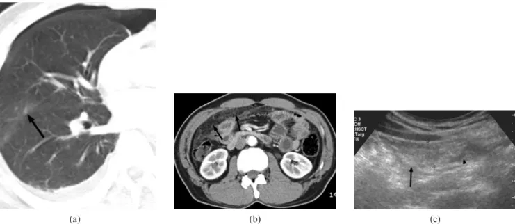
Figure 1. A 23-year-old female with paragonimiasis (Patient 3). An axial chest CT scan (3 mm collimation) obtained in the lung setting (a) shows an irregular cavitary lesion with a branching appearance in the right upper lobe (arrow) and a track-like lesion in left upper lobe (arrowhead) with adjacent pleural thickening. Axial abdominal CT images (b, c) show ill-defined nodules with linear strands in the subcutaneous layer of the anterior rectus abdominis muscle (arrows; b) and hypodense tubular structures (arrow; c) in segment 4 of the liver with ascites in the perihepatic space. Ultrasonography for biopsy of the abdominal wall (d) shows an ill-defined heterogeneous echoic nodule in the subcutaneous layer of the left upper quadrant of the abdominal wall (arrow). A surgical specimen obtained from the abdominal wall shows dense infiltration of eosinophils among adipocytes, forming an eosinophilic abscess (e) and a collapsed adult worm of Paragonimus westermani (f).
Pleuropulmonary and abdominal paragonimiasis
Image analysis
Two abdominal radiologists reached decisions by consensus based on the findings. Two chest radiologists also retrospectively evaluated the chest CT scans. The chest CT features evaluated in our study included those previously reported in patients with pleuropulmonary paragonimiasis: the presence of a nodule or consolida-tion, pleural effusion and cyst formation. Abdominal CT images were evaluated for the presence of any intraper-itoneal or abdominal wall nodules or linear strands. The presence of adjacent bowel thickening (if .5 mm), low-attenuation lesions in the solid organs, intra-abdominal masses and ascites were evaluated as additional findings in the intraperitoneal area.
Results
Clinical features
The clinical course and laboratory data of the patients are summarised in Table 1. All patients presented with
abdominal symptoms as the initial chief complaint, including palpable abdominal wall masses (n53, 75%) and abdominal pain (n52, 50%). The apparent duration of illness ranged from 2 to 3 months. As minor presenting symptoms, one patient had fever and dyspnoea, but the other three patients had no respiratory symptoms (including haemoptysis, cough, dyspnoea, chest pain and fever, which are known as the main presenting symptoms of PW). As the initial diagnostic imaging study, abdom-inal CT was performed in the patient with a painful abdomen, and abdominal wall ultrasound was performed in the patients with palpable abdominal wall masses. Subsequently, in the follow-up period, chest postero-anterior view radiographs and chest CTs were performed in the patients with thoracic manifestations, including hydropneumothorax (Patient 1) detected on simple abdominal radiography, dyspnoea, fever (Patient 2) and mild cough (Patients 3 and 4). Leukocytosis (.10 000 m l–1) and eosinophilia (.500 m l–1) were detected in the periph-eral blood of all patients, and the median degree of eosi-nophilia was 42.7% (range, 31.3–56%) in these 4 patients. Ultrasound-guided omental (Patient 1) and abdominal
(a) (b) (c)
Figure 2. A 50-year-old male with paragonimiasis (Patient 1). An axial chest CT scan (5 mm collimation) ob-tained at the lung setting (a) shows tiny, ill-defined nodules in the fissural area (arrow). Small amounts of pneumothorax and pleural effusion can be seen. An abdominal CT scan (b) at the time of initial hospitalisation showed extensive linear strands and small non-calcified nodules in the greater omentum (arrows). Ultrasonography for biopsy of the abdominal wall (c) showed an ill-defined, approximately 1.57 cm omental thickening (arrow) with an adjacent serpentine low echoic lesion (arrowhead).
(a) (b)
Figure 3. Axial abdominal CT images of abdominal wall nodules in para-gonimiasis patients. Ill-defined nodules in the subcutaneous layer of the left gluteal area (a) (arrows) are seen in Patient 3. (b) A small ring-like nodule (arrowhead) in the left abdominal wall and linear strands with ill-defined nodules in the sub-cutaneous layer of the right lateral abdominal wall (arrow) are noted in Patient 2.
wall (Patients 2–4) nodular biopsies were performed in all patients, and these results showed eosinophil infiltrations with Charcot–Leyden crystals (n53) and eosinophilic abscess with paragonimus organisms (n51; Figure 1e, f). All patients were positive for the PW antibody in the blood.
Abdominal CT and ultrasound findings
The most common findings, observed in all patients, were ascites and intraperitoneal or abdominal wall
nodules with increased fat strands. Peritoneal nodules were seen in two patients as non-specific small nodules (,8 mm in the longest diameter; Figure 2b). Abdominal wall nodules (mean diameter, 19.7 cm; range, 0.9–3.1 cm) in three patients were predominantly heterogeneous, with ring-like lesions (Figures 1b and 3); nodules at different locations in the same patient had a similar pattern, and none of the nodules had calcification. These lesions were predominantly observed in the anterior abdominal wall or greater omentum. The lateral (n52) and posterior (n52) abdominal walls were also involved (Figure 3).
The second most common findings were intrahepatic or intrarenal lesions in two patients. The intrahepatic lesions were seen with tubular and linear low densities that showed a tortuous course, which were presumed to be worm migration tracks (Figures 1c and 4). The intrarenal lesions were ill-defined low-attenuation nodules with adjacent increased fat strands (Figure 5). Circumferential caecal wall thickening, which mimicked tuberculous colitis, was observed in one patient. On initial abdominal images, pleural effusion was seen in all patients (right pleural effusion in two patients and bilateral pleural effusion in two patients). Chest and abdominal CT findings in the four patients are sum-marised in Table 2.
On ultrasonography, all patients showed ill-defined oval-shaped mass lesions, with heterogeneous echogeni-city in the subcutaneous layer of the abdominal wall in three patients (Figure 1d) and in the greater omentum in one patient (Figure 2c), findings that correlated with the abdominal CT findings. Blood flow in these lesions was increased in all patients, as seen on Doppler ultrasound.
Chest CT findings
The main features of the chest CT scans were medias-tinal lymphadenopathy and subfissural or subpleural nodules, which were observed in all patients. Three nodules were seen in two patients, and bilateral multiple nodules were seen in two patients. Nodules were seen with surrounding ground-glass opacities in three patients (Figure 2a) and with internal cavity and adjacent
Figure 5. Axial abdominal CT images of intrarenal lesions (Patient 3). A contrast-enhanced axial CT image shows an ill-defined hypodense nodule in the cortical area of the left kidney (arrow) with increased fat strands adjacent to the intrarenal lesion.
(a) (b)
Figure 4. Axial abdominal CT images of intrahepatic lesions (Patient 2). Contrast-enhanced CT of the liver (a, b) shows a cluster of small cysts and serpentine lesions (arrows; a, b) at the right lobe of the liver. A tubular low-attenuated lesion (arrowhead; b) is also noted in the subcapsular area of the right lobe.
Pleuropulmonary and abdominal paragonimiasis
tubular tracks in one patient (Figure 1a). A thin-walled cystic lesion was observed in one patient. There was combined pleural effusion in all patients, and pneu-mothorax in one patient.
Discussion
In areas where paragonimiasis is endemic, humans eat pickled freshwater crab. When humans ingest infected crabs or fresh vegetables that have been in contact with infected cooking implements, the metacercariae excyst in the small intestine and the juvenile flukes then penetrate the wall of the small bowel and enter the peritoneal cavity [3, 4]. The flukes then migrate into the abdominal wall or liver, where they undergo further development. Approximately 1 week later, the adult flukes re-enter the abdominal cavity and penetrate the diaphragm to make their way through the pleura into the lung. If the metacercariae juvenile or adult flukes are diverted during the migration route from the small intestine to the lung, the flukes may excyst elsewhere in the body [5]. The lung and abdomen are the routine locations of migration for the pathway of PW spread, and involve-ment of these sites may mimic tumorous or inflamma-tory conditions on radiological studies. However, few reports have described the abdominal CT features of such patients [5–8]. Moreover, to our knowledge, no CT findings of patients showing chest and abdominal features at the same time have been reported, except for a few case reports describing the disseminated form of paragonimiasis [6, 9–11].
Peritoneal radiological findings were reported by Rha et al [7], who studied the CT findings of intraperitoneal manifestations of parasitic infestations, including para-gonimiasis. Common features are localised hazy omen-tal infiltration and the presence of a peritoneal mass (predominantly a multiseptated cystic, heterogeneous or calcified granulomatous mass), especially in the case of paragonimiasis showing multiple scattered, densely calcified small nodules. Jeong et al [5] reported a similar case of incidentally detected calcified and fibrotic nodules, which were confirmed as being due to PW in the omentum. In our series, however, all of the nodules showed ill-defined small, non-specific masses, a hetero-geneous attenuated mass-like pattern without calcifica-tion or the presence of multiseptated cystic lesions. Ascites, representing one of the most common findings, was observed in all of the patients. We suggest that these inconsistent findings may have resulted from the shorter time interval between acute manifestation and CT imaging in our study. The interval between the time of imaging and suspected time of exposure to the organism was 45–80 days (mean, 63 days). Peritoneal nodules suggestive of granulomas are small in the early stages but can grow into large masses. These peritoneal masses may have varied appearances, depending on the stage of the infectious process; the masses may be solid, cystic (unilocular or multilocular), calcified or non-calcified.
Several cases of early-stage PW have been reported that have presented as a subcutaneous induration or as a mass in the abdominal subcutaneous tissue [9, 12]. Yokogawa [3] investigated the migratory route of juvenile PW in cats and rats. It was found that juvenile worms migrating into
Table 2. CT findings in three patients with pleuropulmonary and abdominal paragonimiasis Patients Chest CT Abdomen C T Number Sex/age (years) Nodule o r consolidation Pleural effusion P neumothorax Cyst Abdominal wall Intraperitoneum Nodule Linear strand Non-specific small nodules Linear strand Adjacent bowel
wall
thickening
Intra- abdominal mass
A scites Intrahepatic lesion Intrarenal lesion 1 M/50 Present Present Present Absent Absent Absent Present Present Absent Absent Present Absent Absent 2 F/35 Present Present Absent Absent Present Present Present Present Present Absent Present Present Present 3 F/23 Present Present Absent Present Present Present Absent Absent Absent Absent Present Present Present F, female; M, male; No., number.
the abdominal cavity immediately entered the inner wall. Lee et al [13] observed subcutaneous air bubbles in dogs at day 30 in the subcutaneous tissue of the abdominal or chest wall on CT images when hydropneumothorax began to be observed. In our study, we found hetero-geneous attenuated or ring-like nodules in the subcuta-neous fat layer and abdominal wall, which were located predominantly in an anterior location and lateral and posterior locations, respectively. All of these nodules presented with multiple increased fat strands, and the mean size of these nodules was larger than the mean size of the intraperitoneal nodules.
Hepatic manifestations of parasites more commonly present in patients with fascioliasis hepatica or echino-coccosis. In a patient with PW, ectopic infection most commonly involves the brain, while involvement of the liver is rare [14]. CT findings of hepatic paragonimiasis have been reported in four cases. One report described the presence of hypodense cystic lesions with peripheral enhancement in the subcapsular area, suggestive of capsular invasion of parasites [11]. A second case des-cribed the presence of a low-attenuation tubular lesion in an unusual central hepatic location [10]. The other reported cases described a multiseptated cystic lesion and multiple small cysts that represented eosinophilic abscesses [7, 8]. Hepatic involvement in our study showed a higher incidence rate (50%) than in previously reported cases, and had a similar pattern of lesions as previous reports that showed cystic, linear and serpen-tine hypodensities in the subcapsular and central areas of the liver.
Concomitant gastrointestinal tract involvement is another feature of parasitic infestation, and usually demonstrates eccentric bowel wall thickening or an intramural, perigastric, peri-enteric or pericolonic mass [15]. However, for PW, the incidence of this involvement is very low and radiological manifestations in affected intestines have been demonstrated in only a few reports. In one case in our study, caecum and small bowel wall thickening with an adjacent smudged pattern of infiltra-tion that mimicked tuberculous peritonitis was seen.
Common abdominal lesions that include nodules with increased fat strands and ascites may closely mimic other infectious conditions, such as tuberculous peritonitis or carcinomatosis peritonei [16]. Two of our cases were presumed to have had tuberculosis or carcinomatosis peritonei, as diagnosed on the first visit. Until recently, distinguishing between paragonimiasis and tuberculosis has frequently proven to be quite difficult, and consti-tutes a diagnostic dilemma in areas where tuberculosis and paragonimiasis coexist. Based on our study and previous reports, nodules with fat strands that are dominantly located in the subcutaneous layer and abdominal wall, as opposed to intraperitoneum and serpentine (linear and tubular) low-attenuation intrahe-patic lesions, might be helpful in differentiating para-gonimiasis from tuberculosis. Although these findings do not distinguish paragonimiasis from other parasitic diseases, concomitant pleural effusion and pulmonary cystic nodules can be helpful in the diagnosis of paragonimiasis.
Our study has some limitations. The major one is that the sample size was small, because of the low incidence of concomitant pleuropulmonary and abdominal PW.
Second, the biopsy specimens may not represent all of the lesions. However, follow-up serial images (chest radiography, chest CT and abdominal CT) of all the mentioned lesions showed serial improvement with chemotherapy, suggesting the same disease process. Despite these limitations, the cases in this study showed unusual findings in the presentation of PW patients and emphasise the following points. First, the disseminated form of PW can occur from the consumption of raw or undercooked food. Two patients in our study with no-dules in the posterior abdominal wall and lesions that affected the liver and kidney frequently ate sashimi and sushi. In these patients, recent heavy infection or recurrent infestation with a short-term interval might have been possible causes of the unusual disseminated presentation. Second, the major complaints in patients who presented with abdominal symptoms without reported typical tho-racic manifestations (chest pain, haemoptysis and cough) might cause a clinician to overlook the possibility of para-gonimiasis. These results correspond with a recent study about the radiological features of recently diagnosed para-gonimiasis, which reported a variety of clinical and radi-ological findings dissimilar to classic presentations [17].
In conclusion, in patients with simultaneous pleuro-pulmonary and abdominal paragonimiasis, ascites and intraperitoneal or abdominal wall nodules are the most common abdominal CT findings. Low-attenuation ser-pentine lesions of the liver were another common and relatively specific feature. Thus, cases with these findings that present especially with pleural effusion and cystic nodules should prompt radiologists to consider the possibility of paragonimiasis, even in non-endemic areas, owing to global dietary patterns and an increase in overseas travel.
References
1. Kim TS, Han J, Shim SS, Jeon K, Koh WJ, Lee I, et al. Pleuropulmonary paragonimiasis: CT findings in 31 patients. AJR Am J Roentgenol 2005;185:616–21.
2. Mukerjee CM, Simpson SE, Bell RJ, Walker JC. Pleuropul-monary paragonimiasis in a Laotian immigrant to Australia. Chest 1992;101:849–51.
3. Yokogawa M. Paragonimus and paragonimiasis. Adv Parasitol 1969;7:375–87.
4. Lee SC, Jwo SC, Hwang KP, Lee N, Shieh WB. Discovery of encysted Paragonimus westermani eggs in the omentum of an asymptomatic elderly woman. Am J Trop Med Hyg 1997;57:615–I8.
5. Jeong WK, Kim Y, Kim YS, Park DW, Park CK, Baek HK, et al. Heterotopic paragonimiasis in the omentum. J Com-put Assist Tomogr 2002;26:1019–21.
6. Jeong MG, Yu JS, Kim KW, Kim JK, Kim SJ, Kim HJ, et al. Retroperitoneal paragonimiasis: a case of ectopic paragoni-miasis presenting as periureteral masses. J Comput Assist Tomogr 1999;23:696–8.
7. Rha SE, Ha HK, Kim JG, Choi BI, Kim PN, Lee MG, et al. CT features of intraperitoneal manifestations of parasitic infestation. AJR Am J Roentgenol 1999;172:1289–92. 8. Singcharoen T, Rawd-Aree P, Baddeley H. Computed
tomography findings in disseminated paragonimiasis. Br J Radiol 1988;61:83–6.
9. Mizuki M, Mitoh K, Miyazaki E, Tsuda T. A case of Paragonimiasis westermani with pleural effusion eight months after migrating subcutaneous induration of the Pleuropulmonary and abdominal paragonimiasis
abdominal wall. [In Japanese] Nihon Kyobu Shikkan Gakkai Zasshi 1992;30:1125–30.
10. Yao A, Hammond N, Alasadi R, Nikolaidis P. Central hepatic involvement in paragonimiasis: appearance on CT and MRI. AJR Am J Roentgenol 2006;187:W236–7.
11. Kim EA, Juhng SK, Kim HW, Kim GD, Lee YW, Cho HJ, et al. Imaging findings of hepatic paragonimiasis: a case report. J Korean Med Sci 2004;19:759–62.
12. Takemasa H, Saito K, Nakayamada S, Kanazawa T, Tanaka Y. A case of Paragonimiasis westermanii complicated with migrating subcutaneous induration and multiple involve-ments in the liver. [In Japanese] Kansenshogaku Zasshi 2002;76:594–9.
13. Lee CH, Im JG, Goo JM, Lee HJ, Hong ST, Shen CH, et al. Serial CT findings of paragonimus infested dogs and the
micro-CT findings of the worm cysts. Korean J Radiol 2007;8:372–81.
14. Choo JD, Suh BS, Lee HS, Lee JS, Song CJ, Shin DW, et al. Chronic cerebral paragonimiasis combined with aneurys-mal subarachnoid hemorrhage. Am J Trop Med Hyg 2003;69:466–9.
15. Kim SY, Ha HK. Peritoneal manifestations of parasitic infection. Abdom Imaging 2008;33:172–6.
16. Rangheard AS, N’Senda P, Dahan H, Tubiana JM, Arrive L. Peritoneal location of fascioliasis mimicking a perito-neal carcinomatosis. J Comput Assist Tomogr 1999;23: 699–700.
17. Jeon K, Koh WJ, Kim H, Kwon OJ, Kim TS, Lee KS, et al. Clinical features of recently diagnosed pulmonary para-gonimiasis in Korea. Chest 2005;128:1423–30.