Safety of Using Matrix Metalloproteinase Inhibitor in
Experimental Glaucoma Filtration Surgery
We evaluated the safety of matrix metalloproteinase (MMP) inhibitor in experimental glaucoma filtration surgery in an animal model. Fifteen New Zealand white rabbits underwent an experimental trabeculectomy and were randomly allocated into 3 groups according to the adjuvant agent: no treatment group (n = 5), 0.02% mitomycin C (MMC) soaking group (n = 5), and MMP inhibitor (ilomastat) subconjunctival injection group (n = 5). Slit lamp examination with Seidel testing, pachymetry, and specular microscopy was performed preoperatively and postoperatively. The conjunctiva and ciliary body toxicity were evaluated with scores according to the pathologic grading systems. Electron microscopy was used to examine the structural changes in cornea, conjunctiva, and ciliary body. In the ilomastat-treated group, there was no statistically significant change in central corneal thickness preoperatively and at 28 days postoperatively (P = 0.655). There were also no significant changes in specular microscopy findings over the duration of the study in the ilomastat-treated group. The conjunctival toxicity score was 1 in the control group, 1.5 in the ilomastat-treated group, and 2 in the MMC-treated group. When assessing ciliary body toxicity scores, the ilomastat-treated group score was 0.5 and the MMC-treated group score was 1.5. Transmission electron microscopy did not show structural changes in the cornea and ciliary body whereas the structural changes were noticed in MMC group. A single subconjunctival injection of MMP inhibitor during the experimental trabeculectomy showed a less toxic affect in the rabbit cornea, conjunctiva, and ciliary body compared to MMC.
Keywords: Glaucoma; Filtration; Matrix Metalloproteinase Inhibitor
Wool Suh,1 Kyung Eun Han,2
and Jae ryong Han1
1Department of Ophthalmology, Hallym University
Dongtan Sacred Heart Hospital, Hallym University College of Medicine, Hwaseong, Korea;
2Department of Ophthalmology, Institute of
Ophthalmology and Optometry, Ewha Womans University Mok-Dong Hospital, Ewha Womans University School of Medicine, Seoul, Korea Received: 25 November 2016
Accepted: 7 January 2017 Address for Correspondence: Jae ryong Han, MD
Department of Ophthalmology, Hallym University Dongtan Sacred Heart Hospital, Hallym University College of Medicine, 7 Keunjaebong-gil, Hwaseong 18450, Republic of Korea E-mail: scarpel@hally.or.kr
Funding: This research was supported by Hallym University
Research Fund 2015 (HURF-2015-22).
https://doi.org/10.3346/jkms.2017.32.4.666 • J Korean Med Sci 2017; 32: 666-671
INTRODUCTION
The most common cause of failure in glaucoma filtering sur-gery is scarring of the filtering bleb, and the increased amount of collagen in the surgical site suggests that proliferation of fi-broblasts with associated production of collagen and glycos-aminoglycans is important (1).
Mitomycin C (MMC) has enhanced the surgical success rate of trabeculectomy through inhibition of Tenon capsule fibro-blast proliferation since its introduction in 1983 (2). However, use of MMC was associated with conjunctival epithelial dam-age, and it is characterized by non-healing leaking blebs and the risk of an avascular bleb and bleb infection (3). Hypotony maculopathy is one of the serious complications. It involves prolonged intraocular pressure (IOP) reduction associated with disc edema, vascular tortuosity, and chorioretinal macular folds, potentially producing marked reduction in visual acuity (1). The suggested causes are excessive filtering blebs and aqueous hy-posecretion, i.e., ciliary body changes (4). In addition to these serious complications, corneal problems are still common (5). Pastor et al. (6) examined corneal endothelial cell densities in
10 patients who underwent trabeculectomies with MMC and found a 4.7% to 20.0% decrease in cell density.
Therefore, many agents that can modulate wound healing and are safe for other ocular tissues simultaneously have been tried. In a previous study, matrix metalloproteinase (MMP), par-ticularly MMP-2/MT1-MMP, was associated with degradation of the extracellular matrix in the wound healing process after glaucoma filtration surgery and it was suggested as an impor-tant target for therapeutic intervention after glaucoma filtration surgery (7). In an animal study, Wong et al. (8) reported that MMP inhibitor can effectively reduce subconjunctival scarring after experimental glaucoma filtration surgery. In another study, the sequential treatment group used the following: Bevacizum-ab (Avastin), a monoclonal, VEGF antibody; Saratin, a 12 kD polypeptide with anti-inflammatory and anti-thrombotic prop-erties; and Ilomastat, an MMP inhibitor. The study demonstrat-ed a significant prolongation of bleb survival compardemonstrat-ed to the controls, which was not significantly different from the MMC positive control group (9). In another study, the MMP inhibitor significantly improved surgical outcomes compared with con-trols and the length of bleb survival was similar to the MMC
group (10).
However, a MMP inhibitor used in previous studies was a nonspecific target agent; i.e., producing general inhibition on MMP. Also, there has been no study on the influence on other ocular tissues. Therefore, we evaluated the safety of a MMP in-hibitor on other ocular tissues in an animal model.
MATERIALS AND METHODS
Fifteen New Zealand white rabbits aged between 12 and 14 weeks and weighing 2.0 to 2.5 kg were used.
Procedures and examination
Glaucoma filtration surgery was performed only on the right eye similarly to a previous report (11). Animals were randomly divided into 3 groups according to the adjuvant agent used dur-ing the surgery: the control group (n = 5), no agent; ilomastat-treated group (n = 5), 0.1 mL of 100 μM ilomastat (Calbiochem-Novabiochem, Nottingham, UK) in a subconjunctival injection; and MMC-treated group (n = 5), a thin cellulose sponge soaked with MMC (0.2 mg/mL). The bleb, conjunctiva, cornea, and an-terior chamber were evaluated with slit lamp biomicroscopy and Seidel test preoperatively and at 7 days, 14 days, and 28 days postoperatively. A pachymeter (Ultrasonic pachymeter; Nidek, Aichi, Japan) was used preoperatively and at postoperative 28 days to check the central corneal thickness (CCT). Specular mi-croscopy (Robo-Specular Microscope; Konan Medical Corpo-ration, Nishinomiya, Japan) was performed preoperatively and 28 days postoperatively. At the postoperative 28th day, the rab-bits in each group were sacrificed in a CO2 chamber after
iso-flurane gas induced anesthesia. The right eye of each rabbit was enucleated to preserve the bleb. Tissues were preserved in 10% formaldehyde, embedded in paraffin, and sequential 3–4 µm sections of the operative wound site were prepared. Hematoxy-lin and eosin staining was evaluated to check the general cellu-larity and inflammatory cells. Based on previous reports, the conjunctiva and the ciliary body toxicity were evaluated and scored according to the grading systems by 2 pathologists (12). The scale used for conjunctival changes was: 0 = no histologic change: no inflammation; 1 = minimal histologic change/con-junctiva epithelium preserved, thickening of the conchange/con-junctiva; 2 = mild histologic change/conjunctiva epithelium preserved, thickening of the conjunctiva, and mild inflammatory cell infil-tration; 3 = moderate histologic change/score 2 with loss of col-lagen fibril organization; and 4 = severe histologic change: loss of the conjunctival epithelium, total disorganization, and ne-crosis of the underlying scleral stroma. The scale used for ciliary body changes was: 0 = no histologic change; 1 = minimal his-tologic change/ciliary epithelium height normal, demonstrat-ing minimal fibroblast proliferation, congestion, and edema, no fibrin; 2 = mild histologic change/ciliary epithelium height
decreased, demonstrating moderate fibroblast proliferation with fibrin, moderate congestion, and edema; 3 = moderate histologic change/score 2 with inflammatory cell infiltration; 4 = severe histologic change/desquamation of the ciliary epi-thelium, total disorganization, and necrosis of the ciliary body. The toxicity of the cornea was also assessed with light modifica-tions.
For transmission electron microscopic observations, the cor-nea was obtained from the treated eyes immediately postmor-tem, and fixed in 2.5% glutaraldehyde in 0.1 M phosphate buf-fer for transmission electron microscopic examination. After-wards, the specimens were postfixed in 1% osmium tetroxide solution, dehydrated and embedded in epoxy resin using the usual procedure. Ultrathin sections of approximately 60–70 nm thickness were made using an Ultracut-E Microtome (Reichert-Jung, Buffalo, NY, USA) and were stained with heavy metals, uranylacetate and lead citrate. Stained sections were assessed and photographed under transmission electron microscopy (TEM; H-7650; Hitachi, Tokyo, Japan).
Statistical analysis
Statistical analyses were conducted using IBM SPSS stastics ver-sion 24.0 (IBM Corp., Armonk, NY, USA). The means and stan-dard errors were calculated using descriptive statistics. The Mann-Whitney U test and the Kruskal-Wallis test were used to com-pare the clinical findings of the groups. Significance was con-sidered at P values < 0.05.
Ethics statement
This study was approved by the Institutional Animal Care and Use Committee of Ewha Medical Center (ESM15-0291). All ani-mal procedures and methods used for securing the aniani-mal tis-sue complied with the Association for Research in Vision and Ophthalmology (ARVO) Statement for the Use of Animals in Ophthalmic and Vision Research and our institutional guide-lines.
RESULTS
On postoperative day 7, 14, and 28, fluorescein slit lamp micro-scopic examination revealed no abnormalities in the bleb con-junctiva such as bleb leakage and blebitis in the ilomastat treat-ed group, whereas one MMC treattreat-ed eye showtreat-ed one avascular Table 1. Pachymetric result (µm)
Group Before operation Postoperative 28 days P value Contol group 377.0 ± 18.9 361.5 ± 10.6 0.655 Ilomastat-treated group 382.00 ± 22.69 384.00 ± 8.28 0.655 Mitomycin-treated group 372.40 ± 21.17 368.33 ± 21.12 0.285 Values are presented as mean ± SE.
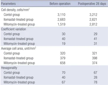
Table 2. Specular microscopic results
Parameters Before operation Postoperative 28 days Cell density, cells/mm2
Contol group 3,110 3,212 Ilomastat-treated group 2,683 2,621 Mitomycin-treated group 1,519 2,812 Coefficient variation Contol group 30 29 Ilomastat-treated group 40 41 Mitomycin-treated group 19 31
Average cell area, unit/mm2
Contol group 320 321 Ilomastat-treated group 379 398 Mitomycin-treated group 658 374 Hexagonality Contol group 70 67 Ilomastat-treated group 40 28 Mitomycin-treated group 67 78
All values are expressed as mean.
Fig. 1. Specular microscopy of corneal endothelial cells of the rabbits on postoperative 28 days. (A) Control group. (B) Ilomastat treated group. (C) Mitomycin-treated group. In the ilomastat-treated group, there was no statistically significant differences in all parameters preoperatively and at postoperative 28 days.
A B C
cystic bleb. In the ilomastat-treated group, cornea problems such as keratitis and corneal ulcer were not noticed vis slit lamp ex-amination. Any other adverse events, such as cataract, anterior chamber cell reaction, and endophthalmitis, were not found during the study.
The CCTs in each group are shown in Table 1. CCT was mea-sured before surgery as follows: 377.0 ± 18.9 in controls, 382.00 ± 22.69 in the ilomastat-treated group and 372.40 ± 21.17 in the MMC-treated group. There was no significant difference among the groups (P = 0.756). On postoperative days, CCTs were 361.5 ± 10.6 in the control group, 384.0 ± 8.28 in the ilomastat-treat-ed group, and 368.33 ± 21.12 in the MMC treatilomastat-treat-ed group and also showed no statistically significant changes among the 3 groups (P = 0.253). In the ilomastat-treated groups, there were no statistically significant changes in CCT between preopera-tive and postoperapreopera-tive 28 days (P = 0.655) and the MMC-treat-ed groups also showMMC-treat-ed no significant change between preoper-ative and postoperpreoper-ative 28 days (P = 0.285; Table 1).
Specular microscopy findings, such as cell density, coefficient of variation, average cell area, and hexagonality for the 3 groups are shown in Fig. 1 and Table 2. In the ilomastat-treated group, the parameters on preoperative and postoperative 28 days were the following: cell density 2,683 and 2,621, coefficient of varia-tion 40 and 41, average cell area 379 and 398, hexagonality 40 and 28, respectively (Table 2). In the ilomastat-treated group, there was no statistically significant difference in all parameters preoperatively and at postoperative 28 days (all P > 0.05). In the MMC-treated group, the coefficient of variation was changed from 19 preoperatively to 31 at postoperative 28 days. The aver-age cell area also decreased from 659 to 374, and hexagonality changed from 67 to 78 in the MMC-treated group. However, when parameters were compared among the 3 groups preop-eratively and at postoperative 28 days, there were no statistically significant differences in all parameters (all P > 0.05).
When conjunctival toxicity was evaluated, the average score was 1 in the control group, 1.5 in the ilomastat-treated group and 2 in the MMC-treated group. When assessing the ciliary body toxicity score, the ilomastat-treated group was 0.5 and the
MMC-treated group was 1.5.
Electron micrographs of corneal structures are shown in Fig. 2. In MMC-treated eyes, variable-sized vacuoles were observed in the cytoplasm of epithelial cells and loss of junctional com-plexes were observed in basal cells. Multiple vacuoles were also observed in the cytoplasm of keratocyte corneal endothelial cells. In ilomastat-treated eyes, stratified squamous non-kera-tinized epithelial cells in the tear film were observed and nor-mal long spindle shaped keratocytes were scattered among the lamellae of the stroma. A thick homogenous noncellular Des-cemet’s membrane lined by a single endothelial cell layer was also noticed. In addition, any other adverse event, such as cata-ract formation, anterior chamber cell reaction, or endophthal-mitis via slit lamp examination, were not found in the ilomastat treated group during the study.
DISCUSSION
Glaucoma is a progressive optic neuropathy that may cause blindness if not properly treated. Many glaucoma patients need
glaucoma surgery during the treatment period and glaucoma filtration surgery is usually first attempted to control IOP (13,14). However, the success rate of trabeculectomy is not perfect and many adjunctive antifibrotic agents to modulate the wound heal-ing process of glaucoma filtration surgery have been tried (15). In addition, MMC is one of the most commonly used agents, but it is an antibiotic derived from Streptomyces caespitosus with al-kylating properties that exert their most profound cellular toxic-ity in the late G1 and early S cellular phases (16). MMC can lead to the development of hypotony with a shallow chamber, hypo-tonic maculopathy, extended choroidal detachment, or a de-crease in visual acuity related to progressive cataracts (17). Dur-ing the many trials to find a more physiologic, alternative agent, a previous study reported that the healing response after sur-gery could be modulated by inhibiting the effects of MMPs in
animal studies. However, a MMP inhibitor is a nonspecific tar-get agent and we did not know the exact mechanism of modu-lating the wound healing process in filtration blebs. In addition, keratocytes are known to be quiescent in the normal cornea but are readily activated and transformed into myofibroblasts that express a-smooth muscle actin in response to various insults (18). Then myofibroblasts produce ECM, collagen-degrading enzymes, MMPs, and cytokines to compensate for the insults (18). If the MMP inhibitor could influence the cornea as with 5-fluorouracil or MMC, it could further disturb the physiologic compensation process of keratocytes. Therefore, we assessed the safety of the MMP inhibitor in glaucoma filtration surgery, especially the conjunctiva, cornea and ciliary body.
When assessing the corneal endothelium with specular mi-croscopy, in MMC-treated group, the coefficient of variation was Fig. 2. Transmission electron microscopic view of cornea. (A, D, G) Control group. (B, E, H) Ilomastat-treated group. (C, F, I) Mitomycin-treated group. (A) The stratified squa-mous nonkeratinized epithelium resting on Bowman’s membrane was noticed. Wing cells and basal low columnar cells with oval nuclei were observed. Interdigitation and junc-tional complexes at the lateral surface of basal epithelial cells were noticed. (B) Variable-sized vacuoles were observed in cytosplsm and loss of juncjunc-tional complexes at lateral side was observed in basal cells. (C) Cellular integrity was maintained and surface damage or cellular desquamation resulting from toxicity was not observed. Microvilli on su-perficial cells were well preserved. (D) Normal long spindle shaped keratocyte are scattered among the lamellae of the stroma and the structures in the cytoplsam were well preserved. (E) Multiple vacuoles were observed in the cytoplasm of keratocyte. (F) A normal-looking spindle-shaped keratocyte was observed. Numerous well-preserved rough endoplasmic reticulum was noticed. (G) A thick homogenous noncellular Descemet’s membrane lined by a single endothelial cell layer with a moderate electron-dense oval nu-cleus. (H) Intracytoplasimc multiple vacuoles are observed in corneal endothelial cell in MMC-treated group.
MMC = Mitomycin C. A D G B E H C F I
changed from 19 preoperatively to 31 at postoperative 28 days, the average cell area also decreased from 659 to 374, and hex-agonality changed from 67 to 78. Electron micrograph of cornea microstructures showed intracytoplasmic multiple vacuoles in epithelial cells, keratocytes, and endothelial cells in the MMC-treated group. Loss of junctional complexes in epithelial layers was observed in the MMC-treated group. In previous human studies, Pastor et al. (6) reported a 4.7% to 20.0% decrease in cor-neal endothelial cells in 10 patients who underwent trabeculec-tomies with MMC. In other studies, the mean corneal endothe-lial cell loss following MMC augmented trabeculectomy was reported to be about 4% to 14% (19). Fukuchi et al. (20) pub-lished a report on 2 patients with moderate to severe guttata who underwent trabeculectomy with MMC. It is difficult to compare the exact results because of variation in the study sub-jects and methods. We used a relatively low dose (0.2 mg/mL) of MMC in a soaking irrigation method that may result in fewer microstructural changes than in previous studies. However, these results showed again that special caution is needed in MMC use regarding corneal microstructures, although there was no definite clinical corneal problem.
There were no adverse corneal problems in the ilomastat treat-ed group via slit lamp microscopy. In the pachymetric results, there was no statistically significant change in the CCT between preoperative and postoperative 28 day measurements in the il-omastat-treated group (P = 0.655). In specular microscopic as-sessments, there were no significant changes in the endothelial cell density, coefficient of variation, average cell area, and hex-agonality between preoperative and postoperative 28 day mea-surements. Through transmission electron micrographs, in the ilomastat-treated group, cellular integrity was maintained and surface damage or cellular desquamation resulting from toxici-ty was not observed in the epithelial layers and there was no ev-ident abnormal change in keratocytes and endothelial cells. In assessing conjunctival toxicity, the conjunctiva epithelium was preserved and minimal histologic changes were found in the ilomastat-treated group compared to the conjunctival chang-es of the MMC-treated group. Slit lamp microscopic examina-tion revealed no abnormalities in bleb conjunctiva such as bleb leakage and blebitis in the ilomastat treated group, whereas one MMC treated eye showed one avascular cystic bleb. An in vivo study showed MMP inhibition significantly reduced matrix con-traction and production without Tenon’s capsule fibroblast tox-icity (21). Similar to our study, Martorana et al. (9) also reported that less thinning of tissues and avascularity were noted in the ilomastat-treated group compared to the MMC group.
Regarding the ciliary body toxicity scores, the ilomastat-treat-ed group was 0.5 and the MMC-treatilomastat-treat-ed group was 1. Previous studies have shown that episcleral MMC damages the ciliary body, and another study reported that a 5-minute exposure of MMC (0.2 mg/mL, 0.1 mL) in rabbit eyes caused the
nonpig-mented epithelial cells of the ciliary body to become swollen, and the intracellular abundant mitochondria were swollen and vacuolized (22). Many studies have been conducted to adjust the concentration and application duration of the drug due to the toxic effects of MMC (23). We used MMC (0.2 mg/mL) soak-ing for 2 minutes in blebs and found mild histologic changes, congestion and edema in the ciliary body. However, the iloma-stat-treated group showed less histologic changes compared with the MMC-treated group.
There were some limitations in this study. First, our study was performed in a rabbit model and it is hard to apply our results clinically considering the proliferative capacity of the rabbit en-dothelium. Second, the number of rabbit eyes used in our study was too low, so there are some limitations in statistical analysis. Therefore, larger scale cases will be needed to confirm the safe-ty of the drug. Third, the authors did not demonstrate drug lev-els in the ocular tissues. However, the 0.2 mg/mL MMC injection procedure was reported to result in higher conjunctiva, sclera, and aqueous body concentrations and there has been no report on ilomastat intraocular concentrations (24). Forth, we applied a single injection of the MMP inhibitor, so the toxic effect of the drug may have been underestimated whereas previous studies used multiple injections (8-10). Finally, we did not assess the long-term potential toxic effects of the drug.
In conclusion, a single subconjunctival injection of a MMP inhibitor in experimental trabeculectomy showed a less toxic effect on the rabbit corneas, conjunctiva and ciliary bodies com-pared to MMC. However, the toxicity in human use may be dif-ferent and more detailed long term studies are needed to con-firm the safety of the drug.
DISCLOSURE
The authors have no potential conflicts of interest to disclose.
AUTHOR CONTRIBUTION
Conceptualization: Suh W, Han JR. Data curation: Suh W, Han KE. Investigation: Suh W, Han KE, Han JR. Writing - original draft: Suh W, Han KE. Writing - review & editing: Suh W, Han KE, Han JR.
ORCID
Wool Suh http://orcid.org/0000-0002-1953-5684 Kyung Eun Han http://orcid.org/0000-0001-8484-6086 Jae ryong Han http://orcid.org/0000-0003-1663-1440
REFERENCES
Shaf-ranov G, Shields MB, editors. Shields’ Textbook of Glaucoma. 5th ed. Phil-adelphia, PA, Lippincott Willliams & Wilkins, 2005, p568-609.
2. Jampel HD. Effect of brief exposure to mitomycin C on viability and pro-liferation of cultured human Tenon’s capsule fibroblasts. Ophthalmology 1992; 99: 1471-6.
3. Smith S, D’Amore PA, Dreyer EB. Comparative toxicity of mitomycin C and 5-fluorouracil in vitro. Am J Ophthalmol 1994; 118: 332-7.
4. Nuyts RM, Felten PC, Pels E, Langerhorst CT, Geijssen HC, Grossniklaus HE, Greve EL. Histopathologic effects of mitomycin C after trabeculecto-my in human glaucomatous eyes with persistent hypotony. Am J Oph-thalmol 1994; 118: 225-37.
5. Hau S, Barton K. Corneal complications of glaucoma surgery. Curr Opin Ophthalmol 2009; 20: 131-6.
6. Pastor SA, Williams R, Hetherington J, Hoskins HD, Goodman D. Corneal endothelial cell loss following trabeculectomy with mitomycin C. J Glau-coma 1993; 2: 112-3.
7. Shima I, Katsuda S, Ueda Y, Takahashi N, Sasaki H. Expression of matrix metalloproteinases in wound healing after glaucoma filtration surgery in rabbits. Ophthalmic Res 2007; 39: 315-24.
8. Wong TT, Mead AL, Khaw PT. Matrix metalloproteinase inhibition mod-ulates postoperative scarring after experimental glaucoma filtration sur-gery. Invest Ophthalmol Vis Sci 2003; 44: 1097-103.
9. Martorana GM, Schaefer JL, Levine MA, Lukowski ZL, Min J, Meyers CA, Schultz GS, Sherwood MB. Sequential therapy with saratin, bevacizumab and ilomastat to prolong bleb function following glaucoma filtration sur-gery in a rabbit model. PLoS One 2015; 10: e0138054.
10. Wong TT, Mead AL, Khaw PT. Prolonged antiscarring effects of ilomastat and MMC after experimental glaucoma filtration surgery. Invest Ophthal-mol Vis Sci 2005; 46: 2018-22.
11. Lim DH, Kim TE, Kee C. Evaluation of adenovirus-mediated down-regu-lation of connective tissue growth factor on postoperative wound healing after experimental glaucoma surgery. Curr Eye Res 2016; 41: 951-6. 12. Polak MB, Valamanesh F, Felt O, Torriglia A, Jeanny JC, Bourges JL, Rat P,
Thomas-Doyle A, BenEzra D, Gurny R, et al. Controlled delivery of 5-chlo-rouracil using poly (ortho esters) in filtering surgery for glaucoma. Invest
Ophthalmol Vis Sci 2008; 49: 2993-3003.
13. Weinreb RN, Khaw PT. Primary open-angle glaucoma. Lancet 2004; 363: 1711-20.
14. Spaeth G, Walt J, Keener J. Evaluation of quality of life for patients with glaucoma. Am J Ophthalmol 2006; 141: S3-14.
15. Suh W, Kee C. The effect of bevacizumab on the outcome of trabeculec-tomy with 5-Fluorouracil. J Ocul Pharmacol Ther 2013; 29: 646-51. 16. Dorr RT. New findings in the pharmacokinetic, metabolic, and
drug-re-sistance aspects of mitomycin C. Semin Oncol 1988; 15: 32-41.
17. Parikh CH, Edelhauser HF. Ocular surgical pharmacology: corneal endo-thelial safety and toxicity. Curr Opin Ophthalmol 2003; 14: 178-85. 18. Nishida T, Saika S. Basic science: cornea, sclera, ocular adnexa anatomy,
physiology and pathophysiologic responses. In: Krachmer JH, Mannis MJ, Holland E, editors. Cornea. 3rd ed. St. Louis, MO, Mosby/Elsevier, 2011, p3-24.
19. Zarei R, Zarei M, Fakhraie G, Eslami Y, Moghimi S, Mohammadi M, Ab-dollahi A. Effect of mitomycin-C augmented trabeculectomy on corneal endothelial cells. J Ophthalmic Vis Res 2015; 10: 257-62.
20. Fukuchi T, Hayakawa Y, Hara H, Abe H. Corneal endothelial damage after trabeculectomy with mitomycin C in two patients with glaucoma with cornea guttata. Cornea 2002; 21: 300-4.
21. Daniels JT, Cambrey AD, Occleston NL, Garrett Q, Tarnuzzer RW, Schultz GS, Khaw PT. Matrix metalloproteinase inhibition modulates fibroblast-mediated matrix contraction and collagen production in vitro. Invest Oph-thalmol Vis Sci 2003; 44: 1104-10.
22. Xia X, Jiang Y, Huang P, Wu Z, Zeng Q, Wen J. Cytotoxic effect of mitomy-cin C on the nonpigmented epithelium of ciliary body in rabbit eyes. Zhon-ghua Yan Ke Za Zhi 1998; 34: 190-3.
23. Cetinkaya A, Akman A, Take G, Bilezikci B, Akova YA. Ciliary body toxici-ty of subconjunctival suramin compared with mitomycin-C in the rabbit eye: determining the toxic concentration. Ophthalmic Res 2009; 41: 91-7. 24. Kawase K, Matsushita H, Yamamoto T, Kitazawa Y. Mitomycin concen-tration in rabbit and human ocular tissues after topical adminisconcen-tration. Ophthalmology 1992; 99: 203-7.