저작자표시-비영리-변경금지 2.0 대한민국 이용자는 아래의 조건을 따르는 경우에 한하여 자유롭게 l 이 저작물을 복제, 배포, 전송, 전시, 공연 및 방송할 수 있습니다. 다음과 같은 조건을 따라야 합니다: l 귀하는, 이 저작물의 재이용이나 배포의 경우, 이 저작물에 적용된 이용허락조건 을 명확하게 나타내어야 합니다. l 저작권자로부터 별도의 허가를 받으면 이러한 조건들은 적용되지 않습니다. 저작권법에 따른 이용자의 권리는 위의 내용에 의하여 영향을 받지 않습니다. 이것은 이용허락규약(Legal Code)을 이해하기 쉽게 요약한 것입니다. Disclaimer 저작자표시. 귀하는 원저작자를 표시하여야 합니다. 비영리. 귀하는 이 저작물을 영리 목적으로 이용할 수 없습니다. 변경금지. 귀하는 이 저작물을 개작, 변형 또는 가공할 수 없습니다.
Thesis for the Degree of Master
Hydrangenol inhibits lipopolysaccharide-induced nitric oxide
production in BV2 microglial cells by suppressing the NF-κB
pathway and activating the Nrf2-mediated HO-1 pathway
Hee Ju Kim
Department of Marine Life Science
Graduate School
Jeju National University
Hydrangenol inhibits lipopolysaccharide-induced nitric oxide
production in BV2 microglial cells by suppressing the NF-κB
pathway and activating the Nrf2-mediated HO-1 pathway
Hee Ju Kim
Department of Marine Life Science
Graduate School
Jeju National University
CONTENTS
LIST OF FIGURES
1. ABSTRACT
...1
2. INTRODUCTION
...3
3. MATERIALS AND METHODS
...6
3.1. Reagents and antibodies ... 6
3.2. Cell culture and viability ... 6
3.3. Flow cytometric analysis ... 7
3.4. NO production ... 7
3.5. Isolation of total RNA and RT-PCR ... 8
3.6. Western blot analysis ... 8
3.7. Electrophoretic mobility assay (EMSA) ... 9
3.8. Transient knockdown of Nrf2 ... 10
4. RESULTS
...11
4.1. Hydrangenol has no influence on viability of BV2 microglial cells ... 11 4.2. Hydrangenol inhibits NO production and iNOS expression inLPS-stimulated BV2 microglial cells ... 13 4.3. Hydrangenol inhibits LPS-induced iNOS expression by suppressing
NF-κB activation and nuclear translocation in BV2 microglial cells ... 15 4.4. Hydrangenol decrease NO release through expression of HO-1 in
BV2 microglial cells ... 17 4.5. Hydrangenol-induced Nrf2 is an important regulator for HO-1 induction ... 19
5. DISCUSSION
...23
LIST OF FIGURES
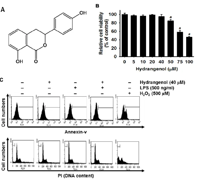
Figure 1. Effects of Hydrangenol on viability of BV2 microglial cells.
...12
Figure 2. Effect of Hydrangenol on LPS-induced NO production and
iNOS expression in BV2 microglial cells.
...14
Figure 3. Effect of Hydrangenol on NF-κB activity in LPS-stimulated
BV2 microglial cells.
...16
Figure 4. NO production by Hydrangenol-induced HO-1 expression in
BV2 microglial cells.
...18
Figure 5. Effect of Hydrangenol-induced Nrf2 in LPS-stimulated
BV2 microglial cells.
...20
Figure 6. Anti-inflammatory effect of Hydrangenol in LPS-stimulated
- 1 -
1. Abstract
We previously demonstrated the anti-inflammatory effects of water extract of Hydrangea macrophylla in lipopolysaccharide (LPS)-stimulated macrophage cells. Here, we investigated whether hydrangenol, one of bioactive component from H. macrophylla, attenuates the expression of nitric oxide (NO) and its regulatory gene, inducible NO synthase (iNOS), in LPS-stimulated BV2 microglial cells. Low dosages of hydrangenol inhibited LPS-stimulated NO release and iNOS expression without any accompanying cytotoxicity. Hydrangenol also suppressed LPS-induced nuclear translocation of the nuclear factor-κB (NF-κB) subunits by inhibiting IκBα phosphorylation and consequently inhibited DNA-binding activity of NF-κB. Additionally, the NF-κB inhibitors, pyrrolidine dithiocarbamate (PDTC) and proteasome inhibitor (PSI), diminish LPS-induced iNOS expression, indicating that hydrangenol-induced NF-κB inhibition might be a key regulator of iNOS expression. Furthermore, our data also showed that hydrangenol suppresses NO production by inducing heme oxygenase-1 (HO-1) and the presence of cobalt protoporphyrin (CoPP), a specific HO-1 inducer, potently suppressed LPS-induced NO. Additionally, hydrangenol promoted nuclear translocation of nuclear factor erythroid 2-related factor 2 (Nrf2) and subsequently increased its binding activity in specific ARE sites. Transient knockdown of Nrf2 remarkably downregulated hydrangenol-induced HO-1 expression, indicating that hydrangenol-induced Nrf2 is an upstream molecule of HO-1. Taken together, these data indicate that hydrangenol attenuates
- 2 -
production of proinflammatory NO and iNOS in LPS-stimulated BV2 microglial cells by inhibiting NF-κB activation and stimulating the Nrf2/HO-1 signal pathway. Therefore, hydrangenol might be a good therapeutics in LPS-mediated inflammatory diseases.
- 3 -
2. Introduction
Microglia normally function to perform host protection in the brain acting as phagocytes to swamp dead cells and tissue debris [1]. In response to injury or infection, microglia become readily activated and secrete proinflammatory mediators such as nitric oxide (NO), cyclooxgenase-2 (COX-2), interleukin-1 beta (IL-1β), and IL-6, which are essential to regulate cellular signal involved in protecting organ disorder such as ischemic damage [2]. Nevertheless, recent studies proved that excessive and abnormal production of these mediators results in systemic inflammatory syndrome, severe tissue damage, atherosclerosis and septic shock [3,4]. NO is one of proinflammatory mediator secreted by microglia, act as effector molecules in the non-specific defense and also act as a signaling molecule to control the inflammatory reaction [5]. NO normally regulates resting blood flow to function neuroprotective and pathophysiological processes [6] however, excessive NO release promotes early blood-brain barrier disruption [7] as well as exerts oxidative injury in microglia, but not astrocytes [8]. In particular, iNOS is a main stimulant for peroxynitrite to
promote protein radical formation during microglia-mediated neurodegenerative disorders [9]. Li et al. [10] potentially highlighted that silencing iNOS expression protects
neurodegeneration of nigrostriatal dopaminergic neurons in the animal model of Parkinson’s disease. Therefore, many researchers have recently attempt to identify phytochemicals to suppress the aberrant expression of NO and iNOS in therapeutic aspects of neurodegenerative
- 4 -
disease.
The Nuclear factor-κB (NF-κB) is known as a main proinflamatory pathway, based on the role of NF-κB in the expression of proinflamatory genes to regulate many proinflammatory mediators, regulatory genes such as iNOS containing NF-κB binding sites [11,12]. In the inflammatory response, IκB is degraded through its phosphorylation and ubiquitination, and consequently free NF-κB is released and translocated to the nucleus to promote proinflammatory genes such as iNOS [13]. Therefore, NF-κB has been thought as a good strategic target for inflammatory responses or diseases. So far, many researchers reported that variety of natural and designed molecules, including proteasome inhibitors, small molecules, active polypeptides, and flavonoids which are targeting NF-κB, suppresses iNOS-mediated inflammatory diseases [12,14]. Camuesco et al reported the treatment of Quercitrin to experimental colitis induced rat’s down regulated the iNOS expression correlated with the inhibition of NF-κB activity [15]. An another report was shown the pioglitazone, a peroxisome proliferator-activated receptor (PPARᵧ) agonist, may offers as good therapeutic strategy for the treatment of neurodegenerative diseases such as Parkinson’s disease because of inhibition of NF-κB activation, iNOS induction and NO-mediated cytotoxicity in Parkinson’s disease model mice [16]. Additionally, recent studies found that heme oxygenase-1 (HO-1) is induced in most tissues by a variety of oxidative stimuli and involved in the protection against different types of oxidant-induced tissue and cellular injury; however, HO-2 is constitutively expressed in tissues [12,13]. Therefore, HO-1 has also been
- 5 -
shown to have important immune-modulatory and anti-inflammatory functions by regulating the pro-inflammatory mediators, NO and iNOS [14]. Nuclear transcription factor erythroid 2-related factor-2 (Nrf2) is a redox-sensitive transcription factor responsible for the induction of anti-oxidant enzymes and recent report showed that HO-1 regulates major immunomodulatory and anti-inflammatory properties via Nrf2 induction [17,18].
Hydrangea macrophylla is currently used as medicinal plant for an oral refrigerant and as a sweetener for diabetic patients [19]. Additionally, H. macrophylla is highlighted because febrifugine and its analogues target the cytoplasmic prolyl-tRNA synthetase of the malaria parasites [20,21]. Our previous study also showed that water extract of processed H. macrophylla leaf possesses anti-inflammatory effect in lipopolysaccharide (LPS)-stimulated RAW264.7 macrophages [22]. In the present study, we investigated the inhibition of NO production and iNOS expression by hydrangenol isolated from H. macrophylla via suppression of NF-κB activity in LPS-stimulated BV2 microglial cells. In addition, antagonistic regulation of hydrangenol is associated with the induction of Nrf2-mediated HO-1 induction.
- 6 -
3. Materials and methods
3.1. Reagents and antibodies
Rabbit anti-mouse antibodies against iNOS, p65, p50, C-23, HO-1, and Nrf2 were purchased from Santa Cruz Biotechnology (Santa Cruz, CA). The antibody against β-actin, LPS, and 3-(4,5-dimethylthiazol-2-yl)-2,5-diphenyl-tetrazolium bromide (MTT) were obtained from Sigma (St. Louis, MO). Peroxidase-labeled goat anti-rabbit immunoglobulin was purchased from KOMA Biotechnology (Seoul, Republic of Korea). Pyrrolidine dithiocarbamate (PDTC) and proteasome inhibitor (PSI) were purchased from Calbiochem (San Diego, CA). Cobalt protoporphyrin (CoPP) was purchased from Tocris Bioscience (Bristol, UK). Dulbecco's Modified Eagle's medium (DMEM), fetal bovine serum (FBS), and antibiotic mixtures were obtained from WelGENE Inc. (Daegu, Republic of Korea). Other chemicals were purchased as Sigma grades. hydrangenol was isolated in our previous study [22] and structure of hydrangenol is illustrated in Fig. 1A.
3.2. Cell culture and viability
BV2 microglial cells were cultured in DMEM medium containing antibiotic mixtures in the 5% FBS and incubated at 37°C and 5% CO2 conditions. MTT assays were performed to
determine relative cell viability. Briefly, BV2 microglial cells (1 × 10⁵ cells/ml) were treated with various concentrations of hydrangenol 2 h before treatment with LPS (500 ng/ml). After
- 7 -
24 h incubation, the cells were incubated with MTT solution (0.5 mg/ml) for 15 min at 37°C. Supernatant was removed and the formation of formazan was observed by monitoring the signal at 540 nm using a microplate reader (Thermo Electron Corporation, Marietta, OH).
3.3. Flow cytometric analysis
BV2 microglial cells were pretreated with hydrangenol for 2 h and then administered with 500 ng/ml of LPS for 24 h. After harvesting, the cells were washed two times with phosphate buffer saline (PBS). The cells were fixed with 1 U/ml RNase A (DNase free) and 10 μg/ml of propidium iodide (PI, Sigma) for 1 h at room temperature in the dark. For annexin V staining, live cells were incubated with annexin V (R&D systems, Minneapolis, MN) according manufacturer's instructions. A FACSCalibur flow cytometer (Becton Dickenson, San Jose, CA) was used to analyze the level of apoptotic cells containing sub-G1 DNA content and
annexin V+ population.
3.4. NO production
BV2 microglial cells (1 × 10⁵ cells/ml) were dispensed on to 24 well plates and pretreated with the indicated various concentrations of hydrangenol 2 h prior to stimulation with 500 ng/ml of LPS for 24 h. Supernatants were collected and assayed for NO production by Griess reaction. Briefly, the samples were mixed with equal volume of Griess reagent (1% sulfanilamide in 5% acetic acid and 0.1% naphthylethylenediamine dihydrochloride) and then
- 8 -
incubated at room temperature for 10 min. The absorbance was measured at 540 nm on a microplate reader. Sodium nitrite dilution series were used as a standard to determine the nitrite concentration in the supernatants.
3.5. Isolation of total RNA and RT-PCR
Total RNA was extracted using an easy-BLUE kit (iNtRON Biotechnology, Sungnam, Republic of Korea) according to the manufacturer's instruction. One microgram RNA was reverse-transcribed using moloney murine leukemia virus (MMLV) reverse transcriptase (Promega, Madison, WI). cDNA was amplified by PCR using specific primer, iNOS (forward 5′-CCT CCT CCA CCC TAC CAA GT-3′ and reverse 5′-CAC CCA AAC TGC TTC AGT CA-3′), HO-1 (forward 5′-TCG CCA GAA AGC TGA GTA TAA-3′ and reverse 5′-ATT GCC AGT GCC ACC ACC AAG TTC AAG-3′), and β-actin (forward 5′-TGT GAT GGT GGG AAT GGG TC-3′ and reverse 5′-TTT GAT GTC ACG CAC GAT TT-3′). The following PCR conditions were applied: iNOS and HO-1, 25 cycles of denaturation at 94°C for 30 s, annealing at 59°C for 30 s and extended at 72°C for 30 s; β-actin, 23 cycles of denaturation at 94°C for 30 s, annealing at 57°C for 30 s and extended at 72°C for 30 s.
3.6. Western blot analysis
Total cell extracts were prepared using PRO-PREP protein extraction kit (iNtRON Biotechnology). Cytoplasmic and nuclear extracts were prepared using NE-PER nuclear and
- 9 -
cytosolic extraction reagents (Pierce, Rockford, IL). Briefly, lysates were centrifuged at 14,000 × g and 4°C for 10 min to obtain the supernatants. The supernatants were collected and protein concentrations determined using a Bio-Rad protein assay kit (Bio-Rad, Hercules, CA). The samples were stored at -80°C or immediately used for western blot analysis. The proteins were separated on SDS-polyacrylamide gels and transferred to nitrocellulose membranes (Schleicher & Schuell, Keene, NH). Proteins were detected using an enhanced chemiluminescence detection system (Amersham, Arlington Heights, IL).
3.7. Electrophoretic mobility assay (EMSA)
EMSA was performed with the nuclear extract. Synthetic complementary NF-κB (5′-AGT TGA GGG GAC TTT CCC AGG C-3′) binding oligonucleotides (Santa Cruz Biotechnology) and anti-oxidant response elements (ARE) consensus (5′-TMA NNR TGA YNN NGC RWW WW-3′) were 3′-biotinylated using the biotin 3′-end DNA labeling kit (Pierce) according to the manufacturer′s instructions, and annealed for 30 min at room temperature. Assays were loaded onto native 4% polyacrylamide gels pre-electrophoresed for 1 h in 0.5 × Tris borate/EDTA before being transferred onto a positively charged nylon membrane (HybondTM-N+) in 0.5 × Tris borate/EDTA at 100 V for 30 min. The transferred DNAs were cross-linked to the membrane at 120 mJ/cm2. Horseradish peroxidase-conjugated streptavidin was used according to the manufacturer's instructions to detect the transferred DNA.
- 10 -
3.8. Transient knockdown of Nrf2
Cells were seeded on a 24 well plate at a density of 1 × 10⁵ cells/ml and transfected Nrf2-specific silencing RNA (siRNA, Santa Cruz Biotechnology) for 24 h. For each transfection, 450 μl of growth medium was added to 20 nM siRNA duplex with the transfection reagent G-Fectin (Genolution Pharmaceuticals Inc., Seoul, Republic of Korea) and the entire mixture was added gently to the cells.
3.9. Statistical analysis
The images were visualized with Chemi-Smart 2000 (Vilber Lourmat, Marine, Cedex, France). Images were captured using Chemi-Capt (Vilber Lourmat) and transported into Photoshop. Statistical analyses were conducted using SigmaPlot software (version 12.0). Values were presented as mean ± S.E. of three experiments. Significant differences between the groups were determined by one-way or two-way ANOVA followed by Bonferroni’s test. Statistical significance was regarded at a and b, P < 0.05.
- 11 -
4. Results
4.1. Hydrangenol has no influence on viability of BV2 microglial cells
First, we determined the cytotoxicity of hydrangenol on cell viability in LPS-stimulated BV2 microglial cells. MTT data showed that cell viability was not significantly altered by hydrangenol up to 40 µM. However, treatment with more than 50 µM concentrations of hydrangenol decreased the viability of BV2 microglial cells in a dose-dependent manner (Fig. 1B). These results suggest that concentrations of hydrangenol treatment below 40 µM were non-toxic to BV2 microglial cells. Thus, for further experiments, the cells were treated with hydrangenol in the concentration range of 5-40 µM. According to data of the annexin V staining, the H2O2-treated BV2 microglial cells were composed of approximately 38 ± 5%
annexin V+ apoptotic cell populations; however, a little annexin V+ population was observed in hydrangenol-pretreated BV2 microglial cells, thereby suggesting that hydrangenol has no influence on apoptotic cell death in BV2 microglial cells (Fig. 1C, top). We then analyzed the effects of hydrangenol on cytotoxicity based on the amount of sub-G1 DNA as assessed by
flow cytometry. There was no apoptotic cell death in any of the panels compared to that of the positive H2O2-treated group (Fig. 1C, bottom). Taken together, these data indicate that
- 12 -
Figure 1. Effects of Hydrangenol on viability of BV2 microglia l cells.
(A) Chemical structure of hydrangenol. (B) BV2 microglial cells (1 × 10⁵ cells/ml) were incubated with the indicated concentrations of hydrangenol (5-100 μM) 2 h before treatment with LPS (500 ng/ml) for 24 h. Cell viability was determined by an MTT assay. (C) The percentage of sub-G1 DNA content and annexin V+-cell population are indicated in each
panel. Each value indicates means ± S.E. and is representative of results obtained from three independent experiments. Statistical significance was determined by one-way ANOVA test (a, P < 0.05 vs. untreated control).
- 13 -
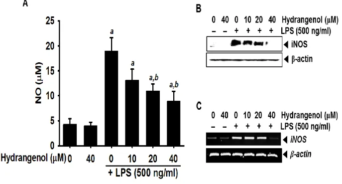
4.2. Hydrangenol inhibits NO production and iNOS expression in LPS-stimulated BV2 microglial cells
In order to analyze the suppressive effect of hydrangenol on NO production, we pretreated with different concentrations of hydrangenol for 2 h, followed by LPS for 24 h, and the levels of NO in culture media were determined by the Griess assay. LPS markedly increased NO production in BV2 microglial cells (18.9 ± 2.7 μM; Fig. 2A). On the other hand, hydrangenol significantly decreased NO production in LPS-stimulated BV2 cells in a concentration dependent manner (13.1 ± 2.2 μM, 10.9 ± 1.5 μM, and 8.8 ± 2.1 μM at 10 μM, 20 μM, and 40 μM of hydrangenol, respectively). In particular, 20 μM and 40 μM of hydrangenol induced statistically significant decreases of LPS-induced NO production; however, the downregulation did not reach to the untreated control (4.2 ± 1.2 μM). Additionally, we investigated whether hydrangenol regulates LPS-stimulated iNOS expression. Western blot analyses showed that treatment with LPS resulted in marked increase of iNOS protein at 24 h, which was significantly suppressed with pretreatment with hydrangenol in a concentration-dependent manner (Fig. 2B). Consistent with the decrease of iNOS protein, hydrangenol also significantly attenuated LPS-induced upregulation of iNOS mRNA expression (Fig. 2C). These results demonstrate that hydrangenol possesses significant inhibitory effects on LPS-induced expression of iNOS, which is considered as a major source of cytotoxic NO.
- 14 -
Figure 2. Effect of Hydrangenol on LPS-induced NO production and iNOS expression in BV2 microglial cells.
BV2 microglia cells (1 × 10⁵ cells/ml) were incubated with the indicated concentrations of hydrangenol 2 h before LPS treatment (500 ng/ml) for 24 h. (A) The amounts of NO were determined using Griess reagent and a standard curve was constructed using NaNO2 in
culture medium. In a parallel experiment, cells (1 × 10⁵ cells/ml) were incubated with indicated concentration of hydrangenol for 2 h before LPS (500 ng/ml) treatment for 24 h (Western blot analyses) and 6 h (RT-PCR). (B) Cell lysates were resolved on SDS-polyacrylamide gels, transferred to nitrocellulose membranes, and probed with antibodies against iNOS. (C) Total RNA was isolated and RT-PCR analysis of iNOS was performed. β-actin was used as an internal control for western blot analyses and RT-PCR, respectively. Each value indicates means ± S.E. and is representative of results obtained from three independent experiments. Statistical significance was determined by two-way ANOVA test (a, P < 0.05 vs. untreated control and b, P < 0.05 vs. LPS-treated group).
- 15 -
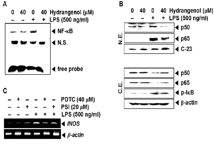
4.3. Hydrangenol inhibits LPS-induced iNOS expression by suppressing NF-κB activation and nuclear translocation in BV2 microglial cells
In order to examine the activity of NF-κB in iNOS expression, we conducted an EMSA and Western blot analyses. EMSA data confirmed that LPS treatment significantly increased the specific DNA-binding activity of NF-κB at 30 min; however, pretreatment with hydrangenol completely suppressed LPS-induced NF-κB activity (Fig. 3A). Additionally, we investigated whether hydrangenol regulates nuclear translocation of NF-κB subunits, p50 and p65 in LPS-treated BV2 microglial cells. Western blot analyses showed that LPS significantly increases total amount of p50 and p65 in the nuclear extracts at 30 min (Fig. 3B, top) and relatively decreases p50 and p65 in the cytosolic extracts (Fig. 3B, bottom), indicating that LPS promotes NF-κB activity by inducing nuclear translocation of NF-κB subunits and degrading IκBα. The current data also displayed that hydrangenol decreases LPS-induced nuclear translocation of p50 and p65 and sustains p50 and p65. Next, we tested the functional effects
of impairing NF-B activity using NF-B inhibitors, PDTC and PSI. Both inhibitors
significantly decreased the expression levels of LPS-induced iNOS mRNA (Fig. 3C),
suggesting that hydrangenol-mediated NF-B regulation is an important factor that mediates
- 16 -
Figure 3. Effect of Hydrangenol on NF-κB activity in LPS-stimulated BV2 microglial cells.
BV2 microglial cells were pre-incubated with the indicated concentration of hydrangenol for 2 h before stimulation with LPS (500 ng/ml) for 30 min. (A) Then the nuclear extracts were assayed for NF-κB activity by electrophoretic mobility shift assay. (B) The nuclear (top) and cytoplasmic (bottom) extracts were prepared to determine the levels of p65 and p50 by Western blot analysis. C-23 and β-actin were used as nuclear and cytosol internal controls for Western blot analysis. (C) In a parallel experiment, BV2 microglial cells (1 × 10⁵ cells/ml) were incubated with pyrrolidine dithiocarbamate (PDTC; 40 μM) and proteasome inhibitor (PSI; 20 μM) 2 h before treatment with LPS (500 ng/ml) for 6 h. Total RNA was isolated and PCR analysis of iNOS was performed. β-actin was used as an internal control for RT-PCR.
- 17 -
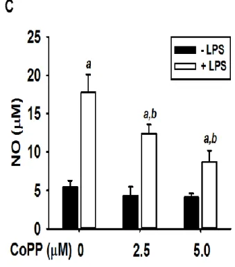
4.4. Hydrangenol decrease NO release through expression of HO-1 in BV2 microglial cells
We examined the effects of hydrangenol on HO-1 expression in BV2 microglial cells and HO-1 activity is associated in hydrangenol-mediated anti-inflammatory response. According to Western blot analyses, hydrangenol induced HO-1 expression in a concentration-dependent manner at 24 h (Fig. 4A). Similar to the expression of HO-1 protein, HO-1 mRNA was significantly expressed by treatment with hydrangenol at 6 h (Fig. 4B). Then, we assessed the level of NO release for the confirmation of the capability of reducing inflammation by induction of HO-1 through the inducer of HO-1, CoPP. Pretreatment with CoPP significantly decreased LPS-induced NO release (17.8 ± 2.3 μM, 12.4 ± 1.2 μM, and 8.7 ± 1.5 μM at 0 μM, 2.5 μM, and 5.0 μM of CoPP, respectively), indicating that one of major role of HO-1 decreases LPS-induced NO release (Fig. 4C). These data indicate that HO-1 reduces LPS-induced inflammatory response by reducing NO release.
- 18 -
Figure 4. NO production by Hydrangenol-induced HO-1 expression in BV2 microglial cells.
(A) BV2 microglial cells (1 × 10⁵ cells/ml) were pretreated with indicated concentration of hydrangenol for 24 h. Equal amounts of cell lysates were resolved on SDS-polyacrylamide gels, transferred to nitrocellulose membranes, and probed with antibodies against HO-1. (B) Total RNA was isolated at 6 h and an RT-PCR analysis of HO-1 was performed. β-actin was used as an internal control for RT-PCR and Western blot analysis. (C) In a parallel experiment, BV2 microglial cells were pretreated with indicated concentration of CoPP for 2 h and then incubated with LPS (500 ng/ml) for 24 h. The amount of NO production in the medium was measured using the Griess reaction. Each value indicates means ± S.E. and is representative of results obtained from three independent experiments. Statistical significance was determined by two-way ANOVA test (a, P < 0.05 vs. untreated control and b, P < 0.05 vs. respective LPS-untreated group).
- 19 -
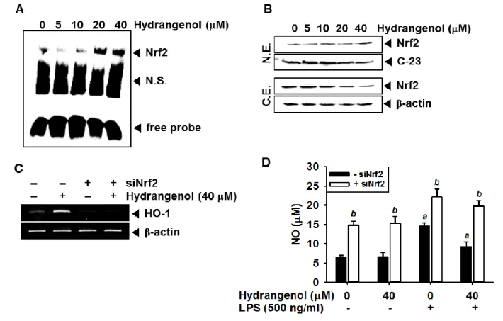
4.5. Hydrangenol-induced Nrf2 is an important regulator for HO-1 induction
Since Nrf2 known as a dominant anti-inflammatory mediator potentially activates the antioxidant stress proteins such as HO-1, we investigated whether hydrangenol regulates Nrf2-mediated HO-1 activation in BV2 microglial cells. According to EMSA data, Nrf2 activity was increased by treating hydrangenol in a concentration dependent manner at 30 min (Fig. 5A). Additionally, Western blot analyses confirmed that hydrangenol decreased the Nrf2 level in cytoplasmic extract and gradually increased in the nuclear extract, indicating that hydrangenol promotes the specific DNA-binding activity of Nrf2 by inducing nuclear translocation of Nrf2 (Fig. 5B). Next, we investigated whether hydrangenol-induced Nrf2 regulates the expression of HO-1 and subsequent production of NO using Nrf2 siRNA (siNrf2). The transient knockdown of Nrf2 significantly decreased hydrangenol-induced HO-1 mRNA expression (Fig. 5C). Finally, we investigated the production of NO in the condition of Nrf2 knockdown. Production of NO in the control (6.5 ± 0.5 μM) was almost similar to that hydrangenol alone treatment (6.6 ± 1.2 μM); however, it was increased more than double in the siNrf2 state (14.8 ± 1.2 μM) and also siNrf2 treatment significantly increased LPS-induced NO (22.1 ± 2.3 μM; Fig. 5D), suggesting that Nrf2 is an upstream regulator of HO-1 expression as hydrangenol-mediated NO inhibition. These data indicate that Nrf2-mediated HO-1 activation is an axis of hydrangenol-mediated anti-inflammatory response in BV2 microglial cells.
- 20 -
Figure 5. Effect of Hydrangenol-induced Nrf2 in LPS-stimulated BV2 microglial cells.
(A) BV2 microglial cells (1 × 10⁵ cells/ml) were incubated with the indicated concentrations of hydrangenol 2 h before LPS stimulation for 30 min. Nuclear extracts were prepared to analyze ARE-binding of Nrf2 by EMSA. (B) In a parallel experiment, equal amounts of nuclear (top) and cytosolic (bottom) lysates were resolved on SDS-polyacrylamide gels, transferred to nitrocellulose membranes, and probed with antibodies against Nrf2. C-23 and β-actin were used as nuclear and cytosol internal controls for Western blot analysis. (C) BV2 microglial cells were transiently transfected with Nrf2 siRNA (siNrf2) and then treated with or without hydrangenol (40 μM) for 24 h. Total RNA was isolated at 6 h and RT-PCR analysis of HO-1 was performed. β-actin was used as an internal control for RT-PCR. (D)
- 21 -
BV2 microglial cells were transiently transfected with siNrf2 for 24 h and then treated the indicated concentration of hydrangenol in the presence or absence of LPS (500 ng/ml). The amount of NO production in the medium was measured using the Griess reaction. Each value indicates means ± S.E. and is representative of results obtained from three independent experiments. Statistical significance was determined by two-way ANOVA test (a, P < 0.05 vs. untreated control and b, P < 0.05 vs. respective siNrf2-untreated group).
- 22 -
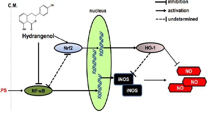
Figure 6. Anti-inflammatory effect of Hydrangenol in LPS-stimulated BV2 microglial cells.
hydrangenol inhibits LPS-induced NF-κB activation by suppressing nuclear translocation of respective subunits, p65 and p50 and simultaneously activates Nrf2 activity to express HO-1. Consequently, hydrangenol-induced HO-1 blocks NO production in LPS-stimulated BV2 microglial cells. LPS, lipopolysaccharide; C.M., cytoplasmic membrane; iNOS, inducible nitric oxide synthase; NO, nitric oxide.
- 23 -
5. Discussion
Since it has known that leaf extract of H. macrophylla possesses anti-malarial, antidiabetes and anti-inflammatory activity [23-26], many scientists have studied to isolate active compounds from H. macrophylla. Beside of medicinal function of H. macrophylla, the processed leaves of H. macrophylla were highlighted to own nutritive and taste values on experimental models of diabetes [24]. Recently, we also showed that water extract of the processed leaves of H. macrophylla inhibits the production of proinflammatory mediators in RAW 264.7 macrophage cells and also possesses two bioactive compounds such as hydrangenol and phyllodulcin [22]. In the current study, we investigated whether one of bioactive compound, hydrangenol isolated from leaves of H. macrophylla has a possibility to use pharmaceutical candidate for inflammatory response in vivo, even though phyllodulcin is still under researching. The present study showed that hydrangenol attenuates NO production and the expression of their respective regulatory gene, iNOS. In addition, we found that hydrangenol regulates iNOS expression and NO release by suppressing NF-κB activity and inducing Nrf2-mediated up regulation of HO-1 expression (Fig. 6).
An accumulated number of studies have shown that NO and iNOS are involved in the pathogenesis of neurodegenerative disorders through the various harmful pathways such as oxidative injury in microglia and early blood-brain barrier disruption [6-8]. Moreover, iNOS plays a pivotal role in producing NO by oxidative deamination and, in contrary, inhibition of
- 24 -
iNOS significantly reverse neuroinflammatory responses in animal models [9,10]. The current data confirm that hydrangenol down regulates NO production and the expression of its respective regulatory gene, iNOS. These data indicate that hydrangenol may be a good therapeutics in LPS-induced inflammatory response. Nevertheless, we need to pay attention to some recent data that NO is also essential for neuroprotection and development of brain [27,28]. In particular, Gulati and Singh [27] certified that pretreatment with a nonselective NOS inhibitor, L-NAME significantly exaggerates neuroprotective effect. Additionally, NO-mediated S-nitrosylation of proteins is prerequisite for neuronal differentiation and maturation [28]. Even though we cannot still make a conclusion about the discrepancy on NO function in neuroinflammation, iNOS inhibition is a main pathway of hydrangenol-induced anti-inflammatory response because LPS-induced iNOS may have neurotoxic effects, but not neuronal NOS [6].
NF-κB is the one of the key transcription factor to regulate proinflammatory genes such as iNOS in neuroinflammatory diseases [6,7]. In particular, even though severe neuroinflammatory processes such as Alzheimer’s disease are complicated to fully understand, widely established theory is that the diseases targets neuronal cell death through activation of NF-κB gene products [29]. Therefore, we have been trying to find new compounds that regulate NF-κB activity [30,31]. In this regard, the current study also showed that hydrangenol isolated from H. macrophylla might be a good therapeutics against neuroinflammatory diseases by suppressing NF-κB activity. Nevertheless, enigmatic problem
- 25 -
still exists concerning the function of NF-κB like a double-edged sword, because the NF-κB signal pathway is indispensable in the process of postnatal neurogenesis in physiological condition [32]. On the contrary, Nrf2 is a main factor of oxidative- or stress-induced endogenous defense system in the cells. Under the stress condition, Nrf2 translocates to the nucleus and binds the specific binding site, ARE to promotes transcription of cytoprotective genes such as HO-1 for acute cerebral insults and neurodegenerative diseases [27]. In particular, genetic deletion and dysfunction of Nrf2 were observed in neurodegenerative diseases whereas up regulated Nrf2 delays onset of neuroinflammation and extends survival in a mouse model by inducing HO-1 [28]. In above respect, our results indicate that hydrangenol increases the expression of Nrf2-mediated HO-1 which responsible for the inhibition of NO in LPS-simulated BV2 microglial cells. Intriguingly, cross-talk between NF-κB and Nrf2 is also characterized that NF-NF-κB subunit, p65, induces ubiquitination-mediated degradation of Nrf2 and consequently diminishes Nrf2 binding to its cognate DNA sequence [29]. Therefore, clinical trials to activate the Nrf2 system are good challenges to cure inflammatory diseases including brain disorder.
In summary, we showed that hydrangenol inhibits NO production in LPS-stimulated BV2 microglial cells through suppression of their regulatory genes. In addition, anti-inflammatory effects of hydrangenol are associated with suppression of NF-κB and enhanced of Nrf2-mediated HO-1 activation. Taken together, we concluded that hydrangenol has potential capacities to be able to become new anti-inflammatory drug.
- 26 -
6. References
[1] Nakagawa Y, Chiba K. Role of microglial m1/m2 polarization in relapse and remission of psychiatric disorders and diseases. Pharmaceuticals 2014;7(12):1028-1048.
[2] Jin R, Yang G, Li G. Inflammatory mechanism in ischemic stroke: role of inflammatory cells. J Leukoc Biol 2010;87(5):779-789.
[3] Noda M, Suzumura A. Sweepers in the CNS: Microglial Migration and Phagocytosis in the Alzheimer Disease Pathogenesis. Int J Alzheimers Dis 2012;2012:891087.
[4] Glodmann T, Prinz M. Role of microglia in CNS autoimmunity. Clin Dev Immunol 2013;2013:208093.
[5] Ghoshal A, Das S, Ghosh S, Mishra MK, Sharma V, Koli P, Sen E, Basu A. Proinflammatory mediators released by activated microglia induces neuronal death in Japanease encephalitis. Glia 2007; 55(5):483-496.
[6] Garry PS, Ezra M, Rowland MJ, Westbrook J, Pattinson KT. The role of the nitric oxide pathway in brain injury and its treatment--from bench to bedside. Exp Neurol 2015;263:235-243.
[7] Jiang Z, Li C, Arrick DM, Yang S, Baluna AE, Sun H. Role of nitric oxide synthases in early blood-brain barrier disruption following transient focal cerebral ischemia. PLoS One 2014;9(3):e93134.
- 27 -
[8] Wang JY, Lee CT, Wang JY. Nitric oxide plays a dual role in the oxidative injury of cultured rat microglia but not astroglia. Neuroscience 2014;281C:164-177.
[9] Kumar A, Chen SH, Kadiiska MB, Hong JS, Zielonka J, Kalyanaraman B, Mason RP. Inducible nitric oxide synthase is a key to peroxynitrite-mediated, LPS-induced protein radical formation in murine microglial BV2 cells. Free Radic Biol Med 2014;73:51-59.
[10] Li M, Dai FR, Du XP, Yang QD, Chen Y. Neuroprotection by silencing iNOS expression in a 6-OHDA model of Parkinson’s disease. J Mol Neurosci 2012;48(1):225-233.
[11] Arias-Salvatierra D, Silbergeld EK, Acosta-Saavedra LC, Calderon-Aranda ES. Role of nitric oxide produced by iNOS through NF-κB pathway in migration of cerebellar granule neurons induced by lipopolysaccharide. Cell Signal 2011; 23(2): 425-435.
[12] de Melo MS, Quintans Jde S, Araújo AA, Duarte MC, Bonjardim LR, Nogueira PC, Moraes VR, de Araújo-Júnior JX, Ribeiro EA, Quintans-Júnior LJ. A systematic review for anti-inflammatory property of clusiaceae family: a preclinical approach. Evid Based Complement Alternat Med 2014;2014:960258.
[13] Verstrepen L, Beyaert R. Receptor proximal kinases in NF-κB signaling as potential therapeutic targets in cancer and inflammation. Biochem Pharmacol 2014;92(4):519-529.
- 28 -
[14] Duarte J, Francisco V, Perez-Vizcaino F. Modulation of nitric oxide by flavonoids. Food Funct 2014;5(8):1653-1668.
[15] Camuesco, D., Comalada, M., Rodríguez-Cabezas, M.E., Nieto, A., D.Laorente, M., Concha, A., et al. The intestinal anti-inflammatory effect of quercitrin is associated with an inhibition in iNOS expression. British Journal of Pharmacology 2004; 143: 908-918.
[16] Dehmer T, T.Heneka M, Sastre M, Dichgans J, B.Schulz J. Protection by pioglitazone in the MPTP model of Parkinson’s disease correlates with IκBα induction and block of NFκB and iNOS activation. Journal of Neurochemistry 2003; 88: 494-501.
[17] Agarwal A, Bolisetty S. Adaptive responses to tissue injury: role of heme oxygenase-1. Trans Am Clin Climatol Assoc 2013;124:111-122.
[18] Wu ML, Ho YC, Lin CY, Yet SF. Heme oxygenase-1 in inflammation and cardiovascular disease. Am J Cardiovasc Dis 2011;1(2):150-158.
[19] Liu J, Nakamura S, Zhuang Y, Yoshikawa M, Hussein GM, Matsuo K, Matsuda H. Medicinal flowers. XXXX . Structures of dihydroisocoumarin glycosides and inhibitory effects on aldose reducatase from the flowers of Hydrangea macrophylla var. thunbergii. Chem Pharm Bull 2013;61(6):655-661.
- 29 -
[20] Jain V, Yogavel M, Oshima Y, Kikuchi H, Touguet B, Hakimi MA, Sharma A. Structure of Prolyl-tRNA Synthetase-Halofuginone Complex Provides Basis for Development of Drugs against Malaria and Toxoplasmosis. Structure 2015;23(5):819-829.
[21] Keller TL, Zocco D, Sundrud MS, Hendrick M, Edenius M, Yum J, Kim YJ, Lee HK, Cortese JF, Wirth DF, Dignam JD, Rao A, Yeo CY, Mazitschek R, Whitman M. Halofuginone and other febrifugine derivatives inhibit prolyl-tRNA synthetase. Nat Chem Biol 2012;8(3):311-317.
[22] Dilshara MG, Jayasooriya RG, Lee S, Jeong JB, Seo YT, Choi YH, Jeong JW, Jang YP, Jeong YK, Kim GY. Water extract of processed Hydrangea macrophylla (Thunb.) Ser. leaf attenuates the expression of pro-inflammatory mediators by suppressing Akt-mediated NF-κB activation. Environ Toxicol Pharmacol 2013;35(2):311-319.
[23] Kamei K, Matsuoka H, Furuhata SI, Fujisaki RI, Kawakami T, Mogi S, Yoshihara H, Aoki N, Ishii A, Shibuya T. Anti-malarial activity of leaf-extract of Hydrangea macrophylla, a common Japanese plant. Acta Med Okayama 2000;54(5):227-232.
[24] Nakamura S, Matsuda H, Yoshikawa M. Search for antidiabetic constituents of medicinal food. Yakugaku Zasshi 2011;131(6):909-915.
[25] Ishih A, Miyase T, Terada M. Comparison of antimalarial activity of the alkaloidal fraction of Hydrangea macrophylla var. Otaksa leaves with the hot water extract in ICR mice infected with Plasmodium yoelii 17 XL. Phytotherapy Research 2003; 17: 633-639.
- 30 -
[26] Sagar A, Joshi M, Srivastava B. Study on antifungal activity of Hydrangea macrophylla (Thunb.) Ser and Allium cepa Linn. Against pathogenic fungi. Plant Archives 2011; 11: 37-41.
[27] Gulati P, Singh N. Pharmacological evidence for connection of nitric oxide-mediated pathways in neuroprotective mechanism of ischemic postconditioning in mice. J Pharm Bioallied Sci 2014;6(4):233-240.
[28] Okamoto SI, Lipton SA. S-Nitrosylation in neurogenesis and neuronal development. Biochim Biophys Acta 2015;1850(8):1588-1593.
[29] Zhou W, Hu W. Anti-neuroinflammatory agents for the treatment of Alzheimer’s disease. Future Med Chem 2013;5(13):1559-1571.
[30] Dilshara MG, Lee KT, Lee CM, Choi YH, Lee HJ, Choi IW, Kim GY. New compound, 5-O-isoferuloyl-2-deoxy-D-ribono-γ-lacton from Clematis mandshurica: Anti-inflammatory effects in lipopolysaccharide-stimulated BV2 microglial cells. Int Immunopharmacol 2015;24(1):14-23.
[31] Dilshara MG, Lee KT, Choi YH, Moon DO, Lee HJ, Yun SG, Kim GY. Potential chemoprevention of LPS-stimulated nitric oxide and prostaglandin E2 production by
α-L-rhamnopyranosyl-(1→6)-β-D-glucopyranosyl-3-indolecarbonate in BV2 microglial cells through suppression of the ROS/PI3K/Akt/NF-κB pathway. Neurochem Int 2014;67:39-45.
- 31 -
[32] Bortolotto V, Cuccurazzu B, Canonico PL, Grilli M. NF-κB mediated regulation of adult hippocampal neurogenesis: relevance to mood disorders and antidepressant activity. Biomed Res Int 2014;2014:612798.
[33] Jazwa A, Cuadrado A. Targeting heme oxygenase-1 for neuroprotection and neuroinflammation in neurodegenerative disease. Curr Drug Targets 2010;11(12):1517-1531.
[34] Vargas MR, Johnson DA, Sirkis DW, Messing A, Johnson JA. Nrf2 activation in astrocytes protects against neurodegeneration in mouse models of familial amyotrophic lateral sclerosis. J Neurosci 2008;28(50):13574-13581.
[35] Yu M, Li H, Liu Q, Liu F, Tang L, Li C, Yuan Y, Zhan Y, Xu W, Li W, Chen H, Ge C, Wang J, Yang X. Nuclear factor p65 interacts with Keap1 to repress the Nrf2-ARE pathway. Cell Signal 2011;23(5):883-892.