Cho
Dong-Ho Shin, Hyo-Eun Park, Sang Il Choi, Kay-Hyun Park, Jung Yun Choi and Goo-Yeong
Atrial Septal Defect With Total Anomalous Pulmonary Venous Return in an Adult
Print ISSN: 0009-7322. Online ISSN: 1524-4539
Copyright © 2011 American Heart Association, Inc. All rights reserved.
is published by the American Heart Association, 7272 Greenville Avenue, Dallas, TX 75231
Circulation
doi: 10.1161/CIRCULATIONAHA.110.986752
2011;123:e612-e613
Circulation.
http://circ.ahajournals.org/content/123/21/e612
World Wide Web at:
The online version of this article, along with updated information and services, is located on the
http://circ.ahajournals.org/content/suppl/2011/05/26/123.21.e612.DC1.html
Data Supplement (unedited) at:
http://circ.ahajournals.org//subscriptions/
is online at:
Circulation
Information about subscribing to Subscriptions:
http://www.lww.com/reprints
Information about reprints can be found online at: Reprints:
document. Permissions and Rights Question and Answer
this process is available in the
click Request Permissions in the middle column of the Web page under Services. Further information about Office. Once the online version of the published article for which permission is being requested is located,
can be obtained via RightsLink, a service of the Copyright Clearance Center, not the Editorial
Circulation
in
Requests for permissions to reproduce figures, tables, or portions of articles originally published Permissions:
at CONS KESLI on October 14, 2013
http://circ.ahajournals.org/
Downloaded from http://circ.ahajournals.org/ at CONS KESLI on October 14, 2013 Downloaded from http://circ.ahajournals.org/ at CONS KESLI on October 14, 2013 Downloaded from
Images in Cardiovascular Medicine
Atrial Septal Defect With Total Anomalous Pulmonary
Venous Return in an Adult
Dong-Ho Shin, MD; Hyo-Eun Park, MD; Sang Il Choi, MD, PhD; Kay-Hyun Park, MD, PhD;
Jung Yun Choi, MD, PhD; Goo-Yeong Cho, MD, PhD
A
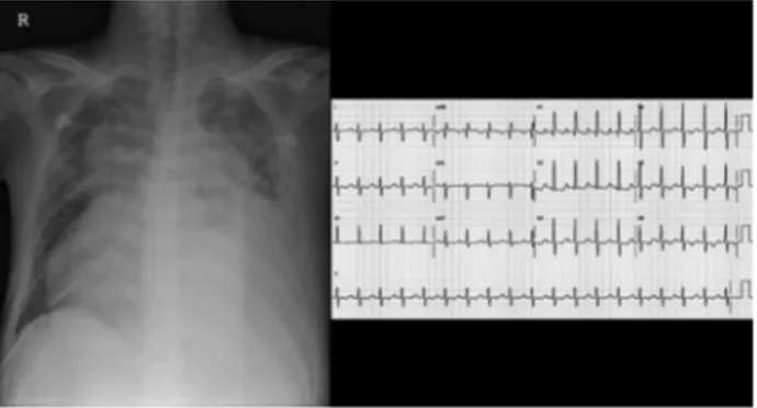
37-year-old man presented at the emergency depart-ment with recent-onset dyspnea (New York Heart As-sociation functional class IV) and abdominal distention. He had no prior history of exercise intolerance, shortness of breath, cyanosis, or palpitation. In the emergency department, tachycardia and engorged jugular veins were observed, and breath sounds were decreased on the left lower lung field, with no crackles. There was no cardiac murmur. The liver was palpable in the distended abdomen.Chest radiography demonstrated marked cardiomegaly with a snowman appearance of increased pulmonary vascular markings (Figure 1, left). The ECG showed sinus tachycardia with evidence of right ventricular hypertrophy (Figure 1, right). Laboratory tests revealed hypoxia (PaO255.9 mm Hg,
Sao289.1%) and elevated levels of N-terminal prohormone of
B-type natriuretic peptide (15 125 pg/mL). A computed tomographic angiogram confirmed type I total anomalous pulmonary venous return (supracardiac type; Figure 2, left), with massive thromboembolism of the left pulmonary arteries (Figure 2, right). A transthoracic echocardiogram demon-strated a large atrial septal defect, a dilated and hypertrophied right ventricle compressing the left ventricle with biventric-ular dysfunction (ejection fraction 34%), loss of drainage of pulmonary venous flow into the left atrium, and large amounts of pericardial effusion (Figure 3 and online-only Data Supplement Movies I through IV). Pulmonary hyper-tension was suggested from the tricuspid regurgitation flow and inferior vena cava plethora (pulmonary arterial systolic pressure was estimated at 60 mm Hg with the modified Bernoulli equation). The pulmonary veins were traced and
were found to have a connection into the superior vena cava through the vertical and innominate veins (online-only Data Supplement Movies II and III).
After pericardiocentesis for symptom relief, a corrective operation was performed, with subsequent procedures of thromboendarterectomy, direct anastomosis of left atrium– pulmonary venous trunk, atrial septal defect closure, and ligation of the vertical vein. The patient was discharged 2 weeks after the operation and has been followed up for 1 year without any discomfort (online-only Data Supplement Mov-ies V and VI).
Disclosures
None.Figure 1. Left, Chest radiograph revealed marked cardiomegaly
with snowman appearance of increased pulmonary vascular markings. Right, ECG indicating sinus tachycardia with evi-dence of right ventricular hypertrophy.
From the Department of Cardiology, Yonsei University College of Medicine, Seoul, Korea (D.-H.S.); and the Division of Cardiology, Department of Internal Medicine (H.-E.P., G.-Y.C.), Division of Radiology (S.I.C.), Department of Thoracic and Cardiovascular Surgery (K.-H.P.), and Department of Pediatrics (J.Y.C.), Cardiovascular Center, Seoul National University Bundang Hospital, Seoul National University, Seoul, Korea.
The online-only Data Supplement is available with this article at http://circ.ahajournals.org/cgi/content/full/123/21/e612/DC1.
Correspondence to Goo-Yeong Cho, MD, PhD, 166 Gumi-ro, Bundang-gu, Seongnam-si, Gyeonggi-do, 463-707, Seoul National University Bundang Hospital, Seoul National University, Seoul, Republic of Korea. E-mail [email protected]
(Circulation. 2011;123:e612-e613.)
© 2011 American Heart Association, Inc.
Circulation is available at http://circ.ahajournals.org DOI: 10.1161/CIRCULATIONAHA.110.986752
Figure 2. CT angiogram. Left, The
verti-cal vein (VV) drained into the superior vena cava (SVC) via the innominate vein (IV). Right, A large amount of pericardial effusion (PE) and massive thromboem-bolism (arrow) of left pulmonary arteries (PA) was seen.
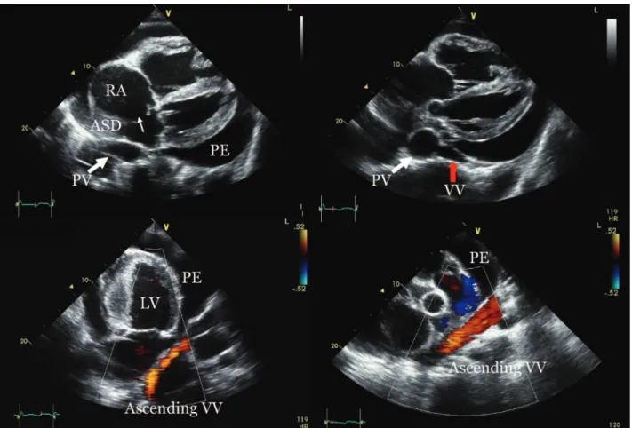
Figure 3. Transthoracic echocardiography revealed a dilated right ventricle and right atrium (RA) and a large amount of pericardial
effu-sion (PE), along with an atrial septal defect (ASD). The pulmonary veins (PV, white arrow) drained into the vertical vein (VV, red arrow), not into the left atrium. LV indicates left ventricle.