서 론
Duchenne 및 Becker 근이영양증(DMD/BMD)은 진행
성 근력저하를 보이고 성염색체 열성으로 유전되는 근
육 질환이다. 1987-1988년
1,2에 근막에 존재하는 디스트
로핀(dystrophin) 유전자와 디스트로핀단백이 발견되어
이 유전자 결손으로 디스트로핀단백이 완전 또는 부분 손
실되어 근섬유의 괴사 및 재생을 보이면서 근력저하가 나
타나는 근육병으로 밝혀져 DMD/BMD를
dystrophino-pathy 또는 dystrophin-deficient muscular dystrophy라
고 한다.
3이러한 dystrophinopathy의 임상 표현형은 대
부분 DMD/BMD로 발현되나 이 외에도 DMD/BMD의
중간형, 그리고 낮은 발생률이지만 증상이 발현된 여
자 유전자 보유자, X 연관 심근병증(X-linked
cardio-myopathy) 등으로 발현되기도 한다. 발병률은 DMD의
경우 출생한 남아 약 3300명 당 1명 정도로 매우 높아 유
전성 근육질환의 약 90%를 차지한다.
4디스트로핀 유전
자의 변이는 결손(deletion)이 가장 흔하여 전체 환자의
55-65%에 달한다.
5,6결손 부위가 mRNA의 translational
reading frame을 이동시킬 경우(out of-frame deletion)
Received March 19, 2004 Accepted May 28, 2004*Address for correspondence Young-Chul Choi, M.D., Ph.D. Department of Neurology, Yondong Severance Hospital 146-92 Dogok-dong, Gangnam-gu, Seoul, 135-720, Korea Tel:+82-2-3497-3323 Fax:+82-2-3462-5904
E-mail:[email protected]
Dystrophinopathy 환자의 임상적, 면역조직화학적 및 유전학적 분석
연세대학교 의과대학 신경과학교실, 재활의학과학교실*, 병리학교실†나상준 강성웅* 김원주 김태승
†최영철
Clinical, Immunohistochemical, and Genetic Analysis in Dystrophinopathy
Sang-Jun Na, M.D., Seong-Woong Kang, M.D.*, Won-Joo Kim, M.D.,
Tai-Seung Kim, M.D.
†, Young-Chul Choi, M.D., Ph.D.
Departments of Neurology, Rehabilitation Medicine*, Pathology†, Yonsei University College of Medicine, Seoul, Korea
Background: Dystrophin deficient muscular dystrophies (dystrophinopathies) are the most common form of muscular
dystrophy with variable clinical phenotypes from the severe Duchenne to the milder Becker forms (DMD/BMD). Dystrophinopathies are X-linked recessive diseases caused by the mutation of the dystrophin gene. Western blot and immunohistochemical staining for dystrophin, and exon deletion analysis by multiplex polymerase chain reaction (PCR) are important diagnostic tools. We investigated the relationship between the clinical characteristics, immunohistochemistry for dystrophin, and the pattern of exon deletions in patients with dystrophinopathy. Methods: We reviewed the clinical and laboratory findings of 35 male patients diagnosed as DMD/BMD. Genomic DNA of the 35 patient was analyzed by multiplex PCR using 19 primer sets of dystrophin gene. Immunohistochemistry for dystrophin of muscle biopsy tissue was performed in all cases. Results: The mean age of symptom onset in 35 patients was 4.6±2.7 years [range, 2-15 years]. Twenty-four of 35 (68.6%) patients showed complete loss (C-, Rod-, N terminal), and 11 of 35 (31.4%) patient showed incomplete loss of dystrophin in immunohistochemistry. Of the 35 patients, 20 had deletions (57%) by multiplex PCR analysis. Sixteen of 20 patients (80%) had exon deletions between exon 45 and 52. Conclusions: Immunohistochemistry of biopsied muscle specimen is an important diagnostic method for expression and localization of dystrophin. The exon deletion analysis by multiplex PCR using peripheral blood is also a simple and useful test for the diagnosis of dystrophinopathies, although it has limited sensitivity.
J Korean Neurol Assoc 22(5):508~515, 2004
전사가 조기에 종식하게 되며 디스트로핀 단백이 형성
되지 않아 증상이 심한 DMD형이 발현되게 된다.
7그러
나 결손이 있더라도 reading frame이 보존될 경우
(in-frame deletion)에는 분자량이 작거나, 비정상적인
단백질이 생성되어 DMD에 비해 증상이 경미한 BMD
형이 발현하게 된다. 과거에는 근육생검에 의한 병리
학적 소견과 임상적인 소견에 의해 진단할 수밖에 없었
으나 최근에는 디스트로핀 단백에 대한 면역조직화학
염색, western blot 및 유전자 검사(multiplex
poly-merase chain reaction) 등에 의해서 확진할 수 있게 되
었다. 이에 본 연구는 dystrophinopathy로 진단된 환자
에서 면역조직화학 염색에 의한 디스트로핀단백 발현
및 디스트로핀 유전자 결손을 분석하여 임상적 특징과
의 연관 관계를 연구하고자 하였다.
대상과 방법
2000년 1월부터 2002년 12월까지 영동세브란스병원에
서 ENMC (Europian neuromuscular center)
8에서 제시
한 DMD/BMD의 임상 기준을 만족하는 환자로서 근육
생검 소견상 근이영양증의 소견을 보이고 면역조직화학
염색에서 디스트로핀 염색의 이상 소견을 보였던 35명
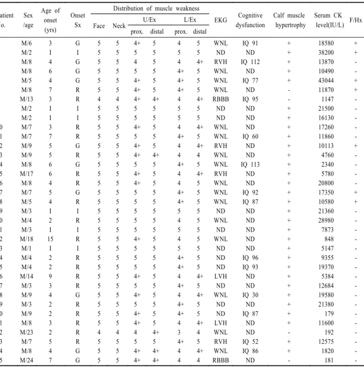
Table 1. Clinical characteristics of 35 patients with dystrophinopathy
Patient No. Sex /age Age of onset (yrs) Onset Sx
Distribution of muscle weakness
EKG Cognitive dysfunction Calf muscle hypertrophy Serum CK level(IU/L) F/Hx
Face Neck U/Ex L/Ex
prox. distal prox. distal 1 2 3 4 5 6 7 8 9 10 11 12 13 14 15 16 17 18 19 20 21 22 23 24 25 26 27 28 29 30 31 32 33 34 35 M/6 M/2 M/8 M/8 M/5 M/8 M/13 M/2 M/2 M/7 M/7 M/9 M/9 M/8 M/17 M/8 M/7 M/5 M/3 M/4 M/3 M/18 M/1 M/4 M/4 M/14 M/3 M/9 M/3 M/9 M/8 M/23 M/7 M/8 M/24 3 I 4 6 4 7 3 I I 3 7 5 5 6 6 4 5 4 I 2 I 15 I 2 2 9 3 4 2 2 3 2 5 4 7 G I G G G R R I I R R G R G R R G R I R I R I R R R R G R R R R R G G 5 5 5 5 5 5 4 5 5 5 5 5 5 5 5 5 5 5 5 5 5 5 5 5 5 5 5 5 5 5 5 4 5 5 5 5 5 5 5 5 5 4 5 5 5 5 5 5 5 5 5 5 5 5 5 5 5 5 5 5 5 5 5 5 5 5 4 5 5 5 4+ 5 4 5 4+ 4+ 4+ 5 5 4+ 5 4+ 4+ 5 4+ 4+ 5 5 5 5 5 4+ 5 5 5 4+ 5 4+ 5 4+ 4+ 4 5 4+ 4+ 5 5 5 5 5 5 4+ 5 5 5 5 5 4+ 5 5 5 5 5 5 5 5 5 5 5 5 5 5 5 5 5 5 4+ 5 4+ 4+ 4 5 4 4+ 4+ 4+ 4 5 5 4 4+ 4 4 4+ 4 4 4+ 4+ 5 4 5 4 5 4+ 4+ 4 4+ 4 4+ 4+ 4 3 4+ 4 4 5 5 4+ 5 5 5 4+ 5 5 4+ 5 4+ 4 5 4+ 5 5 5 5 5 5 5 5 5 5 4+ 5 4+ 5 5 4+ 4 5 4+ 4 WNL ND RVH WNL WNL WNL RBBB ND ND WNL WNL RVH WNL WNL RVH WNL WNL WNL ND WNL ND WNL ND ND ND LVH ND WNL ND ND LVH WNL RVH WNL RBBB IQ 91 ND IQ 112 ND IQ 77 ND IQ 95 ND ND ND IQ 60 ND ND IQ 113 ND ND IQ 92 IQ 87 ND ND ND ND ND IQ 96 IQ 93 ND ND IQ 30 ND IQ 87 ND ND IQ 52 IQ 86 ND + + + + + -+ + + + + + + + + + + + + + + + + + + + + + + + -+ + -18580 38200 13870 10490 43044 11870 1147 21500 16130 17260 11860 10113 4760 2340 5780 20800 17350 10580 21360 28980 7873 848 5147 9355 19370 5384 12684 19580 21380 179 11600 192 12575 1820 181 + + -+ + -+ -+ + -R; difficulty in rising from the floor, G; waddling gait or frequent fall, ND; not done, WNL; within normal limit, I; incidental, U/Ex; upper extremities, L/Ex; lower extremities, prox.; proximal
을 대상으로 하였고, 이들 모두에서 디스트로핀 유전자
(multiplex polymerase chain reaction)검사를 하였다.
발병 시 연령, 내원 시 주 증상, 가족력, 비복근의 가성비대
유무, 심전도, 혈청 creatine kinase (CK) 수치, 인지기능
등을 검사하였다. 환자의 인지기능에 대한 평가는 한국교
육개발원 개인지능검사(Korean Educational
Develop-mental Institute Wechsler Intelligence Scale for
Children; KEDI-WISC)를 사용하였고 5세 이상 14세 미
만의 검사 가능한 14명에서 시행하였다. 35명의 환자에
서 삼각근 내측 원위부 또는 일부에서는 비복근에서 근
육생검을 하여 일반적인 조직화학염색 및 디스트로핀
항체(NCL DYS1, DYS2, DYS3, Novocastra, Newcastle
upon Tyne, UK)를 이용하여 면역조직화학 염색을 하
였다.
9또한 35명의 말초혈액을 이용하여 디스트로핀
유전자 분석을 하였다. 환자의 말초혈액에서 페놀추출
(phenol extraction)방법에 의해 genomic DNA를 추출한
후 1990년 Chamberlain 등
10이 개발한 9종의 시발체
(exon 4, 8, 12, 17, 19, 44, 45, 48, 51)와 1990년 Beggs
등
11이 개발한 10종(exon 3, 6, 13, 43, 47, 49, 50, 52, 60,
muscle specific promoter)의 시발체를 합성하여
Multi-plex PCR법으로 결손 부위를 검사하였다.
12결 과
대상 환자 35명의 평균 연령은 7.9세(1-24세)였고 증
상이 처음 발견된 연령은 평균 4.6±2.7세(2-15세)였다.
첫 증상은 일어서기 힘들거나 계단을 오르기 힘든 경우
등의 하지 근력저하를 보이는 경우가 19명(54.3%), 이상
보행 10명(28.6%), 혈액검사에서 간 효소의 상승이 우연
히 발견된 경우가 6명(17.1%)으로 주로 하지의 근력저
하를 보인 경우가 전체의 83% 이상을 차지하였다. 전
체 35명 중 7명(20%)에서 가족력이 있었다. 비복근의
가성비대는 32명(91.4%)에서 있었으며, 혈청 CK는 평
균 12,976(179-43,044, 정상치; 35-232) IU/L였다. 심전도
를 시행한 28명 중 정상은 20명(71.4%), 우심실 비대 4명
(14.3%), 우측 다발 가지 차단(right bundle branch
block) 2명(7.1%), 좌심실 비대 2명(7.1%) 등이었다. 인
지기능 평가를 시행한 14예에서 평균 intelligence quotient
(IQ)는 83.6이었고 평균이하(below average)를 보인 경
우는 전체 14명 중 7명(50%)이고, 69점 이하의 지능저하
가 3명(21.4%)에서 보였다(Table 1).
근육생검을 한 35명에서 근이영양증의 특징적인 소
견인 다양한 근섬유 크기, 근섬유의 괴사 및 재생, 근세
포 내부의 핵, 간질의 섬유화 등이 관찰되었다(Fig. 1,
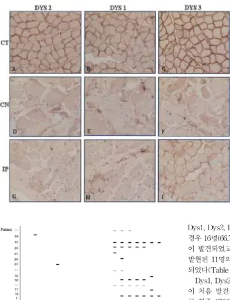
Table 2). 디스트로핀 항체를 이용하여 면역조직화학염
색을 한 35명 중, 24명(68.6%)에서 DYS1, 2, 3 (mid
Rod-, C-, N terminal)에서 모두 디스트로핀이 완전히
발현되지 않았으며(complete negative), 11명(31.4%)에
서 불완전/부분적으로(incomplete/partial) 발현되었다.
11명 중 3명에서 DYS3는 정상 발현되었으나 DYS1,
DYS2는 부분적으로 발현되었다. 3명에서는 DYS3,
DYS1가 부분적으로 발현되었고 DYS2는 완전히 발현되
지 않았다. 2명에서는 DYS3가 부분적으로 발현되었고
DYS1, DYS2는 완전히 발현되지 않았다. 3명에서는
Figure 1. Muscle biopsy findings.
Hematoxylin-eosin (A) and modified Gomori trichrome stain (B) in dystrophinopathy shows fiber size variation, increased internal nuclei, degenerating fibers, and regenerating fibers.
Table 2. Histopathologic findings of muscle biopsy in 35
patients with dystrophinopathies
Muscle fiber Small angulated Atrophic Atrophic 0 (0%) 9/35 (26%) 26/35 (74%) Internal nuclei 29/35 (83%) Degeneration Necrosis Phagocytosis 32/35 (91%) 28/35 (80%) Regeneration fiber 33/35 (94%) Cellular response Inflammatory Fibrosis 7/35 (20%) 30/35 (86%) Architectural change Target fiber Targetoid fiber Moth-eaten fiber Ring fiber 0 0 0 0
Figure 3. The distribution of exon deletions by multiplex PCR
for dystrophin gene in Duchenne muscular dystrophy (DMD) Becker muscular dystrophy (BMD) (black; DMD, gray; BMD).
DYS3, DYS1, DYS2가 모두 부분적으로 발현되었다
(Fig. 2).
Multiplex PCR을 이용한 35명의 유전자 분석 결과는
Fig. 3과 같다. 20명(57%)에서 결손이 발견되었고 이 중
16명(80%)은 다빈도 결손 부위로 알려진 exon 44와 exon
55 사이에서 결손이 발견되었다. Exon 45, exon 47의 결
손이 각각 9명(45%)으로 가장 흔했고 다음으로 흔한 부
위는 exon 48로 8명(40%)에서 발견되었다. 결손이 확인
된 모든 환자에서 exon 6, 12, 44 및 60의 결손은 보이지
않았다.
면역조직화학염색 소견과 multiplex PCR을 이용하여
얻은 exon 결손 결과와의 연관 관계를 분석한 결과
Dys1, Dys2, Dys3이 모두 완전히 발현되지 않은 24명의
경우 16명(66.7%)에서 multiplex PCR을 통한 exon 결손
이 발견되었고 Dys1, Dys2, Dys3이 불완전/부분적으로
발현된 11명의 경우 exon 결손은 4명(36.4%)에서 발견
되었다(Table 3).
Dys1, Dys2, Dys3이 완전히 발현되지 않은 군의 증상
이 처음 발견된 연령은 평균 3.9±1.3세(2-15세)였고 평
균 혈중 CK값은 15,918 (IU/L)이었으며 불완전/부분적
으로 발현된 군의 증상이 처음 발견된 연령은 평균 6.2±
4.2세(2-7세)였고 평균 혈중 CK값은 6,569 (IU/L)였다
(Table 4). Dys1, Dys2, Dys3이 완전히 발현되지 않은
군의 exon 결손율은 24명 중 16명으로 66.7%였고 불완
전/부분적으로 발현된 군에서는 11명 중 4명으로 36.4%
였다. 두 군에서 근육의 근위부 근력저하, 가성비대의 빈
도 등에는 차이가 나지 않았다. 심전도 이상이 있는 환자
는 완전히 발현되지 않은 군에서 17명 중에 5명이었고
불완전/부분적으로 발현된 군에서는 7명 중에 3명이었
다. 인지기능 평가에서 완전히 발현되지 않은 군에서 12
명 중에 3명이 지능저하가 관찰되었고 불완전/부분적으
로 발현된 군에서는 인지기능을 평가한 2명 모두에서 지
능저하는 관찰되지 않았다.
고 찰
디스트로핀 유전자는 약 240만 bp이고 79개의 exon으
로 이루어져 있으며 X 염색체의 약 1%를 차지한다.
6,13이 유전자는 디스트로핀단백을 코드하는데 이 단백은
근세포막을 유지하는 중요한 역할을 하고 있다. 디스
Figure 2. The immunohistochemical staining
of muscle specimen for dystrophin. Normal immunoreactivity against DY1 (B), DY2 (A), and DY3 (C) were noted in normal control. However, complete negative immunoreactivity against DY1 (E), DY2 (D), DY3 (F) and incomplete/partial immunoreactivity against DY1 (H), DY2 (G), DY3 (I) were noted in dystrophinopathy.
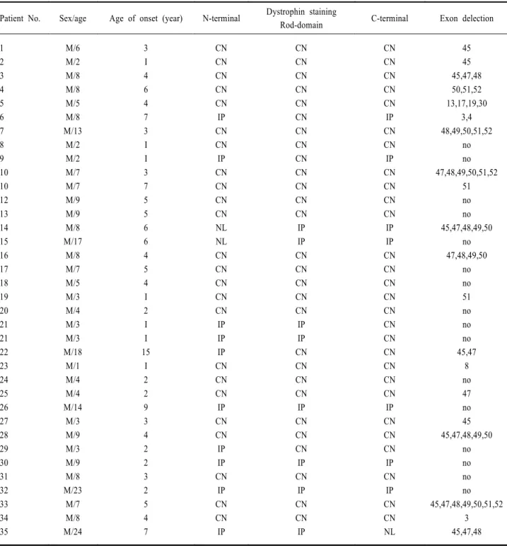
Table 3. Immunohistochemistry and exon deletion patterns in 35 patients with dystrophinopathies
Patient No. Sex/age Age of onset (year) N-terminal Dystrophin staining
Rod-domain C-terminal Exon delection 1 2 3 4 5 6 7 8 9 10 10 12 13 14 15 16 17 18 19 20 21 21 22 23 24 25 26 27 28 29 30 31 32 33 34 35 M/6 M/2 M/8 M/8 M/5 M/8 M/13 M/2 M/2 M/7 M/7 M/9 M/9 M/8 M/17 M/8 M/7 M/5 M/3 M/4 M/3 M/3 M/18 M/1 M/4 M/4 M/14 M/3 M/9 M/3 M/9 M/8 M/23 M/7 M/8 M/24 3 I 4 6 4 7 3 I I 3 7 5 5 6 6 4 5 4 I 2 I I 15 I 2 2 9 3 4 2 2 3 2 5 4 7 CN CN CN CN CN IP CN CN IP CN CN CN CN NL NL CN CN CN CN CN IP IP IP CN CN CN IP CN CN IP IP CN IP CN CN IP CN CN CN CN CN CN CN CN CN CN CN CN CN IP IP CN CN CN CN CN IP IP CN CN CN CN IP CN CN CN IP CN IP CN CN IP CN CN CN CN CN IP CN CN IP CN CN CN CN IP IP CN CN CN CN CN CN CN CN CN CN CN IP CN CN CN IP CN IP CN CN NL 45 45 45,47,48 50,51,52 13,17,19,30 3,4 48,49,50,51,52 no no 47,48,49,50,51,52 51 no no 45,47,48,49,50 no 47,48,49,50 no no 51 no no no 45,47 8 no 47 no 45 45,47,48,49,50 no no no no 45,47,48,49,50,51,52 3 45,47,48 CN; complete negative, IP; incomplete/partial, NL; normal, I; incidental
Table 4. Comparison with clinical characteristics between patients with complete negative and incomplete/partial dystrophin staining
Patient number (%) CN staining, n=24 (68.6%) IP staing, n=11 (31.4%) Total, n=35 (100%) Mean age of onset (range, yrs)
Mean CK level (range, IU/L) Hypertrophy of calf muscle Exon deletion (multiplex PCR)
3.9 (2-15) 15918 (1147-43044) 23/24 (95.8%) 16/24 (66.7%) 6.2 (2-7) 6569 (179-21380) 9/11 (81.8%) 4/11 (36.4%) 4.6 (2-15 ) 12976 (179-43044) 32/35 (91.4%) 20/35 (57.1%) CN; complete negative, IP; incomplete/partial, CK; creatine kinase
트로핀단백은 근육세포 내
횡문근형질막하(subsar-colemma) 부위에 존재하는 400,000달톤 크기의 단백으
로서 3,685개의 아미노산으로 이루어져 있고 아미노 말
단부위(amino terminal domain), 중간 막대 부위(mid
rod domain), 시스틴이 풍부한 부위(cystein-rich
domain) 및 카르복시 말단부위(carboxy terminal domain)
로 구성된다. 이 유전자의 결손 및 변이로 단백질이 소실
되면 근섬유막의 안정성에 장애를 일으켜 근섬유의 괴
사 및 재생을 통해서 혈중 CK가 증가되고, 괴사가 더 진
행하면 근력저하가 생겨 상하지 근위축 및 보행장애가
심해지고, 호흡장애로 사망하게 된다.
14최근에는 디스
트로핀단백의 근육세포막에서 발현 유무 및 정도를
western blot이나 면역조직화학 염색 등을 이용하여 직
접 확인하는 방법과 multiplex PCR을 이용하여 디스트
로핀 유전자 결손을 확인하는 방법이 보편화되어 비교
적 용이하게 진단을 할 수 있게 되었다.
면역조직화학염색을 한 35명 중 24명(68.6%)에서는
DYS1 (중간 막대 도메인 부위), DYS2 (카르복시 말단
부위), DYS3 (아미노 말단부위)에서 모두 디스트로핀이
완전히 발현되지 않았으며 임상적으로는 대부분 DMD
양상을 보였고, 디스트로핀 염색에 불완전하거나 부분적
으로 발현한 11명(31.4%) 중에 임상적으로 BMD인 환자
들이 다수 포함되어 있었다(Table 4).
Multiplex PCR을 이용한 유전자 결손 분석에서 35명
의 DMD/BMD 환자 중 20명(57%)에서 유전자 결손을
보였다. 중국에서는 약 62%,
15이스라엘에서는 약 37%,
16일본에서는 약 40%
17등 대부분의 연구에서는 약 65%의
디스트로핀 유전자 결손율이 보고되고 있다.
6,18,19나머지
점 돌연변이, 미세결손, 중복 등에 의한 약 35%의 경우
정확한 진단을 위해서는 디스트로핀 단백 발현을 직접
확인하는 western blot이나 면역조직화학검사가 반드시
필요하다. 디스트로핀 유전자 결손은 두 곳의 hot spot이
있는데, 5' 끝부분에서 500 kb 이내에 위치한 최초 20개
의 exon부위와 첫번째 exon으로부터 약 1,200 kb 위치
인 exon 44번과 exon 55번 사이에 있다. 이 중에서도
1,200 kb위치인 유전자 중간 부위에서 결손 빈도가 가장
높다고 보고되고 있다.
20본 연구에서 디스트로핀 유전자
결손을 보인 20명 중 16명에서 다빈도 결손 부위로 알려
진 exon 44와 exon 55 사이에서 결손이 발견되었고 이
는 Liu 등의
15결과 및 Baranzini 등의
21보고와 일치한다.
또한 최근 국내에서 80가계의 82명의 환자를 대상으로
19개의 시발체를 사용하여 43명(52%)의 환자에서 유전
자 결손이 확인되었고 exon 45와 exon 52 사이에서 가
장 많은 결손이 있었음을 보고한 것과 유사하나,
229명의
환자를 대상으로 16개의 시발체를 사용하여 8명의 환자
에서 결손을 확인하고 이 중 exon 3의 결손이 6명의 환
자에서 있었음을 보고한 것과는 차이가 있었다.
23아미노 말단 부위 및 카르복시 말단 부위에서의 디스
트로핀 유전자 결손이 중간 막대 부위에서 일어나는 결
손보다 임상적으로 더 심한 표현형을 보인다는 보고가
있었지만
24이러한 법칙에 항상 일치하지 않는다.
25-27또한 동일한 exon 결손을 보이는 환자들에게서도 임상
적으로 그 표현형의 중증도가 다양하게 나타나기 때문
에 dystrophinopathy의 임상 표현형을 결정하는 요인으
로 exon 결손 부위뿐만 아니라 다른 요인이 있을 것으로
추론된다.
26Dystrophinopathy 환자에서 디스트로핀 유전자의 결
손 양상 즉 결손의 부위, 결손의 개수 등과 임상적 소견
의 중증도와는 유의한 상관 관계가 없다는 연구가 있었
고
28,29본 연구에서도 이와 비슷한 경향을 보였다(Table
1, 3).
디스트로핀단백에 대한 면역조직화학염색과
multi-plex PCR에 의한 디스트로핀 유전자 결손 사이의 관계
를 고찰해보면 디스트로핀 염색에서 완전히 발현되지
않은 24명 중 16명(66.7%), 불완전하거나 부분적으로 발
현한 11명 중 4명(36.4%)에서 exon 결손이 확인되었다.
또한 디스트로핀단백에 대한 면역조직화학염색을 했을
때 완전히 발현되지 않은 경우가 불완전/부분적으로 발
현된 경우보다 exon 결손이 빈번한 경향이 있었다. 또한
결손의 위치는 두 경우 모두 hot spot부위인 exon 44와
exon 55 사이에서 대부분의 결손이 일어나는 것으로 확
인되었다.
인지기능 평가를 한 14예에서 IQ가 69 이하인 정신지
체가 3명(21.4%)이었고 이는 약 30%에서 정신지체가 동
반된다는 Proser 등의 보고와 비슷하다.
30정신지체를 보
인 3명 중 2명은 exon 45, 한 명은 exon 51 결손이 있었고
이는 exon 44-55의 결손이 있는 경우 지능저하의 확률이
높다는 Hodgson의 보고와 일치하였다.
31Dystrophino-pathy 환자에서 정신지체의 기전은, 디스트로핀이 배아
기에 신경관에 표현되어 신경형성, 신경세포이주, 세포
분화 등에 관여하고, 성숙 두뇌에서는 신경접합부위의
원위부 막과 관련되어 신경전도의 통합에 참여하는 기
능을 갖는 것과 연관하여 해석되고 있다.
32결론적으로 디스로핀단백이 소실된 경우 DMD, 부분
결손된 경우 BMD로 발현되지만 어떤 경우에는 디스트
로핀단백이 완전 소실되었어도 증상이 경미한 BMD로
표현되기도 하고, 불완전 발현되더라도 DMD으로 표현
되기도 하기 때문에 디스트로핀 면역염색 결과만으로
임상 표현형을 완전하게 구분하기 어렵다. 따라서 디스
트로핀 유전자의 변이가 in-frame인지 out-frame인지가
중요하고, 또한 디스트로핀의 발현량(amount), 이 단백
의 질(quality), 이 단백의 근섬유의 전반적인 분포의 정
도(evenness), 이차적인 utrophin 단백의 발현증가 정도
및 유전자 변이의 exon 부위 등도 중요한 요인으로 작용
할 것이다. 그러므로 dystrophinopathy 진단을 위해 디
스트로핀 단백에 대한 면역조직화학적염색 검사법이 진
단에 중요하지만 임상 표현형과 항상 일치하는 것은 아
니다. 또한 multiplex PCR에 의한 디스트로핀 유전자 결
손율은 약 65% 정도이고 나머지 30-40%는 이 방법으로
확진할 수 없기 때문에 multiplex PCR을 대치할 수 있는
조금 더 정확하고, 용이한 유전자 검사법이 개발되어야
할 것으로 생각된다.
REFERENCES
1. Hoffman EP, Monaco AP, Feener CC, Kunkel LM. Conservation of the Duchenne muscular dystrophy gene in mice and humans. Science 1987;238:347-350.
2. Arahata K, Ishiura S, Ishiguro T, Tsukahara T, Suhara Y, Eguchi C, et al. Immunostaining of skeletal and cardiac muscle surface membrane with antibody against Duchenne muscular dystrophy peptide. Nature 1988;333:861-863. 3. Sadoulet-Puccio HM, Kunkel LM. Dystrophin and its
isoforms. Brain Pathol 1996;6:25-35.
4. Smith SA, Swaiman KF. Muscular dystrophies. In: Swaiman KF, Ashwal S. Pediatric neurology. 3rd ed. St. Louis: Mosby Inc, 1999:1235-1256.
5. Koenig M, Beggs AH, Moyer M, Scherpf S, Heindrich K, Bettecken T, et al. The molecular basis for Duchenne versus Becker muscular dystrophy: correlation of severity with type of deletion. Am J Hum Genet 1989;45:498-506.
6. Den Dunnen JT, Grootscholten PM, Bakker E, Blonden LA, Ginjaar HB, Wapenaar MC, et al. Topography of the Duchenne muscular dystrophy (DMD) gene: FIGE and cDNA analysis of 194 cases reveals 115 deletions and 13 duplications. Am J Hum Genet 1989;45:835-847.
7. Monaco AP, Bertelson CJ, Liechti-Gallati S, Moser H, Kunkel LM. An explanation for the phenotypic differences between patients bearing partial deletions of the DMD locus.
Genomics 1988;2:90-95.
8. Bakker E, Jennekens FG, de Visser M, Wintzen AR. Duchenne and Becker Muscular Dystrophies. In: Emery AE.
Diagnostic Criteria for Neuromuscular Disorders. 2nd ed.
London: Royal Society of Medicine Press,1997;1-4.
9. Bonilla E, Samitt CE, Miranda AF, Hays AP, Salviati G, DiMauro S, et al. Duchenne muscular dystrophy: deficiency of dystrophin at the muscle cell surface. Cell 1988;54:447- 452.
10. Chamberlain JS, Pearlman JA, Muzny DM, Gibbs RA, Ranier JE, Caskey CT. Multiplex PCR for the diagnosis of Duchenne muscular dystrophy. In: Innis MA, Gelfand DH, Sninski JJ, White TJ (eds). PCR protocols: a guide to
methods and applications. 1st ed. New York: Academic
Press. 1990;272-281.
11. Beggs AH, Koenig M, Boyce FM, Kunkel LM. Detection of 98% of DMD/BMD gene deletions by polymerase chain reaction. Hum Genet 1990;86:45-48.
12. Beggs AH, Kunkel LM. Improved diagnosis of Duchenne/ Becker muscular dystrophy. J Clin Invest 1990;85:613-619. 13. Roberts RG, Coffey AJ, Bobrow M, Bentley DR. Exon
structure of the human dystrophin gene. Genomics 1993; 16:536-538.
14. Engel AG, Yamamoto M, Fishbeck KH. Dystrophinopathies. In: Engel AG, Franzini-Armstrong C. Myology. 2nd ed. New
york: McGraw-Hill Inc, 1994:1130-1187.
15. Yuge L, Hui L, Bingdi X. Detection of gene deletions in Chinese patients with Duchenne/Becker muscular dystrophy using CDNA probes and the polymerase chain reaction method. Life Sci 1999;65:863-869.
16. Shomrat R, Gluck E, Legum C, Shiloh Y. Relatively low proportion of dystrophin gene deletions in Israeli Duchenne and Becker muscular dystrophy patients. Am J Med Genet 1994;49:369-373.
17. Kitoh Y, Matsuo M, Nishio H, Takumi T, Nakajima T, Masumura T, et al. Amplification of ten deletion-rich exons of the dystrophin gene by polymerase chain reaction shows deletions in 36 of 90 Japanese families with Duchenne muscular dystrophy. Am J Med Genet 1992;42:453-457. 18. Koenig M, Hoffman EP, Bertelson CJ, Monaco AP, Feener
C, Kunkel LM. Complete cloning of the Duchenne muscular dystrophy (DMD) cDNA and preliminary genomic organization of the DMD gene in normal and affected individuals. Cell 1987;50:509-517.
19. Hodgson SV, Bobrow M. Carrier detection and prenatal diagnosis in Duchenne and Becker muscular dystrophy. Br
Med Bull 1989;45:719-744.
20. Imoto N, Arinami T, Hamano K, Matsumura K, Yamada H, Hamaguchi H, et al. Topographic pattern of the rearrangement of the dystrophin gene in Japanese Duchenne muscular dystrophy. Hum Genet 1993;92:533-536.
21. Baranzini SE, Giliberto F, Herrera M, Bernath V, Barreiro C, Garcia Erro M, et al. Deletion patterns in Argentine patients with Duchenne and Becker muscular dystrophy. Neurol Res 1998;20:409-414.
22. Kang SW, Moon JH , Song KS, Chun SI. Gene Deletion Pattern in Korean Patients with Duchenne or Becker Muscular Dystrophy. J of Korean Acad of Rehab Med 1996;3:583-597.
23. Kang KJ, Han SS, Woo YJ, Kim MH, Choi C. Pattern of Exon Deletions of Dystrophin Gene in Korean Patients with Duchenne Muscular Dystrophy. J of Korean Acad of Rehab Med 2000;24:93-99.
24. Bies RD, Caskey CT, Fenwick R. An intact cysteine-rich domain is required for dystrophin function. J Clin Invest 1992;90:666-672.
25. Hoffman EP, Garcia CA, Chamberlain JS, Angelini C, Lupski JR, Fenwick R. Is the carboxyl-terminus of dystrophin required for membrane association? A novel, severe case of Duchenne muscular dystrophy. Ann Neurol 1991;30:605-610.
26. Gangopadhyay SB, Sherratt TG, Heckmatt JZ, Dubowitz V, Miller G, Shokeir M, et al. Dystrophin in frameshift deletion patients with Becker muscular dystrophy. Am J Hum Genet 1992;51:562-570.
27. Helliwell TR, Ellis JM, Mountford RC, Appleton RE, Morris GE. A truncated dystrophin lacking the C-terminal domains is localized at the muscle membrane. Am J Hum Genet 1992;50:508-514.
28. Beggs AH, Hoffman EP, Snyder JR, Arahata K, Specht L, Shapiro F, et al. Exploring the molecular basis for variability among patients with Becker muscular dystrophy: dystrophin
gene and protein studies. Am J Hum Genet 1991;49:54-67. 29. Arikawa E, Hoffman EP, Kaido M, Nonaka I, Sugita H,
Arahata K. The frequency of patients with dystrophin abnormalities in a limb-girdle patient population. Neurology 1991;41:1491-1496.
30. Prosser EJ, Murphy EG, Thompson MW. Intelligence and the gene Duchenne muscular dystrophy. Arch Dis Child 1969:44:221-230.
31. Hodgson SV, Abbs S, Clark S, Manzur A, Heckmatt JZ, Dubowitz V, et al. Correlation of clinical and deletion data in Duchenne and Becker muscular dystrophy, with special reference to mental ability. Neuromuscul Disord 1992;2:269- 276.
32. Mehler MF. Brain dystrophin, neurogenetics and mental retardation. Brain Res Rev 2000;32:277-307.