Fas-Associated Factor 1 Negatively Regulates the Antiviral Immune
Response by Inhibiting Translocation of Interferon Regulatory Factor
3 to the Nucleus
Soonhwa Song, Jae-Jin Lee, Hee-Jung Kim, Jeong Yoon Lee, Jun Chang, Kong-Joo Lee Graduate School of Pharmaceutical Sciences, College of Pharmacy, Ewha Womans University, Seoul, South Korea
This study is designed to examine the cellular functions of human Fas-associated factor 1 (FAF1) containing multiple
ubiquitin-related domains. Microarray analyses revealed that interferon-stimulated genes ubiquitin-related to the antiviral response are significantly
increased in FAF1-knockdown HeLa cells. Silencing FAF1 enhanced the poly(I·C)- and respiratory syncytial virus (RSV)-induced
production of type I interferons (IFNs), the target genes of interferon regulator factor 3 (IRF3). IRF3 is a key transcription factor
in IFN-
 signaling responsible for the host innate immune response. This study also found that FAF1 and IRF3 physically
associ-ate with IPO5/importin-
3 and that overexpression of FAF1 reduces the interaction between IRF3 and IPO5/importin-3.
These findings suggest that FAF1 negatively regulates IRF3-mediated IFN-
 production and the antiviral innate immune
re-sponse by regulating nuclear translocation of IRF3. We conclude that FAF1 plays a novel role in negatively regulating
virus-in-duced IFN-
 production and the antiviral response by inhibiting the translocation of active, phosphorylated IRF3 from the
cyto-sol to the nucleus.
T
he innate immune system, in contrast to the adaptive immune
response present only in immune cells, is present in all cells
and plays key roles in the host defense against viral infections by
sensing and immediately responding to the invading pathogens
(
1
,
2
). Intracellular pattern recognition receptors (PRRs),
includ-ing Toll-like receptors (TLRs), retinoic acid-inducible gene I
(RIG-I)-like receptors (RLRs), and nucleotide-binding
oligomer-ization domain containing (NOD)-like receptors (NLRs),
recog-nize pathogen-associated molecular patterns (PAMPs) and
acti-vate innate immune signaling pathways, leading to the production
of type I interferons (IFN-
␣/) and other cytokines. Type I IFNs
play a crucial role in limiting viral replication and priming the
adaptive immune response (
3
,
4
). IFN-
 can be produced in most
cell types, and when the cells are infected with a virus, IFN-
expression rapidly increases due to the activation of transcription
factors (
5
). Transcription factor complexes, including interferon
regulatory factor 3 (IRF3), nuclear factor kappa B (NF-B), and
AP1, are bound to the regulatory domains of the IFN-
 promoter
and cooperatively regulate the transcription of IFN-
 (
6
). IFN-

secreted from infected cells binds to type I IFN receptors 1 and 2
(IFNAR1/2) on adjacent cells and then activates the JAK/STAT
signaling pathway, which results in the expression of
interferon-stimulated genes (ISGs). Some ISGs, such as Mx1, OAS1, and
IFIT1, directly interfere with viral replication, while others,
in-cluding RIG-I, MDA5, and IRF7, indirectly do so by enhancing
IFN-
 production (
7
).
The transcription factor IRF3 plays the most critical role in the
regulation of virus-induced IFN- activation. IRF3 is
constitu-tively expressed and localized in the cytoplasm in a latent form.
Single-stranded or double-stranded viral RNAs accumulated
in-side cells after infection are recognized by RLRs and TLR3, which
recruit the adaptor proteins mitochondrial antiviral signaling
protein (MAVS) and TRIF, respectively (
8
,
9
). These adaptor
pro-teins, MAVS and TRIF, recruit the kinases TBK1 and I
B kinase ε
(IKK
ε), which activate IRF3 by phosphorylating the C-terminal
region of IRF3 at seven Ser/Thr residues (Ser385, -386, -396, -398,
-402, and -405 and Thr404). Phosphorylated IRF3 forms dimers
which shuttle into the nucleus, where they interact with the
co-activator CBP/p300 and initiate transcription of target genes,
in-cluding IFN-
 (
10
,
11
). It has been reported that phosphorylation
of IRF3 at Ser386 induces dimerization and interaction with CBP
(
11
) and that phosphorylation at Ser396 occurs in response to
viral infections (
10
). Mutation studies confirmed that
phospho-rylations at Ser386 and Ser396 are important for IRF3 activation
and interaction with CBP (
12
).
The production of IFN- is essential for protecting cells from
virus infection, and aberrant activation of IFN- production can
trigger diseases, such as multiple sclerosis and systemic lupus
ery-thematosus (SLE) (
13
,
14
). Therefore, IFN-
 production needs to
be tightly regulated. Several positive and negative regulators have
been identified. Studies of mechanisms in IRF3 activation as well
as in the negative regulation of transcriptional activity of IRF3 are
still ongoing. The two negative-regulatory mechanisms so far
identified, as already noted, are degradation of IRF3 following its
phosphorylation by the ubiquitin proteasome system and
post-translational modifications of IRF3, which inhibit its activity.
RAUL, a major ubiquitin E3 ligase, ubiquitinates IRF3
regard-less of its phosphorylation status (
15
), while the E3 ubiquitin
Received 29 July 2015 Returned for modification 17 August 2015 Accepted 16 January 2016
Accepted manuscript posted online 25 January 2016
Citation Song S, Lee J-J, Kim H-J, Lee JY, Chang J, Lee K-J. 2016. Fas-associated factor 1 negatively regulates the antiviral immune response by inhibiting translocation of interferon regulatory factor 3 to the nucleus. Mol Cell Biol
36:1136 –1151.doi:10.1128/MCB.00744-15.
Address correspondence to Kong-Joo Lee, [email protected].
Supplemental material for this article may be found athttp://dx.doi.org/10.1128
/MCB.00744-15.
Copyright © 2016 Song et al. This is an open-access article distributed under the
terms of theCreative Commons Attribution 4.0 International license.
on October 31, 2016 by Ewha Womans Univ
http://mcb.asm.org/
ligase RBCK1 and cytoplasmic peptidyl-prolyl-isomerase Pin1
ubiquitinate only phosphorylated IRF3 and trigger its
degrada-tion (
16
,
17
). The second negative-regulation mechanism
reported to change IRF3 activity is posttranslational
modifica-tion of IRF3. Protein phosphatase 2A (PP2A) and
mitogen-activated protein kinase (MAPK) phosphatase 5 (MKP5) are
known to dephosphorylate IRF3 and decrease the IFN response
(
18
,
19
). SUMOylations of IRF3 are another known
mecha-nism to decrease IRF3 activity (
20
).
Thus, phosphorylation is an indispensable step for IRF3
acti-vation, and phosphorylated IRF3 is translocated into the nucleus
to bind the IFN- promoter. However, the mechanism
underly-ing the translocation process remains elusive. Previous studies
demonstrated that IRF3 has an active nuclear localization signal
(NLS) which is recognized by importin-
␣ receptors and
trans-ported to the nucleus (
21
,
22
). IRF3 also has an active nuclear
export signal (NES); it is exported from the nucleus via the
CRM1-mediated pathway to localize mainly in the cytoplasm in
unstimu-lated cells (
23
). Following infection, IRF3 resides in the nucleus
and interacts with CBP (
21
,
24
). This study reports that FAF1 as a
negative regulator of virus triggered the IFN-
 signaling pathway
by inhibiting the nuclear translocation of phosphorylated IRF3.
Fas-associated factor 1 (FAF1) was first identified as a
compo-nent of the apoptosis signaling pathway (
25
). FAF1 is a ubiquitin
receptor containing multiple ubiquitrelated domains that
in-clude a ubiquitin-associated (UBA) domain and three domains
with ubiquitin-like folds, UBL1, UBL2, and ubiquitin-regulatory
X (UBX) (
26
). The N-terminal UBA domain (47 amino acids
long) recruits Lys
48-linkage polyubiquitinated proteins required
for FAF1-mediated apoptosis and the stress response (
26
,
27
). The
UBL1 domain binds to Hsp70 and regulates its chaperone activity
by promoting Hsp70 degradation (
28
,
29
). The C-terminal UBX
domain interacts only with valosin-containing protein (VCP;
AAA ATPase p97) complexed with the Npl4-Ufd1 heterodimer.
This interaction regulates the binding of the polyubiquitinated
proteins via the N-terminal UBA domain. FAF1 promotes the
degradation of the endoplasmic reticulum-associated degradation
(ERAD) substrate in a VCP-Npl4-Ufd1-dependent manner (
26
,
30
). To further understand the cellular functions of FAF1, we
investigated the target genes of FAF1 using microarray analysis.
This microarray result reveals that FAF1 is involved in negative
regulation of a virus-triggered IFN- signaling pathway, in a novel
manner by inhibiting the nuclear translocation of phosphorylated
IRF3 and subsequently IFN-
 production and thereby inhibiting
the cellular antiviral response.
MATERIALS AND METHODS
Reagents and plasmids. The following are the sources of antibodies used
in this study: mouse monoclonal Flag antibody was from Sigma (St. Louis, MO); rabbit anti-IRF3, mouse anti-MAVS, mouse antitubulin, rabbit an-ti-histone deacetylase 1 (anti-HDAC1), and mouse antiactin antibodies were from Santa Cruz Biotechnology (Santa Cruz, CA); rabbit anti-Mx1 and rabbit anti-phospho-IRF3 (Ser386) were from Abcam (Cambridge, United Kingdom); rabbit anti-FAF1 and rabbit anti-glyceraldehyde-3-phosphate dehydrogenase (anti-GAPDH) were from AbFrontier (Seoul, South Korea); rabbit antibodies specific for IRF3, phospho-IRF3 (Ser396), TBK1, TRIF, ISG15, and STAT1 were from Cell Signaling Tech-nology (MA); mouse anti-green fluorescent protein (anti-GFP) antibody was from Life Technologies (CA); mouse anti-RIG-I antibody was from Adipogen AG (CA); mouse antibodies specific for IPO5 and FAF1 were
from Abnova (CA). Cycloheximide (CHX; C7698) and leptomycin B (LMB; L2913) were purchased from Sigma (St. Louis, MO).
pISRE-Luc was provided by Greg Barton (University of California— Berkeley, Berkeley, CA). pIFNB-GL3 and pIFNA4-GL3 were provided by John Hiscott (Vaccine and Gene Therapy Institute of Florida, USA), and pCMV-beta-galactosidase (pCMV-beta-Gal, where CMV is cytomegalo-virus) was from Eunsuk Hwang (Ewha Womans University, South Ko-rea). The expression plasmids for TRIF and GFP-tagged wild-type IRF3 (IRF3-WT-GFP) and IRF3 with five Ser/Thr residues replaced with ASP (IRF3-5D-GFP) were kindly provided by Joo Young Lee (Catholic Uni-versity, South Korea) with kind permission from Katherine A. Fitzgerald (University of Massachusetts Medical School, MA) and John Hiscott (Vaccine and Gene Therapy Institute of Florida, FL). Flag-RIG-I N was from Takashi Fujita (Tokyo, Japan). Flag-MAVS and Flag-TBK1 were from Glen N. Barber (University of Miami Miller School of Medicine, FL). Flag-IKKε was from Ki-Sun Kwon (KRIBB, South Korea).
pFlag-CMV-2-FAF1 WT, pFlag-CMV-pFlag-CMV-2-FAF1(82– 650), pFlag-CMV-pFlag-CMV-2-FAF1⌬UBX
(deletion of amino acids [aa] 569 to 650), pFlag-CMV-2-FAF1⌬UBL1-2 (deletion of aa 100 to 270), pFlag-CMV-2-FAF1⌬UAS (deletion of aa 352 to 487), and pFlag-CMV-2-FAF1(1–201) were prepared as previously de-scribed (26,27). pFlag-CMV-2-FAF1(1–351) was generated by cloning. All plasmid constructs were verified by DNA sequencing.
Cell culture and transfection. HeLa cells were purchased from the
ATCC and cultured in Eagle’s minimal essential medium (EMEM) sup-plemented with 10% fetal bovine serum, 100 units/ml of penicillin G, and 100g/mM streptomycin at 37°C in a 5% CO2-containing humidified incubator. HEK293T cells and Raw264.7 cells were cultured in Dulbecco’s modified Eagle’s medium (DMEM) supplemented under the same con-ditions. HEp-2 cells were cultured in minimal essential medium (MEM) supplemented under the same conditions. For transient overexpression of specific proteins, cells were transfected using LT-1 reagent and analyzed at 24 h or 48 h posttransfection. For gene silencing, FAF1 small interfering RNAs (siRNAs) were obtained from Dharmacon (ON-TARGETplus SMARTpool siRNA; Dharmacon, IL) and Bioneer (Daejeon, South Ko-rea). FAF1 siRNA 1 was from Dharmacon (L-009106-00-0005) and FAF1 siRNA 2 (catalog number 1049605) and a control siRNA were from Bion-eer. siRNA 2 targets the consensus sequence in both human and mouse FAF1 (hFAF1 and mFAF1, respectively) proteins. Cells were transfected with siRNAs using DharmaFECT1 according to the manufacturer’s pro-tocol at a final concentration of 100 nM.
Microarray analysis. HeLa cells were transfected with an FAF1 or
control siRNA and collected at 48 h posttransfection. Total RNA was extracted using TRIzol reagent (Invitrogen Life Technologies, CA) and purified using RNeasy columns (Qiagen, CA) according to the manufac-turer’s protocol. RNA purity and integrity were evaluated by denaturing gel electrophoresis and the ratio of the optical densities at 260 and 280 nm (OD260/280) analyzed with a 2100 Bioanalyzer (Agilent Technologies, CA). Total RNA was amplified and purified using an Ambion Illumina RNA amplification kit (Ambion, CA) according to the manufacturer’s instruc-tions to yield biotinylated complementary RNA (cRNA). Briefly, 550 ng of total RNA was reverse transcribed to cDNA using a T7 oligo(dT) primer. Second-strand cDNA was synthesized, in vitro transcribed, and labeled with biotin-nucleoside triphosphate (NTP). After purification, the cDNA was quantified using an ND-1000 Spectrophotometer (NanoDrop, Wil-mington, DE). A total of 750 ng of labeled cDNA samples was hybridized to each HumanHT-12, version 4, expression bead array for 16 to 18 h at 58°C, according to the manufacturer’s instructions (Illumina, Inc., San Diego, CA). Array signals were detected using Amersham Fluorolink streptavidin-Cy3 (GE Healthcare Bio-Sciences, Little Chalfont, United Kingdom). Arrays were scanned with an Illumina BeadArray Reader con-focal scanner according to the manufacturer’s instructions. The quality of hybridization and overall chip performance were monitored by visual inspection of both internal quality control checks and the raw scanned data. Raw data were extracted using the software provided by the manu-facturer (Illumina GenomeStudio, version 2011.1; Gene Expression
on October 31, 2016 by Ewha Womans Univ
http://mcb.asm.org/
ule, version 1.9.0). Probe signal values were transformed by logarithm and normalized by the quantile method. Statistical significance of the expres-sion data was determined using a local-pooled-error (LPE) test and fold change in which the null hypothesis was that no difference exists among two groups. The false discovery rate (FDR) was controlled by adjusting P values using the Benjamini-Hochberg algorithm.
Luc reporter assay. The cells were transfected with reporter genes and
pCMV-beta-Gal and then, after 24 h, treated with poly(I·C) (10g/ml) by transfection. Transfected cells were harvested, and luciferase (Luc) activ-ity and beta-galactosidase activactiv-ity were measured using a luciferase assay system (Promega, WI) and a Galacto-Light Plus system (Applied Biosys-tems, CA), respectively, on a luminometer (Luminoskan TL plus, Ther-moFisher Scientific, MA). Each experiment was repeated in triplicate, and firefly luciferase activities were normalized to beta-galactosidase activities.
Reverse transcription-quantitative PCR (RT-qPCR). Cells were
har-vested at 48 h posttransfection. Total RNA from these cells was isolated using an RNeasy minikit (Qiagen, CA) and then reverse transcribed using SuperScript II RT (Invitrogen Life Technologies, CA) according to the manufacturer’s protocol. Synthesized cDNA was subjected to real-time PCR (AB7300; Applied Biosystems, CA) for amplification in triplicate. PCRs were performed using SYBR green qPCR master mix (Applied Bio-systems, CA) and the following specific primers: for hIFN- (31), 5=-CAA CAA GTG TCT CCT CCA AAT-3= (sense) and 5=-TCT CCT CAG GGA
TGT CAA AG-3= (antisense); mIFN-, 5=-CAT CAA CTA TAA GCA
CCA-3=(sense) and 5=-TTC AAG TGG AGA GCA CTT GAG-3= (anti-sense); hFAF1, 5=-ATT GGG ACT TAG TGG CAG CT-3= (sense) and 5=-GCA TTA CAG GTC GAA ACG CT-3= (antisense); hIFIT1, 5=-CCT CCT TGG GTT CGT CTA CA-3= (sense) and 5=-GGC TGA TAT CTG GGT GCC TA-3= (antisense); hIFIH1, 5=-TGG TCT CGT CAC CAA TGA AA-3= (sense) and 5=-CTC CTG AAC CAC TGT GAG CA-3= (antisense); hGAPDH, 5=-AAG GTC ATC CCT GAG CTG AA-3= (sense) and 5=-TGC TGT AGC CAA ATT CGT TG-3= (antisense); mGAPDH, 5=-AGA ACA TCA TCC CTG CAT CC-3= (sense) and 5=-CAC ATT GGG GGT AGG AAC AC-3= (antisense). Relative mRNA expression was calculated ac-cording to the comparative threshold cycle (CT) method (⌬⌬CT), and the
GAPDH gene was used as an endogenous control gene.
Native PAGE. HEK293T cells were lysed in buffer containing 50 mM
Tris-HCl, pH 7.4, 150 mM, NaCl, 1 mM EDTA, 1% NP-40, 5 mM Na3VO4, 5 mM NaF, and protease inhibitor cocktail (Sigma, St. Louis, MO). Native gels (Bio-Rad, CA) were prerun with 25 mM Tris-HCl and 192 mM glycine, pH 8.3, with 0.4% deoxycholate (DOC) in the cathode chamber for 30 min at 40 mA on ice before samples were loaded. Samples in native buffer (10g of protein, 62.5 mM Tris-HCl, pH 6.8, 25% glyc-erol, and 1% DOC) were loaded, and native gels were run at 15 mA for 60 min on ice. Gels were soaked in SDS running buffer for 30 min, trans-ferred to polyvinylidene difluoride (PVDF) membranes, and then ana-lyzed by Western blotting.
Immunoprecipitation. Cells were lysed in lysis buffer containing
pro-tease inhibitors [150 mM NaCl, pH 7.4, 50 mM Tris-HCl, 1 mM EDTA, 1 mM phenylmethylsulfonyl fluoride (PMSF), 5g/ml aprotinin, 10 g/ml leupeptin, 10g/ml pepstatin A, 5 mM Na3VO4, 5 mM NaF, 10 mM sodium butyrate, and 1% CHAPS (3-[(3-cholamidopropyl)-dimethyl-ammonio]-1-propanesulfonate)] for 30 min on ice, followed by centrif-ugation at 4,000 rpm for 15 min. The supernatant was incubated with anti-Flag antibody or anti-GFP antibody for 2 h at 4°C, and the lysate-antibody complexes were incubated with protein G-Sepharose 4 Fast Flow beads for another 1 h at 4°C. The precipitated beads were washed six times with lysis buffer to remove nonspecific binding. The immune complex was eluted with gel sample buffer, separated by SDS-PAGE, and analyzed by Western analysis.
Cellular fractionation. HeLa cells (2⫻ 106) were lysed in hypotonic
solution (10 mM HEPES, pH 7.9, 1.5 mM MgCl2, 10 mM KCl, 1 mM
EDTA, 5 mM Na3VO4, 5 mM NaF) containing protease inhibitor cocktail (Sigma, St. Louis, MO) for 30 min at 4°C to swell the cells. Cell lysates were centrifuged at 4,000 rpm for 25 min at 4°C. The pellet was washed and
solubilized with 150l of gel sample buffer and then used as the nuclear fraction. The supernatant was immediately subjected to Western blot analysis as the cytosolic fraction.
Confocal microscopy. Cells were grown on SecureSlip coverslips
(Sigma, St. Louis, MO) and fixed with 4% paraformaldehyde, followed by permeabilization with 0.1% Triton X-100 in Hanks balanced salt solution (HBSS) for 10 min. After cells were washed in HBSS, they were incubated with 3% bovine serum albumin (BSA) in HBSS for 1 h to block nonspe-cific protein adsorption and then incubated with primary antibodies for 2 h at 37°C. After cells were washed three times with HBSS, they were stained for 1 h at 37°C with Alexa Fluor-conjugated secondary antibodies. After samples were washed three times with HBSS, the mounting medium for fluorescence with 4=,6=-diamidino-2-phenylindole (DAPI) was used for staining the nucleus. After being mounted, cells were photographed with a fluorescence confocal microscope (LSM510 META; Zeiss, Ger-many).
Microarray data accession number. The raw and processed
microar-ray data are available in the Gene Expression Omnibus (GEO) database under GEO accession numberGSE71665.
RESULTS
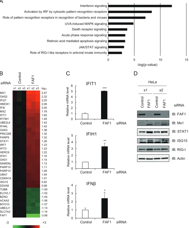
FAF1 is involved in interferon signaling. To investigate the
mo-lecular functions of FAF1, we performed microarray analysis and
compared fold changes in mRNA expression levels between
con-trol and FAF1-knocked-down HeLa cells. A total 150 genes
showed significant changes (fold change of
ⱖ1.5 and P value of
⬍0.05) in FAF1-knocked-down HeLa cells compared to control
levels. Of these, the expression levels of 66 genes increased, and
those of 84 genes decreased in cells in which FAF1 was knocked
down. To examine which cellular pathway was mostly affected by
silencing FAF1, we conducted Ingenuity Pathway Analysis (IPA)
and found that silencing FAF1 significantly raised the expression
levels of genes encoding molecules related to IFN signaling and
IRF activation (
Fig. 1A
). Major upregulated genes in
FAF1-knocked-down cells are ISGs having antiviral activity, such as Mx1
and Mx2 (Mx1/2), OAS1/2/3, IFIT1/2/3, ISG15, ISG20, and IFI6.
Oligoadenylate synthetase (OAS) and Mx genes are the best
stud-ied ISGs in terms of antiviral properties. Unlike OAS genes, Mx
genes are induced exclusively by IFN-
␣/ or IFN-␦ and are not
activated by other cytokines, including interleukin-1 or tumor
necrosis factor alpha (TNF-
␣). Thus, Mx expression has been used
as a specific marker for type I IFN induction in clinical settings (
32
,
33
). As shown in
Fig. 1B
, ISGs increased up to 5.54-fold in
FAF1-knocked-down cells. These results suggest that FAF1 suppresses
the expression of ISGs by interfering with IFN signaling even in
normal cells. To confirm that silencing of FAF1 can induce IFN-
in HeLa cells, we analyzed basal mRNA levels of ISGs and IFN-

using real-time PCR and also measured ISG protein levels using
Western analysis. Results shown in
Fig. 1
demonstrate that
silenc-ing of FAF1 increased the expression levels of the endogenous
IFN-
 and downstream IFIT1 and IFIH1 (
Fig. 1C
) and also
pro-tein levels of ISGs such as Mx1, STAT1, and ISG15 and RIG-I/
DDX58 (
Fig. 1D
). These results suggest that FAF1 inhibits the
expression of IFN- and downstream genes.
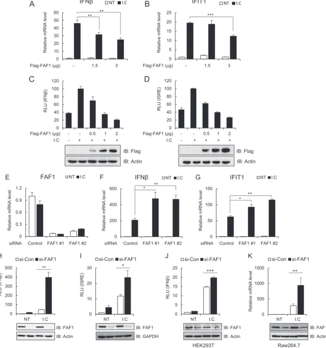
FAF1 negatively regulates poly(I·C)-induced IFN-

activa-tion. IFN-
 is rapidly produced in response to viral infection to
induce a cellular antiviral state (
5
). To investigate whether FAF1 is
involved in virus-triggered IFN- induction, we measured the
mRNA level and the IFN-
 promoter activity after poly(I·C)
transfection in HeLa cells expressing various amounts of FAF1.
Transfection with poly(I·C), a synthetic analogue mimicking
dou-ble-stranded RNA as a stimulant, induces an antiviral response by
on October 31, 2016 by Ewha Womans Univ
http://mcb.asm.org/
0 5 10 15 Role of RIG-I like receptors in antiviral innate immunity
JAK/STAT signaling Retinoic acid mediated apoptosis signaling Acute phase response signaling Death receptor signaling UVA-induced MAPK signaling Role of pattern recognition receptors in recognition of bacteria and viruses Activation by IRF by cytosolic pattern recognition receptors Interferon signaling -log(p-value)
A
IB: FAF1 IB: ActinB
Contro l FAF1 IB: Mx1 IB: STAT1 IB: RIG-I s1 s2 s3 s1 s2 s3 IB: ISG15 -3 Contro l FAF1 HeLa siRNA +3 Difference (log2) s1 s2 Contro l FAF1 siRNAD
C
log2 MX1 2.47 OAS2 2.32 IFI27 2.25 HMOX1 2.08 IFI6 2.00 MX2 1.79 IFIT1 1.60 IFITM1 1.45 UBE2L6 1.42 IFI44L 1.42 OAS3 1.38 PRIC285 1.32 PARP9 1.32 STAT1 1.26 IRF7 1.23 IFIT3 1.23 HERC6 1.22 IFIH1 1.16 OAS1 1.14 SAMD9L 1.12 PARP10 1.11 PARP14 1.09 UBA7 1.04 CDKN1A 1.01 ISG15 0.92 DDX58 0.88 TUBB -1.00 ELOVL1 -1.02 BZW2 -1.04 ACAA2 -1.06 MYO10 -1.07 UBE2J1 -1.13 SLC7A2 -1.14 FAF1 -3.06 0 1 2 3 4 5 R e la ti v e m R N A levelIFIH1
0 1 2 3 4 5 6 R e la ti v e m R N A levelIFIT1
** ***Control FAF1 siRNA
Control FAF1 siRNA
0 1 2 3 4 R e la ti v e m R N A level
IFNβ
*Control FAF1 siRNA
FIG 1 Effects of knocking down FAF1 on the immune signaling pathway. HeLa cells were transfected with a control or FAF1 siRNA 1. After 48 h, cells were
harvested and examined. (A and B) Differentially expressed genes were analyzed using a microarray. Genes (fold change ofⱖ1.5) were analyzed using Ingenuity Pathway Analysis (IPA), and predicted signaling pathways are listed with their P values (A). Heat maps (B) show microarray analysis results. Genes up- or downregulated more than 2-fold in FAF1-knockdown cells and some representative genes are listed (left side of the heat map) with their log2ratios (right side of
the heat map). Columns s1, s2, and s3 indicate biological triplicates of the experiment. (C) mRNA levels of IFIT1, IFIH1, and IFN- were measured using RT-qPCR. The values were normalized to GAPDH mRNA values and represent the means⫾ standard deviations of three experiments. (D) Protein levels of FAF1, Mx1, STAT1, ISG15, and RIG-I were analyzed using Western blot analysis. Actin bands are shown as loading controls. s1 and s2 indicate biological duplicates of the experiment. IB, immunoblot.
on October 31, 2016 by Ewha Womans Univ
http://mcb.asm.org/
binding to TLR3 or RLRs. As shown in
Fig. 2
, overexpression of
FAF1 inhibited poly(I·C)-induced transcription of IFN- and
IFIT1 (
Fig. 2A
and
B
) and significantly blocked poly(I·C)-induced
activation of IFN- and the interferon-stimulated response
ele-ment (ISRE) promoter, which is required for expression of the
IFN-induced gene, in a dose-dependent manner (
Fig. 2C
and
D
).
The results demonstrate that FAF1 is a negative regulator of
poly(I·C)-triggered IFN- induction.
Next, we examined poly(I·C)-induced IFN-
 using real-time
PCR in HeLa cells silencing endogenous FAF1. Two kinds of
siRNA constructs were used to block the expression of FAF1
effi-ciently (
Fig. 2E
). When the endogenous FAF1 was silenced,
tran-scription levels of IFN-
 and IFIT1 increased in response to
poly(I·C) stimulation (
Fig. 2F
and
G
). Consistent with the above
findings, knocking down FAF1 also discernibly increased
poly(I·C)-induced activation of IFN- and the ISRE promoter in
HeLa cells (
Fig. 2H
and
I
). In order to determine whether this
inhibitory effect of FAF1 is cell type specific, we examined FAF1’s
inhibitory effect in HEK293T (human embryonic kidney cells)
and Raw264.7 (mouse macrophage cells) cell lines. Silencing FAF1
in both HEK293T and Raw264.7 cells increased
poly(I·C)-in-duced IFN- promoter activity and IFN- transcription as well as
in HeLa cells (
Fig. 2J
and
K
). These results confirm that FAF1’s
inhibitory effect on poly(I·C)-induced IFN- signaling are
com-mon in many cell lines tested and not cell type specific.
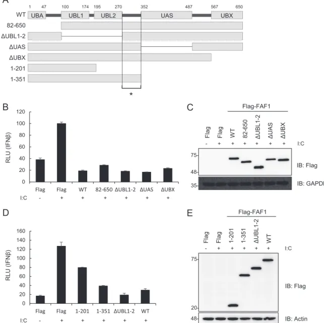
Ubiquitin-related domains of FAF1 are not critical for
inhib-iting IFN-
 activation. In order to identify which of the multiple
domains of FAF1 are involved in the inhibition of
poly(I·C)-in-duced IFN-
 activation, we measured IFN- luciferase activity in
HeLa cells transfected with wild-type FAF1 and various domain
deletion mutants: a deletion of the UBA domain (residues 82 to
650), interacting with polyubiquitinated substrates; a deletion of
the UBL1 and UBL2 (
⌬UBL1-2) domains interacting with Hsp70;
a deletion of the UAS (⌬UAS) domain, whose function is as yet
unknown; and a deletion of the UBX (
⌬UBX) domain interacting
with VCP-Npl4-Ufd1 complex (
Fig. 3A
). Among the mutants
tested, the mutant with a deleted N-terminal UBA domain (aa 82
to 650) and the
⌬UBL1-2, ⌬UAS, and ⌬UBX mutants showed the
same inhibitory effects as did wild-type FAF1 (
Fig. 3B
and
C
).
Since all of these mutants commonly contain two overlapping
regions (aa 271 to 351 or aa 488 to 566), we examined which
region is involved in the inhibition of IFN- promoter activity by
using truncation mutants consisting of aa 1 to 201, aa 1 to 351, and
⌬UBL1-2 and the WT. We found in reporter assays that the
ex-pression of aa 1 to 351 and
⌬UBL1-2 fully blocked
poly(I·C)-induced activation of the IFN- promoter, while the expression of
aa 1 to 201 showed only reduced activation compared to that of
the WT (
Fig. 3D
and
E
). These results show that the linker region
(aa 271 to 351) between UBL2 and the UAS is necessary but not
sufficient to inhibit IFN- promoter activity and that
ubiquitin-related domains of FAF1 are not critical for inhibiting IFN-

ac-tivation. This finding is unexpected because it is well known that
many FAF1 functions are regulated by ubiquitin-related domains.
Knocking down FAF1 induces antiviral ISG. We confirmed
the inhibitory effect of FAF1 on IFN-
 signaling and on
transcrip-tion of antiviral ISGs following poly(I·C) stimulatranscrip-tion. In additranscrip-tion,
silencing FAF1 promoted Mx1 production stimulated by
poly(I·C), while overexpressing FAF1 suppressed Mx1 production
(
Fig. 4A
and
B
). Mx1, a dynamin-like GTPase that broadly inhibits
viral replication by trapping viral nucleocapsids, is one of the most
highly induced ISGs (
34
). We next employed respiratory syncytial
virus (RSV), a negative-sense, single-stranded RNA virus of the
Paramyxoviridae family that causes acute lower respiratory tract
infection in young children (
35
). Like poly(I·C) transfection,
si-lencing FAF1 increased the expression of Mx1 and ISG15 in
re-sponse to RSV infection in HeLa cells (
Fig. 4C
). We also
investi-gated whether FAF1 inhibited IFN-
 signaling in RSV-infected
HEp-2 cells. As shown in
Fig. 4D
, knocking down FAF1 elevated
the transcription of IFN-
 and the expression level of ISG15 after
RSV infection in HEp-2 cells (
Fig. 4E
). Collectively, these results
suggest that FAF1 inhibits the cellular antiviral response by
nega-tively regulating IFN- production. We then compared the effect
of FAF1 on RSV and vesicular stomatitis virus (VSV) replication
by performing plaque formation assays. Silencing FAF1 slightly
reduced replication of the viruses, but this inhibitory effect was
small, although significant, in VSV compared to the induction of
antiviral ISGs (see Fig. S1 in the supplemental material). Further
studies are required to prove the influence of FAF1 on viral
repli-cation.
Targets of the FAF1 inhibitory effect during IRF3 activation.
Recognition of viruses by cytosolic sensors such as RIG-I and
MDA5 leads to activation of downstream signaling molecules like
MAVS, TBK1, IKK
ε, and IRF3 (
8
). To determine the target of the
inhibitory effect of FAF1 in the IRF3 activation signaling cascade,
we conducted an IFN-
 luciferase reporter assay in cells
overex-pressing each signaling molecule, RIG-I N, MAVS, TBK1, IKKε,
or IRF3-5D, together with the IFN-
 promoter in the presence
and absence of FAF1. RIG-I N is a constitutively active form of
RIG-I and is capable of activating IRF3 (
36
). As shown in
Fig. 5A
,
FAF1 suppressed the activation of IFN- promoter mediated by
overexpression of RIG-I N, MAVS, and TBK1. Furthermore,
FAF1 also inhibited IRF3-WT- or IRF3-5D (a constitutively active
mutant of IRF3)-induced activation of IFN-
 and the ISRE
pro-moter (
Fig. 5B
and
C
) even at a higher protein expression level of
IRF3-WT or IRF3-5D. These results were confirmed by silencing
FAF1, potentiating IFN- promoter activity mediated by
overex-pression of all signaling molecules, i.e., RIG-I N, MAVS, TBK1,
IKKε, and IRF3-5D (
Fig. 5D
). The results indicated that FAF1
functions as a negative regulator of IFN-
 signaling after
phos-phorylation of IRF3. Since active IRF3 can bind to the IFN-␣4
promoter as well as to IFN-
 (
37
), we additionally used an
IFN-␣4-Luc plasmid and showed that FAF1 inhibited
IRF3-5D-medi-ated activation of the IFN-
␣4 promoter (
Fig. 5E
). Considering
that IRF3 could be activated by the TLR3 or TLR4 signaling
pathway, we investigated whether FAF1 suppresses the
activa-tion of the IFN- promoter from TLR, employing an IRF3
stimulator, TRIF, an adaptor protein of TLR3 and TLR4. FAF1
also inhibited TRIF-mediated activation of the IFN-
pro-moter in a dose-dependent manner (
Fig. 5F
). These results
confirm that FAF1 negatively regulates activation of IFN- at
or downstream of IRF3.
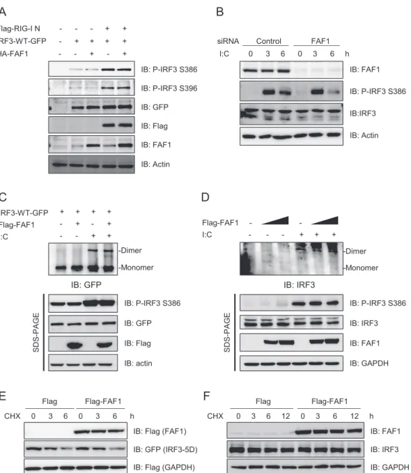
FAF1 does not affect the phosphorylation and dimerization
of IRF3. Upon viral infection, IRF3 is phosphorylated and
acti-vated by active TBK1 or IKKε (
9
). Phosphorylated IRF3
subse-quently forms dimers and translocates to the nucleus, where it
interacts with transcription coactivators and promotes
transcrip-tion of IFN-
 (
10
,
11
). We investigated whether FAF1 affects
phosphorylation and dimerization of IRF3. Using
anti-phospho-IRF3 antibody, we detected phosphorylated forms of anti-phospho-IRF3 in
HeLa cells overexpressing Flag-RIG-I N, IRF3-GFP, and
on October 31, 2016 by Ewha Womans Univ
http://mcb.asm.org/
0 20 40 60 80 100 120 RLU (IFN β ) Flag-FAF1 (μg) - - 0.5 1 2 I:C - + + + + 0 20 40 60 80 100 120 RLU (ISRE) IB: Flag IB: Actin IB: Flag IB: Actin Flag-FAF1 (μg) - - 0.5 1 2 I:C - + + + + 0 10 20 30 40 50 60 Relative m R N A l e v el IFNβ NT I:C ** ** 0 5 10 15 20 25 Relative m R N A l e v el IFIT1 NT I:C *** Flag-FAF1 (μg) - 1.5 3 Flag-FAF1 (μg) - 1.5 3
B
A
D
C
IB: FAF1 IB: Actin NT I:C NT I:C 0 100 200 300 400 500 RLU (IFN β ) si-Con si-FAF1 0 5 10 15 20 25 RLU (IFN β ) si-Con si-FAF1 0 10 20 30 RLU (ISRE) si-Con si-FAF1 J K 0 500 1000 1500 Relative m R N A l e v e l si-Con si-FAF1 IB: FAF1 IB: Actin IB: FAF1 IB: Actin IB: FAF1 IB: GAPDH I HsiRNA Control FAF1 #1 FAF1 #2 siRNA Control FAF1 #1 FAF1 #2
Raw264.7 HEK293T ** * *** ** 0 200 400 600 Relative m R N A l e v el IFNβ NT I:C * ** 0 50 100 150 Relative m R N A l e v el IFIT1 NT I:C * ** 0 0.3 0.6 0.9 1.2 Relative m R N A l e v el FAF1 NT I:C
siRNA Control FAF1 #1 FAF1 #2
G
E F
NT I:C NT I:C
FIG 2 FAF1 inhibits poly(I·C)-induced IFN- activation. (A and B) HeLa cells were transfected with the indicated amount of Flag-FAF1 plasmid. After 24 h, cells were treated with poly(I·C) (I·C; 10g/ml) by transfection for 8 h. mRNA levels of IFN- and IFIT1 were measured using RT-qPCR. The values were normalized to the value for GAPDH mRNA and represent the means⫾ standard deviations of three experiments. **, P ⬍ 0.01; ***, P ⬍ 0.001 (for differences between Flag and Flag-FAF1 values). NT, not treated. (C and D) HeLa cells were cotransfected with the indicated amount of Flag-FAF1 plasmid and IFN--Luc (C) or ISRE-Luc (D) together with a beta-Gal reporter plasmid. After 24 h, cells were treated with poly(I·C) (10g/ml) by transfection for 8 h, and relative luciferase activities were measured. The data represent the means⫾ standard deviations of triplicate experiments. Overexpressed Flag-FAF1 is shown by Western blotting, with actin bands representing loading controls. RLU, relative light units. (E to G) HeLa cells were transfected with two kinds of FAF1 siRNAs, 1 and 2. After 40 h, cells were treated with poly(I·C) (10g/ml) by transfection for 8 h. mRNA levels of FAF1, IFN-, and IFIT1 were measured using RT-qPCR. The values were normalized to the value for GAPDH mRNA. (H and I) HeLa cells were transfected with FAF1 siRNA 2, and 72 h later, cells were transfected with IFN--Luc (H) or ISRE-Luc (I) together with a beta-Gal reporter plasmid. Cells were treated with poly(I·C) (10g/ml) by transfection for 9 h, and relative luciferase activities were measured. Knockdown of endogenous FAF1 was shown by Western blotting, with actin bands and GAPDH bands representing loading controls. si-CON, control siRNA; si-FAF1, siRNA targeting FAF1. (J) HEK293T cells were transfected with FAF1 siRNA 2 for 72 h and transfected with IFN--Luc together with a beta-Gal reporter plasmid. Then cells were treated with poly(I·C) (10g/ml) by transfection for 12 h, and relative luciferase activities were measured. (K) Raw264.7 cells were transfected with FAF1 siRNA 2 for 36 h and treated with poly(I·C) (10g/ml) for 12 h. The mRNA level of IFN- was measured using RT-qPCR. The values were normalized to the value for GAPDH. Knockdown of endogenous FAF1 is shown by Western blotting, with actin bands representing loading controls. All of the data represent the means⫾ standard deviations of three experiments. *, P ⬍ 0.05; **, P ⬍ 0.01; ***, P ⬍ 0.001 (for differences between control siRNA FAF1 siRNA values).
on October 31, 2016 by Ewha Womans Univ
http://mcb.asm.org/
FAF1 or empty vector. As shown in
Fig. 6A
, RIG-I N
overexpres-sion increased the phosphorylation at Ser386 and Ser396 of IRF3;
however, FAF1 overexpression did not affect the phosphorylation
of IRF3. Consistently, poly(I·C) transfection increased the
phos-phorylated IRF3, but silencing of FAF1 did not affect the level of
phospho-IRF3 (
Fig. 6B
). These results accorded with the
observa-tion that FAF1 repressed the activaobserva-tion of IFN-
 promoter
in-duced by the constitutively active mutant IRF3-5D, and thus FAF1
does not affect the phosphorylation of IRF3 in response to
stimu-lation.
Since phosphorylation at Ser386 of IRF3 is required for IRF3
dimerization for nuclear translocation (
11
), we investigated
whether FAF1 inhibits IRF3 dimerization by employing native
PAGE. HEK293T cells overexpressing IRF3-WT-GFP with or
without Flag-FAF1 were stimulated with poly(I·C), cell lysates
were separated by SDS-PAGE or native gel electrophoresis, and
dimerization was evaluated by Western blotting as described
pre-viously (
38
). The IRF3 dimers increased in response to poly(I·C)
transfection on a native gel; however, no discernible change in
dimerization of IRF3 was detected in cells overexpressing FAF1.
Phospho-IRF3 at Ser386 was also detected in cells stimulated by
poly(I·C) regardless of FAF1 expression on SDS-PAGE gels (
Fig.
6C
). We also investigated the effect of FAF1 overexpression on the
dimerization of endogenous IRF3 in response to poly(I·C)
stimu-I:C - + + + + +
UBL1 UBL2 UAS UBX
WT 1-201 1-351 82-650 ΔUBL1-2 ΔUAS ΔUBX I:C - + + + + + + 0 20 40 60 80 100 120
Flag Flag WT 82-650 ΔUBL1-2 ΔUAS ΔUBX
RLU (IFNβ) 0 20 40 60 80 100 120 140 160
Flag Flag 1-201 1-351 ΔUBL1-2 WT
RLU (IFNβ)
IB: Flag
IB: Actin IB: Flag IB: GAPDH
Flag Flag WT 82-650
ΔUBL1-2
ΔUAS ΔUBX
- + + + + + + I:C
Flag-FAF1
Flag Flag 1-201
ΔUBL1-2 WT 1-351 - + + + + + I:C Flag-FAF1
A
B
C
E
D
1 47 100 174 195 270 352 487 567 650*
UBA 35- 48- 20- 75-48-FIG 3 Ubiquitin-related domains of FAF1 are not critical for inhibiting poly(I·C)-induced IFN- activation. (A) Diagram of various domain deletion mutants
of Flag-FAF1. UBA, ubiquitin binding domain; UBL1 and UBL2, ubiquitin-like domains 1 and 2; UAS, upstream activation sequence of unknown function; UBX, ubiquitin regulatory domain. The asterisk indicates the linker region (aa 271 to 351). (B and D) HeLa cells were cotransfected with deletion mutants of Flag-FAF1 plasmid and IFN--Luc together with a beta-Gal reporter plasmid. After 24 h, cells were treated with poly(I·C) (10 g/ml) by transfection for 6 h, and relative luciferase activities were measured. The data represent the means⫾ standard deviations of triplicate experiments. (C and E) Overexpression of Flag-FAF1 deletion mutants was shown by Western blotting, with GAPDH and actin bands representing loading controls.
on October 31, 2016 by Ewha Womans Univ
http://mcb.asm.org/
lation and found that FAF1 did not affect the dimerization of
endogenous IRF3 (
Fig. 6D
). Taken together, these results
demon-strate that FAF1 inhibits neither phosphorylation nor
dimeriza-tion of IRF3 but may inhibit some downstream event after
activa-tion of IRF3.
Since we previously found that FAF1 is a ubiquitin receptor
that facilitates the degradation of Hsp70 and ERAD substrates by
the ubiquitin proteasome system (
26
,
29
,
30
), we investigated
whether FAF1 affects the degradation of IRF3. We monitored the
half-lives of endogenous IRF3 and the active mutant IRF3-5D
after the cells were treated with cycloheximide (CHX), an
inhibi-tor of protein synthesis. Overexpression of FAF1 did not affect the
degradation of IRF3 (
Fig. 6E
and
F
).
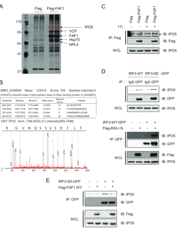
FAF1 interferes the interaction between IRF3 and IPO5. In
the inhibition of IRF3-5D-mediated activation of IFN-

pro-moter by FAF1, no physical interaction between FAF1 and IRF3 in
control or stimulated cells was detected (data not shown). One
explanation for this is that FAF1 may inhibit IRF3-driven IFN-
signaling via an adaptor protein. In order to find the adaptor
mol-ecule of FAF1 in IRF3 inhibition, we performed
immunoprecipi-tation with anti-Flag antibody in HeLa cells overexpressing
Flag-FAF1; immune complexes were separated by SDS-PAGE and
detected with silver staining (
Fig. 7A
). IPO5/importin-
3 was
identified as a protein physically associated with FAF1 by peptide
sequencing with nano-ultraperformance liquid chromatography
with electrospray ionization-quadrupole time of flight
(nano-UPLC-ESI-q-TOF) tandem MS (
Fig. 7B
) and confirmed by
West-ern blotting (
Fig. 7C
). IPO5/importin-3 was first identified as a
binding protein of a small Ran GTPase. It is a member of the
importin- family which binds directly to cargo proteins or to
importin-
␣ and cargo complex to promote the nuclear import of
cargo proteins (
39
). A recent study shows that IPO5 interacts with
the transcription factor
-catenin and plays a role in nuclear
transport of
-catenin (
40
). IRF3 is known to be a subset of
im-portin-
␣ receptors, and FAF1, inhibiting IRF3-mediated IFN-
activation, interacts with IPO5 in control and poly(I ·
C)-stimu-IB: Mx1 IB: Actin IB: FAF1 IB: ISG15 FAF1 siRNA - + - + - + - + - + - + RSV 0 24 30 36 42 48 h
E
FAF1 siRNA - + - + - + - + RSV 0 12 24 36 h IB: FAF1 IB: GAPDH HEp-2 HeLaD
IB: ISG15 0 2 4 6 8 10 Relative m R N A l e v e lIFNβ
si-Con si-FAF1 Mock RSVA
C
*** *** I:C 0 6 12 0 6 12 h Flag Flag-FAF1 IB: Tubulin IB: Mx1 IB: Flag I:C 0 6 12 0 6 12 h si-FAF1 si-Con IB: Tubulin IB: Mx1 IB: FAF1B
FIG 4 Knocking down FAF1 increased expression of ISGs. (A and B) The level of induced Mx1 was detected by Western blotting, with tubulin bands representing
loading controls, in HeLa cells. (A) HeLa cells transfected with a control siRNA or FAF1 siRNA 2 for 40 h were treated with poly(I·C) (10g/ml) by transfection for the indicated times. (B) HeLa cells transfected with Flag or Flag-FAF1 for 24 h were treated with poly(I·C) (10g/ml) by transfection for the indicated times. (C and E) Levels of ISGs and FAF1 were analyzed by Western blotting, with actin bands representing loading controls, in HeLa cells and HEp-2 cells. Cells were transfected with a control siRNA or FAF1 siRNA 2 for 24 h, and cells were incubated with an RSV inoculum (multiplicity of infection, 1) or uninfected. After 2 h, the medium was changed, and cells were harvested at the indicated time points after infection. (D) Levels of IFN- mRNA were measured by RT-qPCR in HEp-2 cells silencing FAF1 after RSV infection and then 24 h of incubation. The values were normalized to the value for GAPDH mRNA and represent the means⫾ standard deviations of three experiments. ***, P ⬍ 0.001 (for differences between control siRNA and FAF1 siRNA values).
on October 31, 2016 by Ewha Womans Univ
http://mcb.asm.org/
C
B
A
0 100 200 300 400 RLU (IFN β ) si-Con si-FAF1 IB: FAF1 IB: GAPDH IB: Flag IB: GAPDH IB: IRF3 0 5 10 15 20 25 30 35 RLU (ISRE) Flag Flag-FAF1 0 50 100 150 RLU (IFN β ) Flag Flag-FAF1 IB: IRF3 IB: Flag IB: Actin IB: Flag IB: IRF3 IB: FAF1 IB: MAVS IB: TBK1 IB: RIG-I 0 1 2 3 4 5 6 RLU (IFN β ) Flag Flag-FAF1 IB: Tubulin IB: RIG-IVec IRF3-WT IRF3-5D
D
TRIF - + + + FAF1 --F
0 20 40 60 RLU (IFN β )Vec IRF3-WT IRF3-5D
* *** *** *** *** *** *** ** ** ** ** * * 35- 75- 48- 75- 35- 75- 35- 75- 63- 75- 35-
75-Vec RIG-I N MAVS TBK1
Vec RIG-I N MAVS TBK1 IKKε IRF3-5D
IB: TRIF IB: Flag IB: Actin IB: IRF3 IB: Flag IB: GAPDH IRF3-5D (μg) - 0.2 0.4 0 2 4 6 8 10 RLU (IFNα4 ) Flag Flag-FAF1
E
*** *** ** ***FIG 5 FAF1 operates at or downstream of IRF3. (A) HeLa cells were transfected with IFN--Luc, a beta-Gal reporter plasmid, Flag-FAF1, or a control plasmid and Flag-RIG-I N, Flag-MAVS, Flag-TBK1, or a control plasmid (Vec). After 24 h, cells were collected, and relative luciferase activities were measured. The data represent the means⫾ standard deviations of three experiments. Overexpressed proteins are shown by Western blotting, with tubulin bands representing loading controls. (B and C) HeLa cells were transfected with IFN--Luc (B) ISRE-Luc (C), beta-Gal, Flag-FAF1, or a control plasmid and IRF3-WT-GFP, IRF3-5D-GFP, or a control plasmid. At 24 h posttransfection, relative luciferase activities were determined as described for panel A. *, P⬍ 0.05; **, P ⬍ 0.01; ***, P ⬍ 0.001 (for differences between Flag and Flag-FAF1 values in panels A to C). (D) HeLa cells were transfected with a control siRNA or FAF1 siRNA 2. At 48 h posttransfection, cells were cotransfected with Flag-RIG-I N, Flag-MAVS, Flag-TBK1, Flag-IKKε, IRF3-5D, or a control plasmid and IFN--Luc together with a beta-Gal reporter plasmid. At 24 h posttransfection, relative luciferase activities were determined as described for panel A. **, P⬍ 0.01; ***, P ⬍ 0.001 (for differences between values for the control siRNA and FAF1 siRNA). (E) HeLa cells were transfected with IFN-␣4-Luc, beta-Gal, Flag-FAF1, or a control plasmid and the indicated amount of IRF3-5D-GFP. At 24 h posttransfection, relative luciferase activities were determined as described for panel A. (F) HeLa cells were transfected with IFN--Luc, beta-Gal, Flag-FAF1, or a control plasmid and TRIF. At 24 h posttransfection, relative luciferase activities were determined as described for panel A. *, P⬍ 0.05; **, P ⬍ 0.01; ***, P ⬍ 0.001 (for differences between values for Flag and Flag-FAF1 in panels E and F).
on October 31, 2016 by Ewha Womans Univ
http://mcb.asm.org/
lated cells (
Fig. 7C
). Hypothesizing that FAF1 interferes with the
translocation of IRF3 to the nucleus by inhibiting the
interac-tion between IRF3 and IPO5, we investigated whether IRF3-5D
binds to IPO5 and found such interaction in HEK293T cells
overexpressing IRF3-5D-GFP. When the cell lysates were
im-munoprecipitated with anti-GFP antibody, we found that
IRF3-5D indeed interacted with IPO5 (
Fig. 7D
, upper panel).
Furthermore, the interaction between IRF3-WT and IPO5 was
increased in response to overexpression of RIG-I N. These
re-sults showed that activated IRF3 more strongly interacted with
IPO5 (
Fig. 7D
, lower panel).
We then tested whether FAF1 inhibits IFN- signaling through
competition with IRF3 for IPO5 binding. HEK293T cells
cotrans-fected with IRF3-5D-GFP and Flag-FAF1 were lysed and
immu-I:C 0 3 6 0 3 6 h siRNA Control FAF1
IB: FAF1 IB: P-IRF3 S386 IB:IRF3 IB: Actin
B
A
HA-FAF1 - - + - + IRF3-WT-GFP - + + + + Flag-RIG-I N - - - + + IB: GFP IB: Flag IB: Actin IB: FAF1 IB: P-IRF3 S386 IB: P-IRF3 S396 Flag-FAF1 I:C IRF3-WT-GFP IB: GFP IB: GFP IB: P-IRF3 S386 IB: actin IB: Flag IB: P-IRF3 S386 IB: IRF3 IB: GAPDH IB: FAF1D
C
-Dimer -Monomer IB: IRF3 -Dimer -Monomer Flag-FAF1 - -I:C - - - + + + SDS-PAG E SDS-PAG E + -+ + -+ -+ + + +E
CHX 0 3 6 0 3 6 h Flag Flag-FAF1IB: Flag (FAF1)
IB: Flag (GAPDH) IB: GFP (IRF3-5D) CHX 0 3 6 12 0 3 6 12 h Flag Flag-FAF1 IB: FAF1 IB: IRF3 IB: GAPDH
F
FIG 6 FAF1 does not affect the phosphorylation and dimerization of IRF3. (A) HeLa cells were transfected with Flag-RIG-I N, IRF3-WT-GFP, HA-FAF1, or a
control plasmid as indicated. At 24 h posttransfection, cell extracts were analyzed by Western blotting. Actin bands represent loading controls. (B) HeLa cells were transfected with a control siRNA or FAF1 siRNA 2 for 72 h and treated with poly(I·C) (10g/ml) by transfection. Cells were then harvested at the indicated time points, and extracts were analyzed by Western blotting, with actin bands representing loading controls. (C) HEK293T cells were cotransfected with IRF3-WT-GFP, Flag-FAF1, or a control plasmid and then stimulated with poly(I·C) (10g/ml) for 12 h. Cell extracts were separated by native gel or SDS-PAGE, and IRF3 dimers were detected by Western blotting, with actin bands representing loading controls. (D) HEK293T cells were transfected with Flag-FAF1 or a control plasmid and stimulated with poly(I·C) (10g/ml) for 12 h. Endogenous IRF3 was analyzed as described for panel C. (E) HeLa cells were transfected with Flag-FAF1, Flag-GAPDH, IRF3-5D-GFP, or a control plasmid. After 24 h, cells were treated with cycloheximide (50g/ml) for the indicated times. Cell lysates were separated by SDS-PAGE and analyzed by Western blotting. (F) HeLa cells were transfected with Flag or Flag-FAF1. After 24 h, cells were treated with cycloheximide (50g/ml) for the indicated times. The level of endogenous IRF3 was analyzed by Western blotting, with GAPDH bands representing loading controls.
on October 31, 2016 by Ewha Womans Univ
http://mcb.asm.org/
noprecipitated with anti-GFP antibody. As shown in
Fig. 7E
,
IRF3-5D physically associates with IPO5, and the amount of IPO5
interacting with IRF3-5D was reduced by FAF1 overexpression. In
Western blot analysis with whole-cell lysates, both IRF3-5D and
FAF1 were well expressed, and the levels of IPO5 were not
differ-ent between the lanes. Although FAF1 and IRF3 did not directly
interact with each other, FAF1 can inhibit IFN-
 activation by
disturbing the interaction between active IRF3 and IPO5.
FAF1 suppresses nuclear translocation of IRF3. FAF1 blocks
IRF3-5D-induced IFN- promoter activation without affecting
B
IMB3_HUMAN Mass: 123419 Score: 104 Queries matched:4
(O00410) Importin beta-3 (Karyopherin beta-3) (Ran-binding protein 5) (RanBP5)
Observed Mr(expt) Mr(calc) Delta mass Mascot
Score Peptide 586.3291 1170.6436 1170.6496 -0.0059 12 SLVEIADTVPK 784.9032 1567.7918 1567.7994 -0.0075 70 FLFDSVSSQNVGLR 787.4034 1572.7922 1572.8003 -0.0081 5 VAAAESMPLLLECAR 893.9479 1785.8812 1785.9043 -0.0230 25 VIAALLQTMEDQGNQR 1567.7918 from (784.9032,2+) intensity(849.7498) IP: Flag Flag Flag-FAF 1 Flag Flag-FAF 1 I:C - - + + IB: IPO5 IB: Flag IB: IPO5
C
WCL IB: IPO5 IB: IPO5 IB: Flag IB: GFP IRF3-5D-GFP Flag-FAF1 WT -+ + -+ + IB: GFP IB: IPO5 IP : IgG GFP IgG GFP WCL IP: GFPD
E
WCL IB: Flag IB: IPO5 IB: GFP IB: IPO5 Flag-RIG-I N - + - + IRF3-WT-GFP - - + + IP: GFP IB: IPO5 WCL IRF3-WT IRF3-5D -GFPA
VCP FAF1 Hsp70 83- 175- 62- 47- 37-Flag 37-Flag-FAF1 NPL4 IPO5 R G V N Q S S V S D F L FFIG 7 FAF1 overexpression abolishes IRF3-IPO5 interaction. (A and B) Silver gel and MS/MS spectrum of IPO5 which was detected in FAF1 immune complex
and identified employing nano-UPLC-ESI-q-TOF tandem MS. (C) HeLa cells transfected with Flag or Flag-FAF1 were treated with poly(I·C) (10g/ml) for 6 h, and immunoprecipitation of FAF1 was performed using anti-Flag antibody. Immune complexes and whole-cell lysates (WCL) were separated by SDS-PAGE and analyzed by Western blotting. (D) HEK293T cells were transfected with IRF3-WT-GFP or IRF3-5D-GFP. After 30 h, cell lysates were immunoprecipitated with anti-GFP antibody or anti-IgG antibody, and immune complexes were analyzed by Western blotting (upper panel). HEK293T cells were transfected with IRF3-WT-GFP, Flag-RIG-I N, or a control plasmid as indicated. After 30 h, cell lysates were immunoprecipitated with anti-GFP antibody, and immune complexes were analyzed by Western blotting (lower panel). (E) HEK293T cells were cotransfected with IRF3-5D-GFP, Flag-FAF1, or a control plasmid as indicated. After 30 h, cells lysates were immunoprecipitated with anti-GFP antibody, and immune complexes were analyzed by Western blotting.
on October 31, 2016 by Ewha Womans Univ
http://mcb.asm.org/
FIG 8 Knocking down FAF1 promotes nuclear translocation of IRF3. HeLa cells were transfected with a control siRNA or FAF1 siRNA 2 for 72 h. (A) Cells were
treated with poly(I·C) (10g/ml) by transfection for the indicated times, and then lysates were divided into cytosolic and nuclear fractions as described in Materials and Methods. Nuclear translocation of IRF3 was assayed using Western blot analysis. Prx6 and lamin B were used as cytosolic and nuclear markers, respectively. (B) Localization of IRF3 in HeLa cells was evaluated by fluorescence confocal microscopy. Control and FAF1-knocked-down HeLa cells were treated with a control and poly(I·C) (10g/ml) for 3 h. Then cells were fixed, permeabilized, and stained with anti-IRF3 antibody (green). Nuclei were detected with DAPI staining (blue). The graph indicates the percentages of cells showing nuclear immunoreactivity for IRF3. Data were calculated after counting the number of cells with nuclear IRF3 from more than 5 fields from the coverslips using ImageJ software (right panel). *, P⬍ 0.05 (for the difference between the values for the control siRNA and FAF1 siRNA). (C) HeLa cells were transfected with FAF1 siRNA 2 for 40 h and transfected with Flag or Flag-FAF1. At 24 h posttransfection, cells were treated with poly(I·C) (10g/ml) by transfection for the indicated times; cell lysates were divided by fractionation and analyzed as described for panel A. (D) Raw264.7 cells were transfected with a control siRNA or FAF1 siRNA 2 for 40 h, and cells were treated with poly(I·C) (10g/ml) for the indicated times. Total samples were obtained before centrifugation, and then cell lysates were divided by fractionation. GAPDH and HDAC1 were used as cytosolic and nuclear markers, respectively.
on October 31, 2016 by Ewha Womans Univ
http://mcb.asm.org/
the phosphorylation or dimerization of IRF3. Since FAF1
inhib-ited IRF3-IPO5 interaction, we investigated whether FAF1 is
in-volved in nuclear translocation of IRF3. We stimulated control
and FAF1-knocked-down HeLa cells with poly(I·C), isolated the
nuclear fraction, and assessed the amount of IRF3 that
translo-cated into the nucleus in response to poly(I·C) using
anti-phos-pho-IRF3 antibody. We found that nuclear IRF3 significantly
in-creased (
Fig. 8A
, right panel), while cytosolic IRF3 slightly
decreased in cells knocking down FAF1 compared to levels in
con-trol cells (
Fig. 8A
, left panel). This translocation to the nucleus was
confirmed with fluorescence microscopy of IRF3 in the
FAF1-knocked-down cells treated with poly(I·C). Image analysis
showed translocation of IRF3 into the nucleus, and counting cells
showing nuclear IRF3 confirmed that silencing FAF1 promoted
translocation of IRF3 to the nucleus in both control and
poly(I·C)-transfected cells (
Fig. 8B
). Next, in order to investigate
whether overexpressed FAF1 could inhibit nuclear translocation
of IRF3, we assessed IRF3 in the nucleus of HeLa cells. To
maxi-mize the effect, HeLa cells knocking down endogenous FAF1 with
an siRNA were transfected with Flag or Flag-FAF1 and stimulated
with poly(I·C). Nuclear and cytosolic fractions were obtained by
fractionation. IRF3 and phospho-IRF3 in the nuclear fraction
were decreased, and levels in the cytosolic fraction increased in
cells overexpressing FAF1 compared to levels in control cells (
Fig.
8C
). The results were also confirmed in mouse Raw 264.7 cells. As
shown in
Fig. 8D
, FAF1 was not completely knocked downed in
the Raw264.7 cells, so the reduction of cytosolic IRF3 was not
dramatic. But nuclear IRF3 was accumulated more, and the
ex-pression of ISG15 was increased in cells knocking down FAF1
compared to levels in control cells.
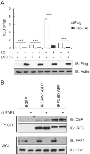
Nuclear accumulation of IRF3 could occur by impairment of
nuclear export. IRF3 has an active nuclear export signal (NES),
and its nuclear export depends on the activity of CRM1 (
23
). To
test whether nuclear export of IRF3 was affected by FAF1, we
examined IFN- promoter activity after overexpressing FAF1 in
the absence or presence of a specific CRM1 inhibitor, leptomycin
B (LMB). As shown in
Fig. 9A
, FAF1 inhibited poly(I·C)-induced
IFN- promoter activity regardless of LMB treatment. These
find-ings indicate that FAF1 regulates IRF3 translocation to the nucleus
by modulating the nuclear import step, not the export step.
In order to investigate whether FAF1 affects the interaction of
active IRF3 with the coactivators CBP/p300, we examined the
in-teraction of CBP with IRF3-WT and IRF3-5D in presence and
absence of FAF1. Interaction of active IRF3-5D with CBP
signifi-cantly increased, but FAF1 did not affect this interaction (
Fig. 9B
).
This indicates that FAF1 does not affect the interaction between
nuclear active IRF3 and CBP; rather FAF1 affects the translocation
of IRF3 from cytosol to nucleus.
DISCUSSION
FAF1, a member of the UBXN family containing the UBA-UBX
domain, plays multiple biological functions, including protein
degradation of Hsp70 and ERAD substrate (
26
,
28
,
30
). However,
the functions of FAF1 remain to be understood. We screened
tar-get genes of FAF1 by employing microarray analysis. Our
mi-croarray studies of FAF1-knocked-down HeLa cells showed that
FAF1 changed the expression levels of 150 genes, including genes
related to apoptosis and proteolysis. The genes showing the most
significant changes by FAF1 depletion were ISGs which encode
proteins repressing viral replication or enhancing type I IFN
pro-duction.
This study shows that overexpression of FAF1 inhibited
poly(I·C)-induced activation of IFN-
 and ISRE promoter as well
as poly(I·C)-induced transcription of IFN- and IFIT1 and ISG
expression. Silencing FAF1 increased IFN-
 production in
re-sponse to poly(I·C) stimulation in all cell lines tested.
Simultane-ously, silencing of FAF1 promoted RSV-induced IFN-

produc-tion and substantial inducproduc-tion of antiviral ISGs such as Mx1 and
ISG15. Reporter assays demonstrated that FAF1 functions at the
downstream step of IRF3 phosphorylation. FAF1 did not alter
phosphorylation and dimerization of IRF3 (
Fig. 6
); rather,
silenc-ing FAF1 augmented its nuclear translocation.
To understand how FAF1 regulates the innate immune system
by modulating IFN- activation, we performed IFN- reporter
assays using molecules in both the RLR and TLR signaling axes.
These studies revealed that FAF1 suppressed activation of the
I:C - - - - + + + + LMB (h) - - + + - - + +
B
si-FAF1 - + - + - + IB: CBP IB: IRF3 IB: FAF1 IB: CBP IP: GFP WCL EGFP IRF3 -W T -G F P IRF3-5D-GF PA
IB: Flag IB: Actin 0 2 4 6 8 10 RLU (IFN β ) Flag Flag-FAF1 *** *** *** ***FIG 9 FAF1 does not affect nuclear export of IRF3 and recruitment of CBP to
IRF3. (A) HeLa cells were cotransfected with IFN--Luc, beta-Gal, Flag-FAF1, or a control plasmid. After 24 h, cells were treated with poly(I·C) (10g/ml) and incubated in the presence or absence of LMB (20 nM). After 6 h of incu-bation, cells were harvested, and relative luciferase activities were measured. The data represent the means⫾ standard deviations of three experiments. ***, P⬍ 0.001 (for differences between values for Flag and Flag-FAF1). (B) HeLa cells were transfected with a control siRNA or FAF1 siRNA 2 for 48 h and then transfected with enhanced GFP, IRF3-WT-GFP, or IRF3-5D-GFP. At 24 h posttransfection, cell lysates were immunoprecipitated with GFP anti-body, and immune complexes were analyzed by Western blotting.
on October 31, 2016 by Ewha Womans Univ
http://mcb.asm.org/
IFN-
 promoter, downstream of IRF3 phosphorylation. Silencing
FAF1 potentiated IFN- activation mediated by overexpression of
RIG-I N, MAVS-, TBK1-, IKK
ε, and IRF3-5D. Neither
overex-pression nor knockdown of FAF1 affected the phosphorylation
and dimerization of IRF3. We also examined whether FAF1
facil-itates the degradation of IRF3 by the ubiquitin proteasome system
because FAF1 is known as a ubiquitin receptor protein. However,
FAF1 overexpression did not increase the degradation of
endoge-nous IRF3 or its active mutant. In addition, a UBA domain
dele-tion mutant which could not interact with polyubiquitinated
sub-strates also inhibited poly(I·C)-induced IFN-
 activation as well
as wild-type FAF1. Then, we examined whether FAF1 affects the
cellular localization of IRF3 and found that silencing FAF1
in-creased the accumulation of active IRF3 in the nucleus induced by
poly(I·C) transfection, while overexpression of FAF1 reduced the
levels of nuclear IRF3 upon poly(I·C) transfection. These results
indicate that FAF1 inhibits IRF3 translocation to the nucleus, not
its phosphorylation or degradation.
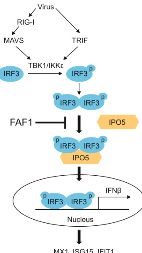
How does FAF1 regulate the nuclear translocation of IRF3?
Previous studies have shown that the IRF3 NLS and nuclear
local-ization of IRF3 are important for IRF3 transcriptional activity (
21
,
22
). IRF3 is known to be phosphorylated by upstream kinases,
forming dimers which are then transported into the nucleus (
8
,
9
).
In order to identify how FAF1 inhibits IRF3 nuclear translocation,
we examined the interacting proteins of FAF1 and found for the
first time that FAF1 constitutively interacted with the nuclear
im-port receptor IPO5/imim-portin-
3 and that active IRF3 also
associ-ated with IPO5/importin-3 (
Fig. 10
).
There are many nuclear import receptors, including 7
impor-tin-␣ genes and 20 importin- genes in human (
41
). Proteins
greater than 40 kDa are known to pass through the nuclear pore
complex using receptors and carrier proteins. Until now, the
cor-relations between cargo proteins and importin receptor have not
been well identified, and many cargoes use more than one import
factor for translocation. IPO5/importin-
3, interacting with
FAF1 and IRF3 (
Fig. 7C
and
D
), is a member of the importin-
family and binds to cargo directly without importin-
␣ adaptor
and mediates nuclear import of ribosomal proteins, histones, and
viral proteins (
39
,
42
). Several viruses are known to escape the
immune response by inhibiting nuclear import of transcription
factors. The Ebola virus VP24 protein inhibits interaction of
STAT1 with importin-␣1 (
43
). Hepatitis B virus polymerase
in-terrupts STAT1/2 binding to importin-
␣5 (
44
). Until the present
study, IRF3 was known to interact with importin-␣3 and
impor-tin-
␣4. This study shows that IRF3 associates with
IPO5/impor-tin-3, and this interaction is increased by overexpression of
RIG-I N, which leads to activation of IRF3 (
Fig. 7D
, lower panel).
Since no direct interaction between IRF3 and FAF1 was observed,
we propose that interaction between FAF1 and IPO5 could affect
nuclear import of IRF3. We found that overexpressing FAF1
sig-nificantly decreased the interaction between IRF3 and IPO5 (
Fig.
5E
). This is a novel inhibitory mechanism by which FAF1
reg-ulates IRF3-mediated IFN-
 induction. Further studies are
needed to understand how FAF1 inhibits innate immunity,
including interacting with nuclear pore complex components
such as Ran GTPase and other importin receptors.
In summary, this study suggests that FAF1 plays a key role as a
negative regulator of the innate immune system in general and of
virus-triggered IFN-
 production in particular, including
inhib-iting the translocation of IRF3 into the nucleus and preventing
antiviral IFN-
 signaling. This is a novel biological function to be
added to the list of cellular functions of FAF1. FAF1 can be a
valuable target for developing therapeutics of autoimmune and
inflammatory diseases caused by IFN-.
ACKNOWLEDGMENT
We are grateful to Joo Young Lee (Catholic University of Korea) for do-nating the signaling molecule plasmids.
FUNDING INFORMATION
This work was supported by the Global Research Lab Program (no. 2012K1A1A2045441) of NRF. S. Song was supported by the Brain Korea 21 Plus (BK21 Plus) Project, and H. J. Kim was supported by a Basic Science Research Program fellowship from NRF (no. 2013R1A1A2061412).
REFERENCES
1. Kawai T, Akira S. 2007. Antiviral signaling through pattern recognition receptors. J Biochem 141:137–145.
2. Hiscott J. 2007. Convergence of the NF-B and IRF pathways in the regulation of the innate antiviral response. Cytokine Growth Factor Rev
18:483– 490.http://dx.doi.org/10.1016/j.cytogfr.2007.06.002.
3. Garcia-Sastre A, Biron CA. 2006. Type 1 interferons and the virus-host relationship: a lesson in detente. Science 312:879 – 882.http://dx.doi.org /10.1126/science.1125676.
4. Gonzalez-Navajas JM, Lee J, David M, Raz E. 2012. Immunomodula-tory functions of type I interferons. Nat Rev Immunol 12:125–135.http: //dx.doi.org/10.1038/nri3133. IPO5 MAVS IFNβ Virus TRIF TBK1/IKKε MX1, ISG15, IFIT1…
FAF1
Nucleus p IRF3 IRF3 p p IRF3 IRF3 IPO5 RIG-I p p IRF3 IRF3 p p IRF3 IRF3FIG 10 Schematic diagram of FAF1 action on the innate-immunity signaling
pathway. Virus infection recruits the kinases TBK1 and IKKε to adaptor pro-teins MAVS and TRIF. These kinases phosphorylate (p) IRF3, and phosphor-ylated IRF3 forms dimers which translocate into the nucleus via interaction with IPO5 to produce IFN-. However, FAF1 inhibits IFN- activation and ISG induction by interfering with the IRF3-IPO5 interaction and represses nuclear translocation of IRF3.