저작자표시-비영리-변경금지 2.0 대한민국 이용자는 아래의 조건을 따르는 경우에 한하여 자유롭게 l 이 저작물을 복제, 배포, 전송, 전시, 공연 및 방송할 수 있습니다. 다음과 같은 조건을 따라야 합니다: l 귀하는, 이 저작물의 재이용이나 배포의 경우, 이 저작물에 적용된 이용허락조건 을 명확하게 나타내어야 합니다. l 저작권자로부터 별도의 허가를 받으면 이러한 조건들은 적용되지 않습니다. 저작권법에 따른 이용자의 권리는 위의 내용에 의하여 영향을 받지 않습니다. 이것은 이용허락규약(Legal Code)을 이해하기 쉽게 요약한 것입니다. Disclaimer 저작자표시. 귀하는 원저작자를 표시하여야 합니다. 비영리. 귀하는 이 저작물을 영리 목적으로 이용할 수 없습니다. 변경금지. 귀하는 이 저작물을 개작, 변형 또는 가공할 수 없습니다.
농학석사학위논문
Isolation and Characterization of
EPN-Degrading Bacteria
from Agricultural Soils
농업 토양으로부터
EPN 분해 세균의
분리 및 특성 규명
2019 년 2 월
서울대학교 대학원
농생명공학부 식물미생물학 전공
A THESIS FOR DEGREE OF MASTER OF SCIENCE
Isolation and Characterization of
EPN-Degrading Bacteria
from Agricultural Soils
BY
SUYEON LEE
ABSTRACT
Isolation and Characterization of
EPN-Degrading Bacteria
from Agricultural Soils
Suyeon Lee
Major in plant Microbiology
Department of Agricultural Biotechnology
The Graduate school of Seoul National University
The phosphonothioate insecticide EPN, O-Ethyl O-(4-nitrophenyl) phenylphosphonothioate, has been used against orchard pests. It is also good to use against European corn borer, rice stem borer, tobacco budworm, and boll weevil. EPN, however, is highly toxic to mammals. EPN easily penetrates the nervous system and inhibits acetylcholinesterase (AChE). EPN is classified as a toxic pesticide to the human body and the registration of pesticide was canceled in 2011. But EPN has been widely used for
cleanup of EPN residues from the environment is important. Objective of this study is to isolate and characterize EPN-degrading bacteria from diverse agricultural soils. In this study, fourteen EPN-degrading bacteria were isolated from agricultural soils. Based on 16S rRNA gene sequence analysis and repetitive extragenic palindromic PCR (REP-PCR), the isolates were related to members of the genera Burkholderia, Cabelleronia, Paraburkhoeria, Pseudoxanthomonas and Pseudomonas. All isolates were able to degrade and utilize EPN as a sole source of carbon and energy. Among these isolates, the isolate SH2-4 degraded EPN most rapidly. The isolate SH 2-4 was also able to degrade various organophosphorus compound such as malathion, fenthion, fenitrothion, and diazinon. The isolate SH 2-4 appeared to transform EPN to p-nitrophenol (4-nitrophenol) and further mineralize the intermediate when analyzed by gas chromatography-mass spectrometry (GC-MS). When analyzed with PCR amplification using previously-reported organophosphorus degradation gene, some isolates showed homology with opdA gene. This is the first time that bacteria involved in EPN mineralization have been isolated. Considering the complete degradation capability of these strains, they might be useful for bioremediation purposes in fields contaminated with EPN.
CONTENTS
Page
ABSTRACT………i
CONTENTS………iii
LIST OF TABLES………v
LIST OF FIGURES………vi
I. INTRODUCTION………1
II. MATERIALS AND METHODS………3
1 . M e d i a a n d c u l t u r e c o n d i t i o n … … … 3
2. Chemicals………3
3. Enrichment and isolation of EPN-degrading bacteria………6
4. Phylogenetic identification by 16S rRNA gene sequence of analysis…9 5. Colony REP-PCR………11
6. Analysis of bacterial growth and degradation of EPN………13
7. Analysis of degradation diversity………14
10. PCR amplification of the genes involved in the hydrolysis of
organophosphorus compound………17
III. RESULTS………19
1. Isoation of EPN-degrading bacteria………19
2. Strain identification by 16S rRNA gene sequence analysis and colony REP-PCR analysis………21 3 . G r o w t h c u r v e p a t t e r n a n d d e g r a d a t i o n p h e n o t y p e … … … 2 5 4 . S u b s t r a t e u t i l i z a t i o n d i v e r s i t y … … … 2 8 5 . P h y s i o l o g i c a l c h a r a c t e r i s i t i c o f i s o l a t e s … … … 3 0 6. Idencification of intermediates and degradation pathway………32
7. PCR amplification of the degradation genes involved in hydrolysis of organophosphate degrading gene………36
IV. DISCUSSION………38
LITERATURE CITED………43
LIST OF TABLES
Page
Table 1. The composition of bacterial culture media………4
Table 2. PCR conditions for 16S rRNA gene amplification……10
T a b l e 3 . P C R c o n d i t i o n s f o r c o l o n y R E P
-P
C
R
…
…
…
…
…
…
…
1
2
T a b l e 4 .
P C R p r i m e r a n d p r o d u c t
s i z e s … … … 1 8
Table 5. Nearest relatives of the EPN-degrading isolates based on
16S rRNA gene sequence analysis………20
Table 6. Substrate utilization patterns of EPN-degrading bacteria
isolates………29
Table 7. Physiological characteristic of the isolate SH 2-4, AS 2-4,
O
P
3
-
3
,
S
M
2
-2 … … … . . . … … . … … . 3 1
LIST OF FIGURES
Page
F i g . 1 .
S t r u c t u r e s o f E P N a n d o t h e r
c h e m i c a l s … … … 5
Fig. 2. Soil sampling sites in South Korea………7
F i g . 3 .
I s o l a t i o n o f E P N - d e g r a d i n g
b a c t e r i a … … . … … … . . . 8
Fig. 4. Phylogenetic relationships constructed by the
neighbor-joining method based on 16S rRNA gene sequences of the
i s o l a t e s a n d c l o s e l y r e l a t e d t y p e
s t r a i n s … . … … … 2 3
Fig. 5. Colony REP-PCR band patterns of isolates………24
Fig. 6. Growth curve pattern of isolates in EPN mineral
medium………26
Fig. 7. Degradation of EPN and cell growth of isolates
…
…
…
…
2
7
Fig. 8. Gas chromatogram of the culture filtrate for EPN mineral
Fig.10. Proposed metabolic pathway of EPN by stain SH 2-4…35
Fig. 11. PCR amplification with the primers specific for the genes
i n vo l ve d i n de gr a d a ti o n o f orga n o ph o s p h or u s
compound………37
I. INTRODUCTION
Organophosphorus are widely used ad pesticides and plant growth regulators. At the present time, 140 different organophosphate compounds are synthesized around the world (Ortiz-Hernández and Sánchez-Salinas, 2010). Chemical belonging to this group are parathion (O,O-Diethyl O-(4-nitrophenyl) phosphorothioate), malathion (Diethyl 2-[(dimethoxyphosphorothioyl) sulfanyl]butanedioate), diazinon (O,O-Diethyl O-[4-methyl-6-(propan-2-yl) pyrimidin-2-yl]phosphorothioate), EPN (O-Ethyl O-(4-nitrophenyl) phenyl phosphonothioate). The EPN was developed as a substitute for parathion, which is very toxic, and commercialized by DuPont in 1949 (Wakabayashi et al., 1983). EPN was an insecticide effective against orchard pests, including apple flea weevil, plum curculio, and coddling moth and for some soil insects. It is also good to use against rice stem borer, pear psylla, European corn borer and many others. EPN, however, is highly toxic to mammal. Organophosphate such as EPN inhibit the activity of acetylcholinesterase (AChE). Irreversible inhibition of this enzyme results in the failure of the nervous system, respiratory paralysis, and immediate death (Sogorb and Vilanova, 2002). EPN is also highly toxic to aquatic organisms
studied (Forsyth and Chambers, 1989). However, microorganisms able to degrade EPN have not been studied deeply, and thus relatively little information is available on the genetic and physiological properties of EPN-degrading bacteria and metabolic pathway of EPN through microbial activities. EPN-degrading bacteria are expected to be used to decompose various organophosphorus pesticide including EPN.
In this study, fourteen EPN-degrading bacteria were isolated from various agricultural soils in South Korea. Species diversity was investigated by 16S rRNA gene sequence analysis and colony REP (repetitive extragenic palindromic)-PCR band patterns of genomic DNA fingerprinting method. Moreover, physiological and genetic properties of the isolates were described, and metabolic biodegradation pathway of EPN by the stain SH 2-4 was proposed based on the GC-MS analysis.
II. MATERIALS AND METHODS
1. Media and culture condition
All isolated bacteria were cultivated on mineral medium (Park and Ka, 2003) containing EPN at a concentration of 100 ppm (μg ml-1).
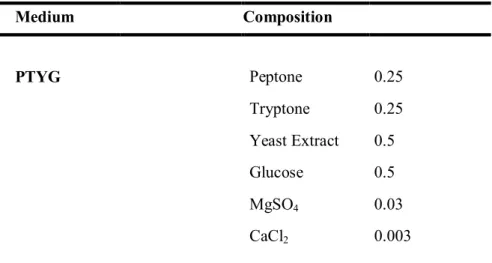
Peptone-tryptone-yeast extract-glucose (PTYG) medium containing (per liter) 0.25 g of peptone (Difco Laboratories, Detroit, USA), 0.25 g of peptone (Difco), 0.5 g of yeast extract (Difco), 0.5 g of glucose, 0.03 g of magnesium sulfate, and 0.003 g of calcium chloride was used to isolate strains and cultivate colonies (Table 1). All cultures were incubated at 28 ℃ and liquid cultures were aerated by shaking at 150 rpm on a rotary shaker (Vision Co., Korea).
2. Chemicals
Analytical grade of EPN (O-Ethyl O-(4-nitrophenyl) phenylphosphonothioate), malathion (Diethyl 2-[(dimethoxyphosphorothioyl) sulfanyl] butanedioate), fenthion (O,O-Dimethyl [3-methyl-4-(methylsulfanyl) phenyl] phosphorothioate), fenitrothion (O,Dimethyl
O-Table 1. The composition of bacterial culture media Medium Composition PTYG Peptone 0.25 Tryptone 0.25 Yeast Extract 0.5 Glucose 0.5 MgSO4 0.03 CaCl2 0.003
MMO Sol. A Na2HPO4 0.71
KH2PO4 0.68 Sol. B (NH4)2SO4 0.3 Sol. C MgSO4·7H2O 0.05 Sol. D CaCl2·H2O 0.001 Sol. E FeSO4·7H2O 0.006 ZnSO4·7H2O 0.0028 MnSO4·7H2O 0.0012 Co(NO3)2·6H2O 0.0017 CuSO4·5H2O 0.0004 (NH4)6Mo7O24· 0.0002
(a) (b)
(c) (d)
(e) (f)
Fig. 1. Structural formulas of EPN (a), malathion (b), fenthion (c),
fenitrothion (d), diazinon (e), 4-nitrophenol (f)
3. Enrichment and isolation of EPN-degrading bacteria
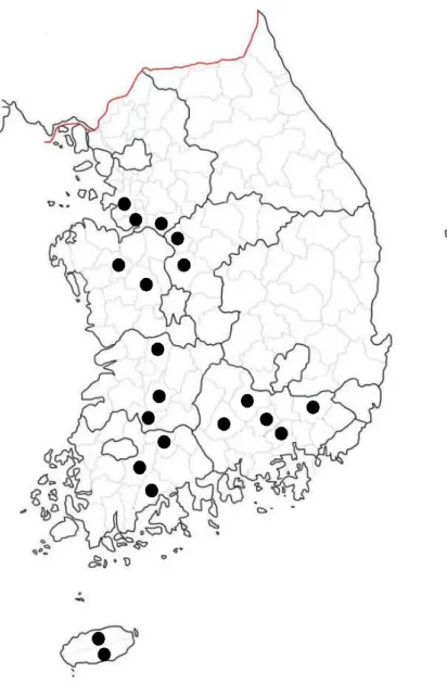
Agricultural soil samples were collected at various locations in South Korea (Fig. 2). Samples from the top 15 cm of soil were taken, sieved through 2-mm-pore size mesh and kept at 4 prior to ℃ use. For enrichment of EPN-degrading bacteria from the agricultural soil, a 20 g of each soil sample was transferred to each sterilized beaker and EPN dissolved in distilled water was treated with a final concentration of 100 μgg-1soil and thoroughly mixed. Theteated soil samples were incubated at room temperature and mixed periodically. Five weeks after the EPN application, a 1 g of soil sample from each beaker was homogenized in 3 ml of a sterilized mineral medium supplemented with EPN of 100μgml-1 (EPN medium) by vortexing. Samples
(0.1 ml) of appropriate 10-fold dilutions were transferred to tube containing 3 ml of EPN medium. For two weeks, the tubes were incubated at 28 ℃ on a rotary shaker at 150 rpm and degradation of EPN was analyzed by NanoDrop 2000c spectrophotometer (Thermo Fisher Scientific, USA). The culture of the final positive tube showing considerable cell growth and less than 10 % of the EPN renaining was diluted and spread onto PTYG agar plates and the plates were incubated at 28 ℃. Each colony was streaked onto PTYG agar and the single colonies were tested for EPN degradation in fresh EPN medium before strain identification (Fig. 3).
Fig. 3. Isolation of EPN-degrading bacteria
Enrichment of EPN
(0.1ul/g, 5 weeks)
Mineral medium with EPN
(0.1ul/ml)
Streaking on
PTYG agar medium
Pure culture on
PTYG agar medium
UV-Vis spectrophotometer
Mineral medium with EPN
4. Phylogenetic identification by 16S rRNA gene sequence
analysis
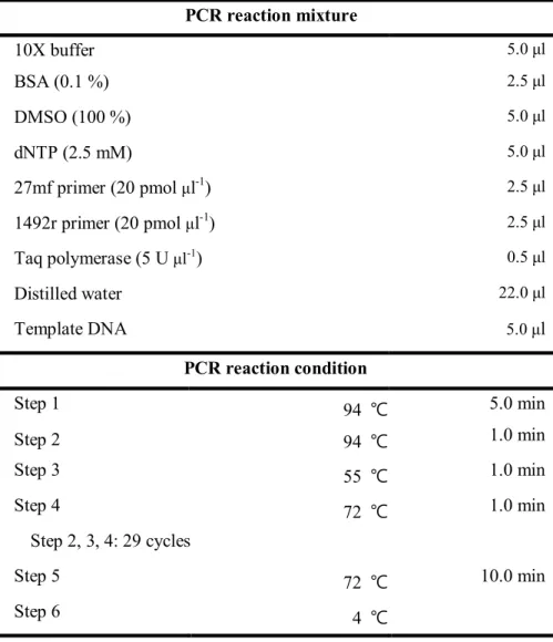
Total genomic DNAs were extracted from the isolates and PCR amplification of 16S rRNA was performed with 27mf and 1492r as previously described (Baker et al., 2003) (Table 2). The amplified 16S rRNA genes were sequenced using an ABI Prism BigDye Terminator Cycle Sequencing Ready Kit according to the manufacturer’s instruction (Perkin-Elmer) with the sequencing primers 515r, 926f and 1055r (Stackebrandt and Goodfellow, 1991). The almost full length 16S rRNA gene sequence was determined as described previously (Kim and Ka, 2017). The 16S rRNA gene sequence was compared with sequences of bacterial type strains from the database of EzBioCloud server (Chun et al., 2003; Yoon et al., 2017) and sequences from isolates and related taxa (retrieved from the NCBI database) were aligned with SINA (v1.2.11) according to the SILVA seed alignment (http://www.arb-silva.de; Pruesse et al., 2012; Quast et al., 2012). Phylogenetic trees were constructed by neighbor-joining method (Saitou and Nei, 1987), maximum-parsimony method (Fitch, 1971) and maximum-likelihood method (Felsenstein, 1981) using the MEGA6 software program (Tamura et al., 2013).
Table 2. PCR conditions for 16S rRNA gene amplification PCR reaction mixture 10X buffer 5.0 μl BSA (0.1 %) 2.5 μl DMSO (100 %) 5.0 μl dNTP (2.5 mM) 5.0 μl 27mf primer (20 pmol μl-1) 2.5 μl 1492r primer (20 pmol μl-1) 2.5 μl Taq polymerase (5 U μl-1) 0.5 μl Distilled water 22.0 μl Template DNA 5.0 μl PCR reaction condition Step 1 94 ℃ 5.0 min Step 2 94 ℃ 1.0 min Step 3 55 ℃ 1.0 min Step 4 72 ℃ 1.0 min Step 2, 3, 4: 29 cycles Step 5 72 ℃ 10.0 min Step 6 4 ℃
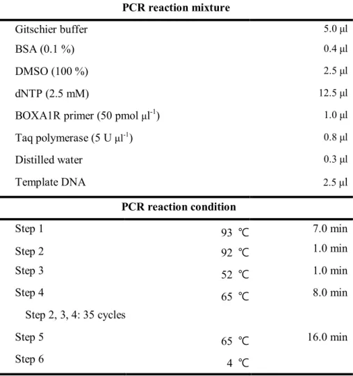
5. Colony REP-PCR
To characterize the isolates at strain level using genomic fingerprints of the isolates, colony repetitive extragenic palindromic (REP)-PCR was performed using BOXA1R primer (5’-CTACGGCAAGGCGACGCTGACG-3’), as described previously (De Bruijn, 1992; Kim and Ka, 2013). Each isolate was grown on PTYG agar plate for 24-48 h, and a small amount of cells was resuspended in 25 μl of PCR mixture containing Gitschier buffer [1 M (NH4)2SO4, 1 M Tris-HCl (pH 8.8), 1 M MgCl2, 0.5 M EDTA (pH 8.8),
14.4 M β-mercaptoethanol], 0.1 % bovine serum albumin, 100 % dimethyl sulfoxide, each deoxynucleotide triphosphate (dNTP) at a concentration of 2.5 mM, and 50 pmol μl-1of DNA (Rademaker et al., 1998). The cycles used were
as follows: 1cycle at 93 ℃ for 7 min, 35 cycles at 92 ℃ for 1 min, 52 ℃ for 1 min, and at 65 ℃ for 8 min;1 cycle at 65 ℃ for 16 min; and a final soak at 4 ℃ (Table 3). After the reactions, PCR products were separated by electrophoresis on 1.2 % agarose gels. After electrophoresis, the image was photographed with UV trans-illumination (306nm).
Table 3. PCR conditions for colony REP-PCR PCR reaction mixture Gitschier buffer 5.0 μl BSA (0.1 %) 0.4 μl DMSO (100 %) 2.5 μl dNTP (2.5 mM) 12.5 μl
BOXA1R primer (50 pmol μl-1) 1.0 μl
Taq polymerase (5 U μl-1) 0.8 μl Distilled water 0.3 μl Template DNA 2.5 μl PCR reaction condition Step 1 93 ℃ 7.0 min Step 2 92 ℃ 1.0 min Step 3 52 ℃ 1.0 min Step 4 65 ℃ 8.0 min Step 2, 3, 4: 35 cycles Step 5 65 ℃ 16.0 min Step 6 4 ℃
6. Analysis of bacterial growth and degradation of EPN
The isolates were cultivated in PTYG broth medium for 48 hours. Cell were collected by centrifugation at 15,000 g for 10 min at 4 and washed ℃ twice with mineral medium. Aliquots of resuspended cells were inoculated into flasks containing 200 ml of mineral medium supplemented with EPN (100 μg ml-1) as the sole carbon source at a final density of OD600=0.005. All
cultures were incubated at 28 in the dark on a rotary shaker (150 rpm). At ℃ specific intervals, aliquots of the cultures were taken out and used to measure cell growth and the concentration of EPN residues. Cell growth was determined at optical density 600 nm with spectrophotometer. For quantification of EPN, 3 ml of cultures were dissolved in an organic solvent and filtered using PTFE syringe filter. Filtered culture was used for the measurement of optical densities at 275 nm for residual EPN. The residue of EPN was calculated using standard curves prepared from the known concentration of EPN in the same medium.
7. Analysis of degradation diversity
Each strain was grown in fresh EPN medium to produce cells induced for EPN metabolism. Cells were harvested, washed, and prepared in the same way as described above. Aliquots of suspended cells were inoculated into culture tubes, with each of which containing 3 ml mineral medium supplemented with malathion, fenthion, fenitrothion, diazinon, and 4-nitrophenol at a concentration of 100 μg ml-1. The tubes were cultured by
shaking 150 rpm at 28 ℃ for two weeks, after which the optical density at 600 nm was measured. To determine the degradation of organophosphorus chemicals, the cultures were centrifuged to remove the cellular material, and UV absorption was measured to monitor degradation of the substrates.
8. Analysis of physiological characteristics
Bacterial growth at 4, 10, 15, 20, 25, 28, 37, 40, and 45 was assessed ℃ after 7 days of incubation in PTYG broth. The pH range for growth was determined after 7 days of incubation at 28 in PTYG broth adjusted pH 5.0 ℃ - 10.0 at intervals of 0.5 pH units by using citrate-NaH2PO4 buffer (pH 5.0
-6.0), NaH2PO4-Na2HPO4buffer (pH 6.5 - 8.0), Tris-HCl buffer (pH 8.5 - 9.0),
and Na2CO3-NaHCO3 buffer (pH 9.2 - 10.0) (Breznak and Costilow, 2007).
Tolerance to NaCl was tested in PTYG broth supplemented with 0 - 8.0 % NaCl (w/v) (at 0.5 % intervals) after 7 days of incubation. Growth of isolates was tested on PTYG agar, R2A agar (R2A; Difco), Luria-Bertani agar (LB; Difco), trypticase soy agar (TSA; Difco), nutrient agar (NA; Difco) and MacConkey agar (MC; Difco) at 28 for 7 days. Growth under anaerobic ℃ conditions was determined with anaerobic atmosphere generation bag (Sigma Chemical). Oxidase and catalase activities were tested using Oxidase Reagent and ID color Catalase Reagent, respectively (bioMérieux).
9. Chemical analysis and identification of intermediates
Incubation condition were same as above. The 50 mL of culture samples were collected and compounds in the samples were extracted and concentrated liquid-liquid extraction. Liquid-liquid extraction was experimented with separatory funnel. Sample 50 ml and dichloromethane 150 ml were added to a separatory funnel was shaken to check for leaks. After 5 min, remove the gas and add saturated NaCl 50ml in separatory funnel and shake it for 5 min. To separate the layers, mixture was leaved at room temperature for 5 minutes. Organic layer containing the compound was transferred to beaker with sodium sulfate for removing moisture and evaporated using a water bath. Lastly, the retained target compounds eluted with 1 ml of methanol were filtered by GH Polypro membrane (Pall Corporaion). Metabolites of EPN were detected by MS analysis. The GC-MS analyses were achieved in electron ionization mode (70 eV) with Perkin-Elmer clarus 680 GC equipped with DB-5MS column (30 m × 0.25 mm id; 0.25 μm film thickness). The column temperature system was programed from 40 °C (1 min hold), at 30 min℃ -1to 130 , at 5 min℃ ℃ -1, to 250 , at ℃ 10 min℃ -1 to 300 , and then held for 5 min℃ . The helium was used as the carrier gas at a constant flow of 1.2 mL min-1. The samples were analyzed in10. PCR amplification of the genes involved in the
hydrolysis of organophosphorus compound
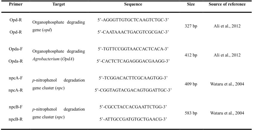
The partial gene sequences specific to the EPN degradation pathway were amplified by PCR with specific primers targeted for the organophosphate degrading gene opd, and opdA, and the 4-nitrophenol degrading gene cluster npc. The most widely studied organophosphate degradation gene is opd gene, encoding the OPH (organophosphorus hydrolase) enzyme. And gene opdA (organophosphate degrading Agrobacterium) was identified in Agrobacterium radiobacter P230 and possessed 88.4 % nucleotide sequence homology with opd gene from Flavobacterium sp. ATCC 27551 (Horne et al., 2002). For the detection of organophosphate degrading genes, the primers designed from conserved regions of previously reported sequences of the opd, opdA genes were used (Ali et al., 2012). The primer for the p-nitrophenol degrading gene cluster npc gene have been reported in Rhodococcus spacus SAO101 (Kitagawa et al., 2004). Among the designed five primer set, set A and set B were used for the detection of the npc gene. The amplification of the oph, opdA, npcA, npcB genes with the corresponding primers was expected to produce 327 bp, 412 bp,
Table 4. PCR primer and product sizes
Primer Target Sequence Size Source of reference
Opd-R Opd-R Organophosphate degrading gene (opd) 5’-AGGGTTGTGCTCAAGTCTGC-3’ 5’-CAATAAACTGACGTCGCGAC-3’ 327 bp Ali et al., 2012 Opda-F Opda-R Organophosphate degrading Agrobacterium (OpdA) 5’-TGTTCCGGTAACCACTCACA-3’ 5’-CACTCTCAGAGGGACGAAGG-3’ 412 bp Ali et al., 2012 npcA-F npcA-R p-nitrophenol degradation gene cluster (npc) 5’-TCGGACACTTCGCAAGTGG-3’ 5’-CGGTAGTACGACAGTGGATTGC-3’ 409 bp Wataru et al., 2004 npcB-F npcB-R p-nitrophenol degradation gene cluster (npc) 5’-CGCCTACCACGAATTCTGG-3’ 5’-ATTGCCGATGTGCTGAACG-3’ 583 bp Wataru et al., 2004
III. RESULTS
1. Isolation of EPN-degrading bacteria
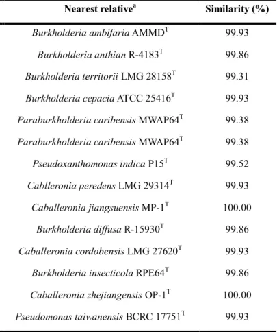
Fourteen EPN-degrading bacteria were isolated through enrichment process from different agricultural soils (Table 5). Among 203 agricultural soil samples, 189 soil samples apparently did not show any degradation of EPN during five weeks incubation period on mineral medium containing EPN 100 μg ml-1. Only 14 enrichment soil samples could completely mineralize EPN as a sole carbon and energy source. Through repeated enrichment and purification steps, fourteen EPN-degrading bacteria were isolated from the enrichment cultures. Most of the EPN-degrading bacteria were found from Jeju-do province.
Table 5. Nearest relatives of the EPN-degrading isolates based on 16S rRNA gene sequence analysis
Isolates Accession number Soil site Nearest relativea Similarity (%)
SH 2-4 Seogwipo-si, Jeju-do Burkholderia ambifaria AMMDT 99.93
AS 2-4 Seogwipo-si, Jeju-do Burkholderia anthian R-4183T 99.86
OP 3-3 Seogwipo-si, Jeju-do Burkholderia territorii LMG 28158T 99.31
SM 2-2 Seogwipo-si, Jeju-do Burkholderia cepacia ATCC 25416T 99.93
SGD Seogwipo-si, Jeju-do Paraburkholderia caribensis MWAP64T 99.38
SGL Jeju-si, Jeju-do Paraburkholderia caribensis MWAP64T 99.38
GJ 3 Anseong-si, Gyeonggi-do Pseudoxanthomonas indica P15T 99.52
GJ 5 Anseong-si, Gyeonggi-do Cablleronia peredens LMG 29314T 99.93
HW Pyeongtaek-si, Gyeonggi-do Caballeronia jiangsuensis MP-1T 100.00
DC 1 Wanju-gun, Jeollabuc-do Burkholderia diffusa R-15930T 99.86
DC 3 Wanju-gun, Jeollabuc-do Caballeronia cordobensis LMG 27620T 99.93
2. Strain identification by 16S rRNA gene sequence
analysis and colony REP-PCR analysis
When analyzed by 16S rRNA gene sequences, the isolates were found to be related to Burkholderia, Caballeronia, Paraburkholderia, Pseudosanthomonas, and Pseudomonas (Table 5), having > 98 % sequence similarity to previoulsy reported species. Fourteen EPN-degrading isolates are composed of five Burkholderia, four Caballeronia, three Paraburkholderia, one Pseudosanthomonas and one Pseudomonas (Table 5). Three genus, Burkholderia, Caballeronia and Paraburkholderia were obligately aerobic, rod-shaped, motile by means single or multiple pollar flagella, Gram-negative and belonged to the beta subgroups of Proteobacteria. The other two genus, Pseudosanthomonas and Pseudomonas were Gram-negative and belonged to the gammaproteobacteria.
The neighbor-joining method was used to establish the phylogenetic relationships among these isolates on the basis of their 16S rRNA gene sequences and also their relationships between closely relate type strains (Fig. 4). Phylogenetic relationships was referred to maximum-parsimony method and maximum-likelihood method as well as neighbor-joining method. The
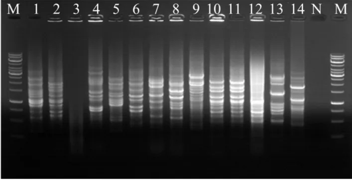
To investigate the genomic relation among the closely related isolates by 16S rRNA sequence analysis, colony REP-PCR experiment was performed by PCR amplification with the BOXA1R primer. One identical REP-PCR band pattern was obtained from strain SGD and SGL and twelve different DNA fingerprinting patterns were obtained (Fig. 5). This result means that strains SGD and SGL were identical.
Fig. 5. Fig. 5. Colony REP-PCR band patterns of isolates. Lane: 1, SH 2-4; 2, AS 2-4; 3, SM 2-2; 4, GJ 5; 5, DC 1; 6, BL; 7, HW; 8, DC 3; 9, ND 1; 10, OP 3-3; 11, SGD; 12, SGL; 13, GJ 3; 14, ND 2; M, Marker; N, Negative control.
3. Growch curve pattern and degradation phenotype
The isolate of each EPN-degrading bacteria was grown on mineral medium supplemented with EPN of 100 μgml-1. All fourteen isolates could
completely degrade and mineralize EPN and grew over period of about two weeks. Isolates were classified into three groups (A, B, and C) according to their growth curve pattern and cell density of peak. There was no significant initial lag phase, growth curve of all isolates shows a gentle slope for two weeks. But cell density of curve peak was different each other. Group A, which showed the highest cell density value (O.D.600peak value of 0.1 to 0.4),
consists of strain SH 2-4, AS 2-4, GJ 3, DC 1. And Group B consists of OP 3-3, SGD, SGL, HW, DC 3-3, with showing O.D.600peak value of 0.03 to 0.05.
Finally, Group C, which has the lowest cell density value (O.D.600peak value
of less than 0.03), consists of SM 2-2, GJ 5, BL, ND 1, ND 2 (Fig. 6).
During the experiment, optical densities and concentration of EPN were measured regularly. Representative isolates of each group were able to completely degrade EPN within five days, and especially strain SH 2-4 showed the fastest degradation. EPN started to be degraded after 12 hours of inoculation (Fig. 7). Also during the sharp decrease of EPN, the concentration
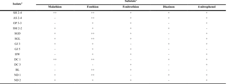
4. Substrate utilization diversity
The isolates were grown on the mineral medium supplemented with EPN of 100μgml-1(EPN medium) to investigate whether these strains are capable
of using other compounds structurally related with EPN. Each strain was inoculated on the mineral medium containing organophosphorus insecticides and 4-nitrophenol. Malathion, fenthion, fenitrothion and diazinon were selected as representative organophosphorus pesticides, and 4-nitrophenol, which is presumed to be and intermediate product of EPN, was selected. The substrate degradation capabilities of the isolates of EPN-degrading bacteria were shown in Table 6.
Strain SH 2-4 was able to fully degrade all substrates and all strains degraded at least two or more substrates (Table 6). Most of EPN-degrading isolates degraded fenthion and fenitrothion. Some strains were capable of degrading nitrophenol, but the others stains were unable to degrade nitrophenol. This means that the strains which could not degrade 4-nitrophenol mineralize EPN through intermediates other than 4-4-nitrophenol. Therefore, it is assumed that there are diverse EPN degradation pathways among the fourteen isolates.
Table 6. Substrate utilization patterns by the EPN-degrading isolates
Isolatea Substate
b
Malathion Fenthion Fenitrothion Diazinon 4-nitrophenol
SH 2-4 ++ ++ + + + AS 2-4 - ++ + + + OP 3-3 - + + - + SM 2-2 + + + + -SGD + ++ + - + SGL + ++ + - + GJ 3 + + - + + GJ 5 - + + - -HW - + + - + DC 1 ++ ++ - + + DC 3 - - + - + BL - ++ + - -ND 1 + ++ - + + ND 2 + + + - +
5. Physiological characteristic of isolates
The strain SH 2-4, AS 2-4, OP 3-3, and SM 2-2 were aerobic and grew at 20 - 37 ℃, pH 5.0 - 8.0, and 0 - 3 % (w/v) NaCl. Optimal growth was observed at 25 - 28 , pH 6.5 and 0 % (w/v) NaCl (Table 7). ℃ Four strains has a similar growth range. Peptone-tryptone-yeast extract-glucose agar (PTYG) was culture medium used to isolate stains from agricultural soils. R2A agar was used for enumerating heterotrophic organisms in treated potable water. PTYG and R2A agar is non-selective culture media. Also Luria-Bertani agar (LB), trypticase soy agar (TSA), and nutrient agar (NA) are a general and non-selective media, providing enough nutrients to support for a various microorganism to grow. MacConkey agar (MC) is a selective and differential medium to isolate Gram-negative and enteric bacilli and differentiate them based on lactose fermentation. Four strains were grown on the PTYG, R2A, LB, TSA, NA, and MC agar media. The strain SH 2-4 and OP 3-3 were positive for catalase, but negative for oxidase. The strain AS 2-4 and SM 2-2 were positive for catalase and oxidase.
Table 7. Physiological characteristics of the isolate SH 2-4, AS 2-4, OP3-3, SM 2-2 Characteristics Isolates SH 2-4 AS 2-4 OP 3-3 SM 2-2 Growth temperature (Optimum) 20 - 37 ℃ (25 - 28 )℃ 15 - 37 ℃ (28 )℃ 20 - 37 ℃ (25℃) 25 - 37 ℃ (25 - 28 )℃ Growth pH range (Optimum) pH 5.0 - 8.5 (pH 6.5 - 7.0) pH 5.0 - 9.5 (pH 6.5 - 7.0) pH 5.0 - 8.0 (pH 6.5) pH 5.0 - 8.0 (pH 7.0) NaCl tolerance ~ 4 % ~ 4 % ~ 3 % ~ 4 %
Aerobic / Anaerobic Aerobic Aerobic Aerobic Aerobic
Catalase + + + +
Oxidase - + - +
Growth media
PTYG + + + +
6. Identification of intermediates and degradation
pathway
To investigate the degradation pathway of EPN, the EPN-degrading strain SH 2-4 was inoculated into EPN mineral medium. After 12 hours incubation with SH 2-4, the intermediate was purified and identified with GC-MS analysis, and then, two peaks A and B were found (Fig. 8). Peak A [Rt
(min) = 24.53] was identified as EPN, which was originally contained in EPN mineral medium(Fig. 9a), and peak B [Rt (min) = 8.76] was identified as
4-nitrophenol (Fig. 9b). This result indicated that strain SH 2-4 transformed EPN to p-nitrophenol. From the results described above, biodegradation pathway of EPN by SH 2-4 was suggested in Figure 11.
Fig. 8. Gas chromatogram of the culture filtrate for EPN mineral medium after 12 hours inoculation of strain SH 2-4. A is EPN with a retention time 24.53 min, B is 4-nitrophenol with a retention time 8.76 min.
Fig. 9. Mass spectrum of gas chromatogram fragmentation and most related standard peak at NIST library. (a) Fragment patterns of the peak A with a retention time 24.53 min and standard peak of EPN in NIST library; (b)
7. PCR amplification of the degradation genes involved in
hydrolysis of organophosphate degrading gene
To investigate genes related with organophosphate degrading genes or 4-nitrophenol degradation gene which were previously reported in other bacterial strain, PCR amplification was performed using PCR primers targeting for previously reported degradative genes, such as opd, opdA (Ali et al., 2012), npc gene (Horne et al., 2002).
All isolates did not show any positive DNA bands of organophosphate gene (opd) and 4-nitrophenol degradation gene cluster (npcB) (Data not show). On the other hand, when PCR amplification was performed using the other organophosphate degradation gene (opdA) and 4-nitrophenol gene (npcA), strains SGD, SGL, and HW showed a positive DNA band of opdA gene (Fig. 11a) and strain SH 2-4, AS 2-4, DC 1, and ND 1 showed a positive DNA band of npcA gene (Fig. 11b)
Fig. 11. PCR amplification with the primers specific for the genes involved in degradation of organophosphorous compound. (a) primer of opdA gene, (b) primer of npcA gene. Lanes: 1, SH 2-4; 2, AS 2-4; 3, OP 3-3; 4, SM 2-2; 5, SGD; 6, SGL; 7, GJ 3; 8, GJ 5; 9, HW; 10, DC 1; 11, DC 3; 12, BL; 13, ND 1; 14, ND 2; M, Maker; N, Negative control.
IV. DISCUSSION
Fourteen bacterial strains capable of utilizing EPN as the sole carbon and energy source were isolated from agricultural soils in Korea. Analysis of 16S rRNA gene sequence showed that all the isolates were phylogenetic related to Burkholderia, Caballeronia, Paraburkholderia, Pseudomonas, and Pseudoxanthomonas. Most of bacterial strains belonging to Burkholderia and Caballeronia (Table 5; Fig. 4).
The genus Burkholderia was proposed by Yabuuchi et al. to accommodate seven species of the genus Pseudomonas homology group II in 1992 (Yabuuchi et al., 1992). Subsequently, parts of the Burkholderia were transferred to
Paraburkholderia and Caballeronia. These four genus are phylogenetically related to
each other(Dobritsa and Samadpour, 2016; Dobritsa et all, 2017). In previous studies, members of Burkholderia have been reported to be able to degrade methyl parathion, fenitrothion (Hayatsu et al., 2000; Ekkhunnatham et al., 2012; Fernández López et al., 2017; Popoca‐ -Ursino et al., 2017). Also, members of Pseudomonas and Pseudoxanthomonas have been known to hydrolyze organophosphorus compound (Serdar and Gibson, 1985; Caldwell and Raushel, 1991; Rani and Lalithakumari, 1994; Liu et al., 2005; Talwar and Ninnekar, 2015).
activity is spite of its adverse effects on mammals including humans as well
as aquatic organisms and birds. For this reason, removal of EPN residues in agricultural soil is important. Many kind of organophosphorus compounds have been studied for biodegradation by microorganisms, but there have been few studies on degradation of EPN. EPN action and enzymatic degradation of EPN in the body had been studied in 1989 (Forsyth and Chambers, 1989). In 1983, study on degradation of EPN in soils under experimental conditions suggested the importance of microbial degradation, but no studies have been conducted since then (Wakabayashi et al., 1983).To our knowledge, this study is the first report on isolation and characterization of EPN-degrading bacteria from agricultural soils.
Fourteen EPN-degrading bacteria were classified into three groups (A, B, and C) according to their growth curve pattern and cell density of peak. All isolates showed a gentle slope growth curve and degradation phenotype for two weeks (Fig. 6; Fig. 7). Group A, which has the highest cell density value had no significant initial lag. Among group A, strain SH 2-4 showed noticeable cell density of peak (Fig. 6). Strain SH 2-4, the representative of group A, degraded EPN entirely within two days (Fig. 7a). Also during the sharp decrease of EPN, the concentration of intermediate increased. Group B,
representative group C, degraded EPN entirely within five days (Fig. 7c). The result of this experiment suggested that EPN was degraded quickly and rapidly by the isolates, and the isolates grew significantly using EPN as the sole source of energy and carbon.
Among the fourteen isolates, Burkholderia ambifastrain SH 2-4 was able to fully degrade all substrates. The others degraded at least two or more substrates (Table 6). Most of EPN-degrading isolates degraded fenthion and fenitrothion. There were many studies on the degradation of fenitrothion by microorganism (Spillner et al., 1979; Lacorte and Barcelo, 1994; Hong et al., 2007). Some strains were able to degrade 4-nitrophenol expected as an intermediate, but the other stains were unable to degrade 4-nitrophenol. This result means that the strains which could not degrade 4-nitrophenol mineralize EPN into intermediate product other than 4-nitrophenol. Thererfor, it is assumed that there diverse EPN degradation pathways among the fourteen isolates.
Four strains has a similar physiological characteristics. The strain SH 2-4, AS 2-4, OP 3-3, and SM 2-2 were aerobic and grew at 20 - 37 ℃, pH 5.0 - 8.0, and 0 - 3 % (w/v) NaCl . Optimal growth was observed at 25 - 28 , pH 6.5℃ , and 0 % (w/v) NaCl (Table 7). Four strains were grown on the agar media; PTYG, R2A, LB, TSA, NA, and MC. The strain SH 2-4 and OP 3-3 were
Previous studies of organophosphate degradation and degradation diversity results of strain SH 2-4 inferred as intermediate products of the EPN degradation process. Burkholderia ambifa strain SH 2-4 could degrade not only EPN but also malathion, fenthion, fenitrothion, diazinon, and even 4-nitropheonl. Nitrophenol was estimated as an intermediate product through the degradation pathway of malathion, fenthion, fenitrothion, and diazinon.
The metabolic degradation pathway of parathion and fenitrothion by Burkholderia sp. NF 100 involved an initial hydrolysis to 4-nitrophenol and 3-methyl-4-nitrophenol, respectively (Chanda et al., 2006;Karpouzas and Singh, 2006; Katsumata te al., 2010). These product were produced by the hydrolysis through the scission of the P-O bond. Diazinon was hydrolysed to diethylthiophosphoric acid (DETP) and 2-isopropyl-4-methyl-6-hydroxypyrimidine (IMHP) (Karpouzas and Singh, 2006; Zhang et al., 2011). Also, this product was produced by the hydrolysis of diazinon through the scission of the O bond (Zhang et al., 2011). These results suggested that P-O bond of EPN was broken and EPN was hydrolyzed to 4-nitrophenol and ethylthiophosphonic acid phenyl ester.
GC-MS analysis of 12 hours samples after inoculation with Burkholderia ambifa strain SH 2-4 showed many kind of peak. EPN degradation by strain
All isolates did not show any positive DNA bands of organophosphate gene (opd) and 4-nitrophenol degradation gene cluster (npcB) (Data not show). On the other hand, when PCR amplification was performed using the other organophosphate degradation gene (opdA) and 4-nitrophenol gene (npcA), strains SGD, SGL, HW showed a positive DNA band of opdA gene (Fig. 11a) and strain SH 2-4, AS 2-4, DC 1, ND 1 showed a positive DNA band of npcA gene (Fig. 11b).
The organophosphate pesticides are concern of environmental pollution because organophosphates are highly toxic to mammals through damage to the nervous system and inhibit acetylcholinesterase (AChE). In this study, the fourteen EPN-degrading bacteria being able to completely mineralize EPN were isolated and characterized in laboratory condition. These isolates may be used as microbial resources for bioremediation purposes in EPN contaminated sites.
LITERATURE CITED
Ali, M., Naqvi, T. A., Kanwal, M., Rasheed, F., Hameed, A., & Ahmed, S. 2012. Detection of the organophosphate degrading gene opdA in the newly isolated bacterial strain Bacillus pumilus W1. Annals of microbiology, 62: 233-239.
Baker, G. C., Smith, J. J., & Cowan, D. A. 2003. Review and re-analysis of domain-specific 16S primers. Journal of microbiological methods, 55: 541-555.
Breznak, J. A., & Costilow, R. N. 2007. Physicochemical factors in growth. In Methods for General and Molecular Microbiology, Third Edition. American Society of Microbiology, 309-329
Caldwell, S. R., & Raushel, F. M. 1991. Detoxification of organophosphate pesticides using an immobilized phosphotriesterase from Pseudomonas diminuta. Biotechnology and bioengineering, 37: 103-109.
Chanda, A., Khetan, S. K., Banerjee, D., Ghosh, A., & Collins, T. J. 2006. Total degradation of fenitrothion and other organophosphorus pesticides by catalytic oxidation employing Fe-TAML peroxide
based on 16S ribosomal RNA gene sequences. International journal of systematic and evolutionary microbiology, 57: 2259-2261.
Cripe, G. M., Goodman, L. R., & Hansen, D. J. 1984. Effect of chronic exposure to EPN and to guthion on the critical swimming speed and brain acetylcholinesterase activity of Cyprinodon variegatus. Aquatic toxicology, 5: 255-266.
De Bruijn, F. J. 1992. Use of repetitive (repetitive extragenic palindromic and enterobacterial repetitive intergeneric consensus) sequences and the polymerase chain reaction to fingerprint the genomes of Rhizobium meliloti isolates and other soil bacteria. Applied and environmental microbiology, 58: 2180-2187.
Dobritsa, A. P., & Samadpour, M. 2016. Transfer of eleven species of the genus Burkholderia to the genus Paraburkholderia and proposal of Caballeronia gen. nov. to accommodate twelve species of the genera Burkholderia and Paraburkholderia. International journal of systematic and evolutionary microbiology, 66: 2836-2846.
Dobritsa, A. P., Linardopoulou, E. V., & Samadpour, M. 2017. Transfer of 13 species of the genus Burkholderia to the genus Caballeronia and reclassification of Burkholderia jirisanensis as Paraburkholderia jirisanensis comb. nov. International journal of systematic and evolutionary microbiology, 67: 3846-3853.
from Burkholderia cepacia capable of degrading organophosphate insecticides. World Journal of Microbiology and Biotechnology, 28: 1739-1746.
Felsenstein, J. 1981. Evolutionary trees from DNA sequences: a maximum likelihood approach. Journal of molecular evolution, 17: 368-376. Felsenstein, J. 1985. Confidence limits on phylogenies: an approach using the
bootstrap. Evolution, 39: 783-791.
Fernández López, M. G., Popoca Ursino, C., Sánchez Sa‐ ‐ ‐ linas, E., Tinoco Valencia, R., Folch Mallol, J. L., Dantán González, E., & ‐ ‐ ‐ Laura Ortiz Hernández, M. 2017. Enhancing methyl parathion ‐ degradation by the immobilization of Burkholderia sp. isolated from agricultural soils. MicrobiologyOpen, 6.
Fitch, W. M. 1971. Toward defining the course of evolution: minimum change for a specific tree topology. Systematic Biology, 20: 406-416.
Forsyth, C. S., & Chambers, J. E. 1989. Activation and degradation of the phosphorothionate insecticides parathion and EPN by rat brain. Biochemical pharmacology, 38: 1597-1603.
Hayatsu, M., Hirano, M., & Tokuda, S. 2000. Involvement of two plasmids in fenitrothion degradation by Burkholderia sp. strain NF100. Applied
61.
Horne, I., Sutherland, T. D., Harcourt, R. L., Russell, R. J., & Oakeshott, J. G. 2002. Identification of an opd (organophosphate degradation) gene in an Agrobacterium isolate. Applied and environmental microbiology, 68: 3371-3376.
Karpouzas, D. G., & Singh, B. K. 2006. Microbial degradation of organophosphorus xenobiotics: metabolic pathways and molecular basis. Advances in microbial physiology, 51: 119-225.
Katsumata, H., Okada, T., Kaneco, S., Suzuki, T., & Ohta, K. 2010. Degradation of fenitrothion by ultrasound/ferrioxalate/UV system. Ultrasonics Sonochemistry, 17: 200-206.
Kim, H., Kim, D. U., Lee, H., Yun, J., & Ka, J. O. 2017. Syntrophic biodegradation of propoxur by Pseudaminobacter sp. SP1a and Nocardioides sp. SP1b isolated from agricultural soil. International Biodeterioration & Biodegradation, 118: 1-9.
Kim, N. H., Kim, D. U., Kim, I., & Ka, J. O. 2013. Syntrophic biodegradation of butachlor by Mycobacterium sp. J7A and Sphingobium sp. J7B isolated from rice paddy soil. FEMS microbiology letters, 344: 114-120.
Kitagawa, W., Kimura, N., & Kamagata, Y. 2004. A novel p-nitrophenol degradation gene cluster from a gram-positive bacterium,
Lacorte, S., & Barcelo, D. 1994. Rapid degradation of fenitrothion in estuarine waters. Environmental science & technology, 28: 1159-1163. Liu, H., Zhang, J. J., Wang, S. J., Zhang, X. E., & Zhou, N. Y. 2005. Plasmid-borne catabolism of methyl parathion and p-nitrophenol in Pseudomonas sp. strain WBC-3. Biochemical and biophysical research communications, 334: 1107-1114.
Ortiz-Hernández, M. L., & Sánchez-Salinas, E. 2010. Biodegradation of the organophosphate pesticide tetrachlorvinphos by bacteria isolated from agricultural soils in México. Revista internacional de contaminación ambiental, 26: 27-38
Pang, G. F., Fan, C. L., Liu, Y. M., Cao, Y. Z., Zhang, J. J., Fu, B. L., ... & Wu, Y. P. 2006. Multi-residue method for the determination of 450 pesticide residues in honey, fruit juice and wine by double-cartridge solid-phase extraction/gas chromatography-mass spectrometry and liquid chromatography-tandem mass spectrometry. Food Additives and Contaminants, 23: 777-810.
Park, H. D., & Ka, J. O. 2003. Genetic and phenotypic diversity of dichlorprop-degrading bacteria isolated from soils. The Journal of Microbiology, 41: 7-15.
367.
Pruesse, E., Peplies, J., & Glöckner, F. O. 2012. SINA: accurate high-throughput multiple sequence alignment of ribosomal RNA genes. Bioinformatics, 28: 1823-1829.
Quast, C., Pruesse, E., Yilmaz, P., Gerken, J., Schweer, T., Yarza, P., ... & Glöckner, F. O. 2012. The SILVA ribosomal RNA gene database project: improved data processing and web-based tools. Nucleic acids research, 41: D590-D596.
Rademaker, J. L. W., Louws, F. J., & De Bruijn, F. J. 1998. Characterization of the diversity of ecologically important microbes by rep-PCR genomic fingerprinting. Molecular microbial ecology manual, 3: 1-27. Rani, N. L., & Lalithakumari, D. 1994. Degradation of methyl parathion by
Pseudomonas putida. Canadian journal of microbiology, 40: 1000-1006.
Saitou, N., & Nei, M. 1987. The neighbor-joining method: a new method for reconstructing phylogenetic trees. Molecular biology and evolution, 4: 406-425.
Serdar, C. M., & Gibson, D. T. 1985. Enzymatic hydrolysis of organophosphates: cloning and expression of a parathion hydrolase gene from Pseudomonas diminuta. Nature Biotechnology, 3: 567. Sogorb, M. A., & Vilanova, E. 2002. Enzymes involved in the detoxification
Spillner, C. J., DeBaun, J. R., & Menn, J. J. 1979. Degradation of fenitrothion in forest soil and effects on forest soil microbes. Journal of Agricultural and Food Chemistry, 27: 1054-1060.
Stackebrandt, E., & Goodfellow, M. 1991. Nucleic acid techniques in bacterial systematics. Wiley.
Talwar, M. P., & Ninnekar, H. Z. 2015. Biodegradation of pesticide profenofos by the free and immobilized cells of Pseudoxanthomonas suwonensis strain HNM. Journal of basic microbiology, 55: 1094-1103.
Tamura, K., Stecher, G., Peterson, D., Filipski, A., & Kumar, S. 2013. MEGA6: molecular evolutionary genetics analysis version 6.0. Molecular biology and evolution, 30: 2725-2729.
Wakabayashi, T., Igarashi, H., & Yamamoto, I. 1983. Degradation of EPN in soils under laboratory conditions. Journal of pesticide science, 8: 155-165
Yabuuchi, E., Kosako, Y., Oyaizu, H., Yano, I., Hotta, H., Hashimoto, Y., ... & Arakawa, M. 1992. Proposal of Burkholderia gen. nov. and transfer of seven species of the genus Pseudomonas homology group II to the new genus, with the type species Burkholderia cepacia (Palleroni and
fruits using solid-phase extraction and gas chromatography–mass spectrometry: Determination of 88 pesticides in berries using SPE and GC–MS. Food Chemistry, 127: 855-865.
Yoon, S. H., Ha, S. M., Kwon, S., Lim, J., Kim, Y., Seo, H., & Chun, J. 2017. Introducing EzBioCloud: a taxonomically united database of 16S rRNA gene sequences and whole-genome assemblies. International journal of systematic and evolutionary microbiology, 67: 1613-1617. Zhang, Y., Hou, Y., Chen, F., Xiao, Z., Zhang, J., & Hu, X. 2011. The
degradation of chlorpyrifos and diazinon in aqueous solution by ultrasonic irradiation: effect of parameters and degradation pathway. Chemosphere, 82: 1109-1115.
농업 토양으로부터
EPN 분해 세균의
분리 및 특성 규명
이수연
초록
살충제 EPN은 유기인계 농약으로 과수 및 농업 해충에 대한 살 충효과가 뛰어나지만 인간 및 생태계 내 비표적 생물과 토양미생물 군집에 영향을 주어 생태계의 구성 및 기능에 부정적인 영향을 미 친다. 포유동물이 EPN에 노출될 경우 EPN이 신경계에 침투하여 acetylcholinesterase를 저해하여 독성을 나타낸다. 인체에 대한 고독성 으로 농촌진흥청은 2011년 EPN의 농약 등록을 취소하였다. 그러나 저렴한 가격, 광범위한 활용 이라는 장점으로 여전히 아시아에서 살 충제로 사용되고 있다. 포유동물뿐만 아니라 수생 생물 및 조류에게내에서 일어나는 대사과정에 관한 연구 결과만 보고되었을 뿐, 미생
물에 의한 EPN의 분해는 보고되지 않았다. 본 연구에서는 EPN을
완전히 분해할 수 있는 14 종의 세균을 농업토양에서 분리하였다.
16S rRNA 유전자 염기서열 분석 및 REP-PCR을 통해 이들 세균이 Burkholderia, Cabelleronia, Paraburkholderia, Pseudoxanthomonas, Pseudomonas 에 속하는 균으로 동정 되었고, 이들 균주는 EPN을 유 일 탄소원으로 사용하여 완전히 분해할 수 있는 것으로 관찰되었다. GC-MS 방법에 의해 분해 중간 산물을 분석한 결과, EPN은 중간물 질 4-nitrophenol로 변형되고 그 후 계속적인 대사를 통해 이 물질이 완전히 분해되는 것으로 관찰되었다. 기존 보고된 유기인계 분해 유 전자의 primer를 이용해 PCR증폭을 한 결과 EPN의 가수분해가 일
부 균주가 가지는 organophosphate degradation 유전자인 opdA에 의한 것임을 확인하였다. 이 연구는 EPN이 미생물에 의해 완전히 분해될 수 있다는 것을 확인한 첫 연구이며 이 연구에서 분리된 균주는 EPN뿐만 아니라 유기인계 살충제에 오염된 토양의 정화를 위한 미 생물 자원으로 이용될 수 있을 것으로 기대된다. 주요어: EPN, 살충제, 생분해, 4-nitrophenol, 세균, 가수분해 학번: 2016-22590