*Corresponding author. Tel: +82-2-2220-2562; Fax: +82-2-2298- 2562; E-mail: incheol@hanyang.ac.kr
https://doi.org/10.5483/BMBRep.2018.51.10.192 Received 20 July 2018
Keywords: Cancer, CCN family, Matricellular protein, Signal trans-duction, Therapeutic target
ISSN: 1976-670X (electronic edition)
Copyright ⓒ 2018 by the The Korean Society for Biochemistry and Molecular Biology
Role of the CCN protein family in cancer
Hyungjoo Kim
1, Seogho Son
1& Incheol Shin
1,2,*
1Department of Life Science, Hanyang University, Seoul 04763, 2Natural Science Institute, Hanyang University, Seoul 04763, Korea
The CCN protein family is composed of six matricellular proteins, which serve regulatory roles rather than structural roles in the extracellular matrix. First identified as secreted proteins which are induced by oncogenes, the acronym CCN came from the names of the first three members: CYR61, CTGF, and NOV. All six members of the CCN family consist of four cysteine-rich modular domains. CCN proteins are known to regulate cell adhesion, proliferation, differentiation, and apoptosis. In addition, CCN proteins are associated with cardiovascular and skeletal development, injury repair, in-flammation, and cancer. They function either through binding to integrin receptors or by regulating the expression and activity of growth factors and cytokines. Given their diverse roles related to the pathology of certain diseases such as fibrosis, arthritis, atherosclerosis, diabetic nephropathy, retino-pathy, and cancer, there are many emerging studies targeting CCN protein signaling pathways in attempts to elucidate their potentials as therapeutic targets. [BMB Reports 2018; 51(10): 486-492]
INTRODUCTION
The extracellular matrix (ECM), which is known to occupy the extracellular space between cells, serve not only as structural supports, but also as modulators of diverse cell functions (1). Of the ECM protein families, matricellular proteins are secreted proteins which do not function primarily as structural proteins but rather by regulating cellular functions through interacting with bioeffector molecules such as cell-surface receptors, growth factors, cytokines, and hormones (2). Matricellular proteins include thrombospondin 1 and 2, osteopontin, tenascin C and X, SPARC, SC1/hevin, and the CCN protein family (2). The CCN protein family plays significant roles in biological processes such as embryonic development, inflammation, and cancer, suggesting their
potential as therapeutic targets (3).
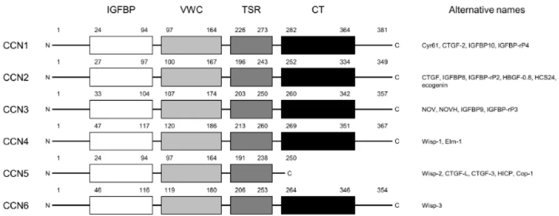
The CCN protein family consists of six members: cysteine-rich angiogenic inducer 61 (Cyr 61; CCN1), connective tissue growth factor (CTGF; CCN2), nephroblastoma over-expressed (NOV; CCN3), and WNT-inducible signaling pathway protein 1, 2, and 3 (WISP 1, 2 and 3; CCN4, 5 and 6). The first three members, Cyr 61, CTGF, and NOV, which gave a name and acronym to the family, were first identified as immediate early gene products of growth factors or tumor transformation related genes (4-6). The six members of the CCN protein family, except for CCN5 which lacks the cysteine-knot (CT) motif, share four conserved protein motifs: the insulin-like growth factor binding protein (IGFBP) motif, von Willebrand factor C-like (VWC) motif, thrombospondin type 1 repeat (TSR) motif, and carboxy-terminal CT motif (Fig. 1). An amino-terminal signal peptide is followed by these four motifs. The VWC and TSR motif is known to be associated in cell-cell interactions and the CT motif is known to be involved in CCN protein dimerization and receptor binding (7). The multimodular structure of the CCN proteins indicate that they may interact with other proteins to exert biological functions. They are known to interact with cell surface integrins, growth factors, cytokines, matrix metalloproteinases (MMPs), and other ECM proteins, such as fibronectin and vitronectin (8).
Since the members of the CCN protein family act as signaling components of the ECM, they are known to be involved in biological processes such as cell adhesion, skeletal development, chondrogenesis, angiogenesis, wound repair, proliferation, and tumorigenesis (8).
NORMAL BIOLOGICAL FUNCTIONS OF THE CCN
PROTEINS
Cell adhesion and migration
As CCN proteins are part of the ECM, one of their primary functions is the regulation of cell adhesion and migration. CCN1 and CCN2 are known to be related to cellular adhesion in diverse cell types. Additionally, CCN2 is required for ECM contraction (9). CCN3 can increase the adhesion of melanocytes to type IV collagen through discoidin domain receptor 1, a receptor tyrosine kinase (10). CCN proteins can also induce adhesion through heparan sulfate proteoglycans (HSPGs) and integrins. In human skin fibroblasts, adhesion to CCN1 and CCN2 through 61-HSPGs induces focal adhesion, actin cytoskeleton rearrangement, and the development of filopodia
Invited Mini Review
Fig. 1. Structure and nomenclature of CCN protein family members. (Left) The amino acid location of the four conserved motifs (IGFBP, VWC, TSR, and CT) are represented as Arabic numerals. (Right) Alternative names for the CCN proteins are indicated. Abbreviations: Cyr61, cysteine rich 61; CTGF-2, connective tissue growth factor 2; IGFBP10, insulin-like growth factor-binding protein 10; IGFBP-rP4, IGFBP-related protein 4; HBGF-0.8, heparin-binding growth factor 0.8; HCS24, hypertrophic chondrocyte specific 24; NOV, nephroblastoma overexpressed gene; NOVH, human nov gene; Wisp, Wnt-inducible secreted protein; Elm-1, expressed in low metastatic cells; HICP, heparin-induced CCN-like protein; and Cop-1, card-only protein 1.
and lamellipodia (11). CCN3 is known to induce adhesion of endothelial cells, vascular smooth muscle cells, and fibroblasts through HSPGs and integrins (8). CCN1, CCN2, and CCN3 all promote cell migration in mesenchymal cells (12-14). CCN4 and CCN5 inhibit cell migration in lung cancer cells and smooth muscle cells, respectively (15, 16).
Cell proliferation
The effects of CCN proteins related to cell proliferation are CCN protein-specific. CCN1 and CCN2 were originally identified as early-response genes related to cell growth (17). In contrast, CCN3 is considered to be an antiproliferative gene (18). Further studies indicated that CCN proteins promote the proliferation of osteoblasts and chondrocytes. CCN1 and CCN2 increases cell proliferation in vascular smooth muscle cells (19, 20). In contrast, CCN3 and CCN5 inhibit cell proliferation in those cells (16, 21). CCN2 is also known to be involved in the mitogen-activated protein kinase/extracellular- signal-regulated kinase (MAPK/ERK) signaling pathway, which is related to cell cycle progression (22).
Osteogenesis and chondrogenesis
All CCN proteins are known to be involved in osteogenesis and chondrogenesis. CCN1 and CCN2 can each promote osteocyte and chondrocyte differentiation (23). Transforming growth factor- (TGF-) increases CCN1, CCN2, and CCN5, but decreases CCN4 mRNA and protein expression in osteoblasts (24). CCN2 and CCN3 can interact with bone morphogenetic protein-2 (BMP-2) and inhibit chondrocyte and osteocyte differentiation, respectively (24, 25). CCN4, on the other hand, increases osteogenesis by enhancing BMP-2 activity (26).
Wound repair and angiogenesis
CCN proteins are well known to have angiogenic activities since they interact with diverse growth factors and integrins (27). CCN1 was first discovered to be related to angiogenesis by using human microvascular endothelial cells (28). Further studies indicated that CCN1, CCN2, and CCN3 can induce angiogenesis in vivo through integrin v3 dependent pathways (14, 29-31). In addition, CCN proteins can inhibit angiogenesis. CCN2 is known to suppress angiogenesis by binding to vascular endothelial growth factor (VEGF), then negatively regulating the angiogenic activity of VEGF (32). The processes of wound healing which include angiogenesis, adhesion, vascularization, and proliferation, are known to be regulated by CCN proteins. CCN1 expression is upregulated in liver regeneration (17, 33). CCN1, CCN2, and CCN3 expression are increased during cutaneous wound repair (17, 34, 35).
CCN PROTEIN FUNCTIONS IN TUMORIGENESIS
In many type of cancers, aberrant CCN protein expression is known to be related to tumorigenesis (36-39). However, although they have similar protein structures, each member of the CCN protein family may play different roles within the same or across different types of cancer.
CCN1
CCN1 expression is known to be upregulated in prostate, ovarian, endometrial, and pancreatic cancer cells (40-43). CCN1 is known to enhance cell migration in prostate cancer (44). In addition, CCN1 expression is elevated in breast cancer, leading to increased invasiveness (43). Tsai et al. (2000) showed that CCN1 acts as a ligand for integrin v3 and is related to breast cancer progression (45). They also
CCN proteins Type of Cancer Role Ref. CCN1 CCN2 CCN3 CCN4 CCN5 CCN6 Prostate cancer Breast cancer Glioma Gastric cancer Breast cancer Pancreatic cancer Glioma Choriocarcinoma Ewing’s sarcoma Melanoma Oral cancer Melanoma Lung cancer Breast cancer Breast cancer
Enhance cell migration Increase invasiveness Related to cancer progression Inhibits apoptosis
Inversely related to MMP-7 expression Increase migration and angiogenesis Increase bone metastasis
Increase tumor growth Decrease cell proliferation Negatively regulate cell proliferation
Decrease cell proliferation and increase migration Decrease proliferation and invasion
Increase cell migration
Attenuates growth and metastasis Decrease migration and invasion Decrease proliferation and invasion Decrease proliferation and invasion
37 36 38 39 41 48 49, 50 46 51 53 54 55 63 64, 65 8 74 76 Table 1. Role of CCN proteins in cancer
demonstrated that CCN1 acts as a downstream of heregulin (HRG) and that CCN1-neutralizing antibodies decreased migration of HRG-positive breast cancer cells (45). One study demonstrated that CCN1 expression is associated with the status of the tumor suppressor gene, p53. They showed that CCN1 was highly expressed in cell lines with mutant and null p53, while low expression of CCN1 was found in cell lines with wild-type p53 (41). In addition, CCN1 is overexpressed in highly tumorigenic glioma cell lines, and forced expression of CCN1 in U343 cells resulted in the activation of the phosphatidylinositol-3-kinase/Akt signaling pathway, leading to the inhibition of the pro-apoptotic protein, Bad (46). On the other hand, CCN1 has been shown to be downregulated in lung and gastric cancer (47, 48). Chien et al. (2004), in contrary to Watari et al. (2009), demonstrated that endometrial cancer cells have lower expressions of CCN1 compared to their normal counterparts, and overexpression of CCN1 decreased endometrial cancer cell growth and induced apoptosis (49).
CCN2
CCN2 overexpression is known to be related to poor prognosis in chondrosarcomas, enchondromas, rhabdomyosarcomas, pancreatic cancer, esophageal cancer, and breast cancer (50-54). Chien et al. (2011) demonstrated that CCN2 overex-pression in a breast cancer cell line resulted in increased migration and angiogenesis, and that the increased migration was dependent of the CT domain of CCN2 protein (55). In contrast, Jiang et al. (2004) analyzed the mRNA and protein expression level of CCN2 in 122 human breast tumors and concluded that CCN2 may act as a tumor suppressor in breast cancer given the results of CCN2 being downregulated in tumor tissues compared to the normal tissues and that CCN2 overexpressing patients have better prognoses than patients
with low CCN2 (52). Additionally, knockdown of CCN2 resulted in decreased pancreatic tumor growth in mouse, indicating that CCN2 may be a good therapeutic target in pancreatic cancer (53). CCN2 overexpression leads to increased breast cancer metastasis to the bone and results in poor-prognosis (56). Shimo et al. (2009) showed that CCN2 is associated with the osteolytic metastasis of breast cancer through the PKA- and PKC-dependent activation of ERK, and that the neutralization of CCN2 using CCN2-specfic antibodies decreased bone metastasis in vivo (57).
CCN3
CCN3 has been shown to have antiproliferative effects in glioma cells (58, 59). Bleau et al. (2007) demonstrated that secreted CCN3 leads to decreased cell proliferation in glioma, and these antiproliferative effects could be neutralized by antibodies that specifically recognize the C-terminal domain of CCN3 (58). They also showed that the CT domain of CCN3 is responsible for the role of CCN3 in cell growth inhibition (58). In addition, CCN3 is able to negatively regulate cell proliferation in choriocarcinoma cells through interacting with a gap junction protein, connexin 43 (60). Benini et al. (2005) showed that CCN3-overexpressing Ewing’s sarcoma cells had reduced cell proliferation but increased migration and invasion (61). In melanoma, CCN3 protein expression is downregulated in invasive cell lines and the forced expression of CCN3 inhibited the proliferation and invasion of melanoma cells (62). In contrast to reports describing the antiproliferative roles of CCN3 in cancer, there have been studies showing the role of CCN3 as an oncogene. CCN3 is overexpressed in rhabdomyosarcoma, cartilage tumors, and prostate cancer (63, 64). Glukhova et al. (2001) demonstrated that CCN3 expression and secretion is increased in nephroblastoma and related to poor prognosis (65). In addition, a study done with
56 paired tissue samples of human cervical cancer and their normal counterparts showed that CCN3 mRNA and protein is overexpressed in cervical cancer (66).
CCN4
CCN4 is known to be overexpressed in colon, colorectal, breast, and lung cancer (47, 67-69). Chuang et al. (2013) demonstrated that CCN4 increased cell migration in oral squamous cell carcinoma through integrin v3 activation and intercellular adhesion molecule-1 expression (70). In contrast, in melanoma, CCN4 expression is inversely correlated to that metastatic potential of K-1735 mouse melanoma cells (71, 72). Additionally, overexpression of CCN4 in highly metastatic K-1735 cells attenuated growth rates and metastasis in vivo (72). Soon et al. (2003) showed that the forced expression of CCN4 in H460 lung cancer cells resulted in decreased cell migration and invasion in vitro and metastasis in vivo through the downregulation of Rac (15). A cohort study done con-ducted on 122 human breast cancer tissues and 32 normal breast tissues indicated that CCN4 mRNA and protein was relatively downregulated in patients with worse prognosis (73). CCN4 expression has been evaluated in chondrosarcomas and enchondromas with various grades and the results found showed that high grade tumors had lower expressions of CCN4 (74).
CCN5
CCN5 is downregulated in human leiomyomas, pancreatic adenocarcinoma, salivary gland tumors, colon tumors, gallbladder cancer, and colorectal cancer (67, 68, 75-78). In hepatocellular carcinoma and adrenocorticotropic hormone- secreting pituitary tumors, CCN5 is upregulated compared to in their normal counterpart tissues (79, 80). In breast cancer, CCN5 expression is low in aggressive breast cancer cell lines (81). The forced expression of CCN5 into MDA-MB-231, an invasive breast cancer cell line, resulted in decreased cell proliferation and invasion (81). Banerjee et al. (2008) demonstrated that the expression profile of CCN5 changes during the course of breast cancer progression (82). In normal breast tissues, CCN5 is undetectable, while its expression is increased in noninvasive breast cancer lesions (82). When noninvasive breast cancer progresses into an invasive type, CCN5 mRNA and protein expression is downregulated by genes such as Snail, MMP-2, and MMP-9 (82).
CCN6
Forced CCN6 expression into an inflammatory breast cancer cell line that resulted in decreased invasion and cell proliferation in vitro and cell growth in vivo (83). Lorenzatti et al. (2011) demonstrated that CCN6 expression level is low in aggressive breast cancer cells, and that recombinant human CCN6 protein attenuates the insulin-like growth factor-1 (IGF-1) signaling pathway and downregulates ZEB1, a transcription factor which is known to be an epithelial-to-
mesenchymal transition activator (84). In addition, chromatin immunoprecipitation assays revealed that the inhibition of CCN6 upregulates Snail and ZEB1 binding to the E-cadherin promoter, which act as transcriptional repressors of E-cadherin in breast cancer (85). In contrast, CCN6 is overexpressed in 63% of human colon tumors and seems to be associated with tumorigenesis in colon cancer (67). In addition, CCN6 was identified as being a novel gene related to colorectal cancers with microsatellite instability (86).
CONCLUSION
CCN family proteins play roles in diverse cellular functions and have different expression profiles among different tissues and organs. Although all six members of the CCN protein family share similar protein structures, their roles are tightly regulated in a spatiotemporal matter rather than playing the redundant roles of other proteins in the same family (7, 18). CCN proteins are known to interact with receptors such as integrins, HSPGs, IGFs, and lipoprotein receptor-related proteins (87, 88). In addition, CCN proteins can bind to other growth factors and cytokines including TGF-, VEGF, fibroblast growth factor 2, and BMPs, altering their biological functions (32, 89, 90). In cancer, the dysregulated expression of CCN proteins is often associated with tumorigenesis and cancer progression (91). Although it differs among various types of cancer, in general, CCN1, CCN2, and CCN4 are known to be related to tumor progression and play roles as oncogenes while CCN3, CCN5, and CCN6 are associated with inhibiting tumor progression and play tumor suppressor roles (Table 1). Since the current literature has certain limitations in clarifying the exact role of CCN proteins in controversial areas, continued studies could help reveal the therapeutic potential of CCN proteins in cancer.
ACKNOWLEDGEMENTS
This work was supported by an NRF grant (2016R1A2 B4011196) from the Korea Research Foundation and by the Bio & Medical Technology Development Program of the NRF funded by the Ministry of Science & ICT (No. 2017M3A9G 8084539).
CONFLICTS OF INTEREST
The authors have no conflicting interests.
REFERENCES
1. Aszodi A, Legate KR, Nakchbandi I and Fassler R (2006) What mouse mutants teach us about extracellular matrix function. Annu Rev Cell Dev Biol 22, 591-621
2. Bornstein P and Sage EH (2002) Matricellular proteins: extracellular modulators of cell function. Curr Opin Cell
Biol 14, 608-616
3. Jun JI and Lau LF (2011) Taking aim at the extracellular matrix: CCN proteins as emerging therapeutic targets. Nat Rev Drug Discov 10, 945-963
4. Joliot V, Martinerie C, Dambrine G, Plassiart G, Brisac M., Crochet J and Perbal B (1992) Proviral rearrangements and overexpression of a new cellular gene (nov) in myeloblastosis-associated virus type 1-induced nephro-blastomas. Mol Cell Biol 12, 10-21
5. O'Brien TP, Yang GP, Sanders L and Lau LF (1990) Expression of cyr61, a growth factor-inducible immediate- early gene. Mol Cell Biol 10, 3569-3577
6. Bradham DM, Igarashi A, Potter RL and Grotendorst GR (1991) Connective tissue growth factor: a cysteine-rich mitogen secreted by human vascular endothelial cells is related to the SRC-induced immediate early gene product CEF-10. J Cell Biol 114, 1285-1294
7. Holbourn KP, Acharya KR and Perbal B (2008) The CCN family of proteins: structure-function relationships. Trends Biochem Sci 33, 461-473
8. Leask A and Abraham DJ (2006) All in the CCN family: essential matricellular signaling modulators emerge from the bunker. J Cell Sci 119, 4803-4810
9. Perbal BV and Takigawa M (2005) CCN proteins : a new family of cell growth and differentiation regulators. Imperial College Press, London ; Hackensack, NJ
10. Fukunaga-Kalabis M, Martinez G, Liu ZJ et al (2006) CCN3 controls 3D spatial localization of melanocytes in the human skin through DDR1. J Cell Biol 175, 563-569 11. Chen CC, Chen N and Lau LF (2001) The angiogenic
factors Cyr61 and connective tissue growth factor induce adhesive signaling in primary human skin fibroblasts. J Biol Chem 276, 10443-10452
12. Grzeszkiewicz TM, Kirschling DJ, Chen N and Lau LF (2001) CYR61 stimulates human skin fibroblast migration through Integrin alpha vbeta 5 and enhances mitogenesis through integrin alpha vbeta 3, independent of its carboxyl-terminal domain. J Biol Chem 276, 21943-21 950
13. Gao R and Brigstock DR (2006) A novel integrin alpha5beta1 binding domain in module 4 of connective tissue growth factor (CCN2/CTGF) promotes adhesion and migration of activated pancreatic stellate cells. Gut 55, 856-862
14. Babic AM, Chen CC and Lau LF (1999) Fisp12/mouse connective tissue growth factor mediates endothelial cell adhesion and migration through integrin alphavbeta3, promotes endothelial cell survival, and induces angiogenesis in vivo. Mol Cell Biol 19, 2958-2966
15. Soon LL, Yie TA, Shvarts A, Levine AJ, Su F and Tchou-Wong KM (2003) Overexpression of WISP-1 down-regulated motility and invasion of lung cancer cells through inhibition of Rac activation. J Biol Chem 278, 11465-11470
16. Lake AC, Bialik A, Walsh K and Castellot JJ Jr (2003) CCN5 is a growth arrest-specific gene that regulates smooth muscle cell proliferation and motility. Am J Pathol 162, 219-231
17. Lau LF and Lam SC (1999) The CCN family of angiogenic regulators: the integrin connection. Exp Cell Res 248,
44-57
18. Perbal B (2001) NOV (nephroblastoma overexpressed) and the CCN family of genes: structural and functional issues. Mol Pathol 54, 57-79
19. Fan WH, Pech M and Karnovsky MJ (2000) Connective tissue growth factor (CTGF) stimulates vascular smooth muscle cell growth and migration in vitro. Eur J Cell Biol 79, 915-923
20. Grzeszkiewicz TM, Lindner V, Chen N, Lam SC and Lau LF (2002) The angiogenic factor cysteine-rich 61 (CYR61, CCN1) supports vascular smooth muscle cell adhesion and stimulates chemotaxis through integrin alpha(6)beta(1) and cell surface heparan sulfate proteoglycans. Endocrino-logy 143, 1441-1450
21. Shimoyama T, Hiraoka S, Takemoto M et al (2010) CCN3 inhibits neointimal hyperplasia through modulation of smooth muscle cell growth and migration. Arterioscler Thromb Vasc Biol 30, 675-682
22. Yosimichi G, Nakanishi T, Nishida T, Hattori T, Takano- Yamamoto T and Takigawa M (2001) CTGF/Hcs24 induces chondrocyte differentiation through a p38 mitogen-activated protein kinase (p38MAPK), and proliferation through a p44/42 MAPK/extracellular-signal regulated kinase (ERK). Eur J Biochem 268, 6058-6065 23. Zuo GW, Kohls CD, He BC et al (2010) The CCN
proteins: important signaling mediators in stem cell differentiation and tumorigenesis. Histol Histopathol 25, 795-806
24. Parisi MS, Gazzerro E, Rydziel S and Canalis E (2006) Expression and regulation of CCN genes in murine osteoblasts. Bone 38, 671-677
25. Maeda A, Nishida T, Aoyama E et al (2009) CCN family 2/connective tissue growth factor modulates BMP signalling as a signal conductor, which action regulates the proliferation and differentiation of chondrocytes. J Biochem 145, 207-216
26. Ono M, Inkson CA, Kilts TM and Young MF (2011) WISP-1/CCN4 regulates osteogenesis by enhancing BMP-2 activity. J Bone Miner Res 26, 193-208
27. Kubota S and Takigawa M (2007) CCN family proteins and angiogenesis: from embryo to adulthood. Angio-genesis 10, 1-11
28. Babic AM, Kireeva ML, Kolesnikova TV and Lau LF (1998) CYR61, a product of a growth factor-inducible immediate early gene, promotes angiogenesis and tumor growth. Proc Natl Acad Sci U S A 95, 6355-6360
29. Lin CG, Leu SJ, Chen N et al (2003) CCN3 (NOV) is a novel angiogenic regulator of the CCN protein family. J Biol Chem 278, 24200-24208
30. Shimo T, Nakanishi T, Nishida T et al (1999) Connective tissue growth factor induces the proliferation, migration, and tube formation of vascular endothelial cells in vitro, and angiogenesis in vivo. J Biochem 126, 137-145
31. Fataccioli V, Abergel V, Wingertsmann L et al (2002) Stimulation of angiogenesis by Cyr61 gene: a new therapeutic candidate. Hum Gene Ther 13, 1461-1470 32. Inoki I, Shiomi T, Hashimoto G et al (2002) Connective
tissue growth factor binds vascular endothelial growth factor (VEGF) and inhibits VEGF-induced angiogenesis. FASEB J 16, 219-221
33. Nathans D, Lau LF, Christy B, Hartzell S, Nakabeppu Y and Ryder K (1988) Genomic response to growth factors. Cold Spring Harb Symp Quant Biol 53 Pt 2, 893-900 34. Grotendorst GR (1997) Connective tissue growth factor: a
mediator of TGF-beta action on fibroblasts. Cytokine Growth Factor Rev 8, 171-179
35. Lin CG, Chen CC, Leu SJ, Grzeszkiewicz TM and Lau LF (2005) Integrin-dependent functions of the angiogenic inducer NOV (CCN3): implication in wound healing. J Biol Chem 280, 8229-8237
36. Dhar A and Ray A (2010) The CCN family proteins in carcinogenesis. Exp Oncol 32, 2-9
37. Perbal B (2001) The CCN family of genes: a brief history. Mol Pathol 54, 103-104
38. Bleau AM, Planque N and Perbal B (2005) CCN proteins and cancer: two to tango. Front Biosci 10, 998-1009 39. Perbal B (2006) NOV story: the way to CCN3. Cell
Commun Signal 4, 3
40. Gery S, Xie D, Yin D et al (2005) Ovarian carcinomas: CCN genes are aberrantly expressed and CCN1 promotes proliferation of these cells. Clin Cancer Res 11, 7243-7254
41. Lv H, Fan E, Sun S et al(2009) Cyr61 is up-regulated in prostate cancer and associated with the p53 gene status. J Cell Biochem 106, 738-744
42. Watari H, Xiong Y, Hassan MK and Sakuragi N (2009) Cyr61, a member of ccn (connective tissue growth factor/cysteine-rich 61/nephroblastoma overexpressed) family, predicts survival of patients with endometrial cancer of endometrioid subtype. Gynecol Oncol 112, 229-234
43. Planque N and Perbal B (2003) A structural approach to the role of CCN (CYR61/CTGF/NOV) proteins in tumourigenesis. Cancer Cell Int 3, 15
44. Schmitz P, Gerber U, Jungel E, Schutze N, Blaheta R and Bendas G (2013) Cyr61/CCN1 affects the integrin-mediated migration of prostate cancer cells (PC-3) in vitro. Int J Clin Pharmacol Ther 51, 47-50
45. Tsai MS, Hornby AE, Lakins J and Lupu R (2000) Expression and function of CYR61, an angiogenic factor, in breast cancer cell lines and tumor biopsies. Cancer Res 60, 5603-5607
46. Xie D, Yin D, Tong X et al (2004) Cyr61 is overexpressed in gliomas and involved in integrin-linked kinase- mediated Akt and beta-catenin- TCF/Lef signaling pathways. Cancer Res 64, 1987-1996
47. Chen PP, Li WJ, Wang Y et al (2007) Expression of Cyr61, CTGF, and WISP-1 correlates with clinical features of lung cancer. PLoS One 2, e534
48. Maeta N, Osaki M, Shomori K et al (2007) CYR61 downregulation correlates with tumor progression by promoting MMP-7 expression in human gastric carcinoma. Oncology 73, 118-126
49. Chien W, Kumagai T, Miller CW et al (2004) Cyr61 suppresses growth of human endometrial cancer cells. J Biol Chem 279, 53087-53096
50. Croci S, Landuzzi L, Nicoletti G et al (2007) Expression of connective tissue growth factor (CTGF/CCN2) in a mouse model of rhabdomyosarcomagenesis. Pathol Oncol Res 13, 336-339
51. Deng YZ, Chen PP, Wang Y et al(2007) Connective tissue growth factor is overexpressed in esophageal squamous cell carcinoma and promotes tumorigenicity through beta-catenin-T-cell factor/Lef signaling. J Biol Chem 282, 36571-36581
52. Jiang WG, Watkins G, Fodstad O, Douglas-Jones A, Mokbel K and Mansel RE (2004) Differential expression of the CCN family members Cyr61, CTGF and Nov in human breast cancer. Endocr Relat Cancer 11, 781-791 53. Bennewith KL, Huang X, Ham CM et al (2009) The role of
tumor cell-derived connective tissue growth factor (CTGF/CCN2) in pancreatic tumor growth. Cancer Res 69, 775-784
54. Shakunaga T, Ozaki T, Ohara N et al (2000) Expression of connective tissue growth factor in cartilaginous tumors. Cancer 89, 1466-1473
55. Chien W, O'Kelly J, Lu D et al (2011) Expression of connective tissue growth factor (CTGF/CCN2) in breast cancer cells is associated with increased migration and angiogenesis. Int J Oncol 38, 1741-1747
56. Kang Y, Siegel PM, Shu W et al (2003) A multigenic program mediating breast cancer metastasis to bone. Cancer Cell 3, 537-549
57. Shimo T, Kubota S, Yoshioka N et al (2006) Pathogenic role of connective tissue growth factor (CTGF/CCN2) in osteolytic metastasis of breast cancer. J Bone Miner Res 21, 1045-1059
58. Bleau AM, Planque N, Lazar N et al (2007) Antiproliferative activity of CCN3: involvement of the C-terminal module and post-translational regulation. J Cell Biochem 101, 1475-1491
59. Gupta N, Wang H, McLeod TL et al (2001) Inhibition of glioma cell growth and tumorigenic potential by CCN3 (NOV). Mol Pathol 54, 293-299
60. Gellhaus A, Dong X, Propson S et al (2004) Connexin43 interacts with NOV: a possible mechanism for negative regulation of cell growth in choriocarcinoma cells. J Biol Chem 279, 36931-36942
61. Benini S, Perbal B, Zambelli D et al (2005) In Ewing's sarcoma CCN3(NOV) inhibits proliferation while promoting migration and invasion of the same cell type. Oncogene 24, 4349-4361
62. Fukunaga-Kalabis M, Martinez G, Telson SM et al (2008) Downregulation of CCN3 expression as a potential mechanism for melanoma progression. Oncogene 27, 2552-2560
63. Maillard M, Cadot B, Ball RY et al (2001) Differential expression of the ccn3 (nov) proto-oncogene in human prostate cell lines and tissues. Mol Pathol 54, 275-280 64. Manara MC, Perbal B, Benini S et al (2002) The
expression of ccn3(nov) gene in musculoskeletal tumors. Am J Pathol 160, 849-859
65. Glukhova L, Angevin E, Lavialle C et al (2001) Patterns of specific genomic alterations associated with poor prognosis in high-grade renal cell carcinomas. Cancer Genet Cytogenet 130, 105-110
66. Zhang T, Zhao C, Luo L et al (2013) The clinical and prognostic significance of CCN3 expression in patients with cervical cancer. Adv Clin Exp Med 22, 839-845 67. Pennica D, Swanson TA, Welsh JW et al (1998) WISP
genes are members of the connective tissue growth factor family that are up-regulated in wnt-1-transformed cells and aberrantly expressed in human colon tumors. Proc Natl Acad Sci U S A 95, 14717-14722
68. Davies SR, Davies ML, Sanders A, Parr C, Torkington J and Jiang WG (2010) Differential expression of the CCN family member WISP-1, WISP-2 and WISP-3 in human colorectal cancer and the prognostic implications. Int J Oncol 36, 1129-1136
69. Xie D, Nakachi K, Wang H, Elashoff R and Koeffler HP (2001) Elevated levels of connective tissue growth factor, WISP-1, and CYR61 in primary breast cancers associated with more advanced features. Cancer Res 61, 8917-8923 70. Chuang JY, Chang AC, Chiang IP, Tsai MH and Tang CH
(2013) Apoptosis signal-regulating kinase 1 is involved in WISP-1-promoted cell motility in human oral squamous cell carcinoma cells. PLoS One 8, e78022
71. Hashimoto Y, Shindo-Okada N, Tani M, Takeuchi K, Toma H and Yokota J (1996) Identification of genes differentially expressed in association with metastatic potential of K-1735 murine melanoma by messenger RNA differential display. Cancer Res 56, 5266-5271
72. Hashimoto Y, Shindo-Okada N, Tani M et al (1998) Expression of the Elm1 gene, a novel gene of the CCN (connective tissue growth factor, Cyr61/Cef10, and neuroblastoma overexpressed gene) family, suppresses In vivo tumor growth and metastasis of K-1735 murine melanoma cells. J Exp Med 187, 289-296
73. Davies SR, Watkins G, Mansel RE and Jiang WG (2007) Differential expression and prognostic implications of the CCN family members WISP-1, WISP-2, and WISP-3 in human breast cancer. Ann Surg Oncol 14, 1909-1918 74. Yu C, Le AT, Yeger H, Perbal B and Alman BA (2003)
NOV (CCN3) regulation in the growth plate and CCN family member expression in cartilage neoplasia. J Pathol 201, 609-615
75. Mason HR, Lake AC, Wubben JE, Nowak RA and Castellot JJ Jr (2004) The growth arrest-specific gene CCN5 is deficient in human leiomyomas and inhibits the proliferation and motility of cultured human uterine smooth muscle cells. Mol Hum Reprod 10, 181-187 76. Dhar G, Mehta S, Banerjee S et al (2007) Loss of WISP-2/
CCN5 signaling in human pancreatic cancer: a potential mechanism for epithelial-mesenchymal-transition. Cancer Lett 254, 63-70
77. Kouzu Y, Uzawa K, Kato M et al (2006) WISP-2 expression in human salivary gland tumors. Int J Mol Med 17, 567-573
78. Yang Z, Yang Z, Zou Q et al (2014) A comparative study of clinicopathological significance, FGFBP1, and WISP-2 expression between squamous cell/adenosquamous
carcinomas and adenocarcinoma of the gallbladder. Int J Clin Oncol 19, 325-335
79. Tomimaru Y, Koga H, Yano H, de la Monte S, Wands JR and Kim M (2013) Upregulation of T-cell factor-4 isoform- responsive target genes in hepatocellular carcinoma. Liver Int 33, 1100-1112
80. Colli LM, Saggioro F, Serafini LN et al(2013) Components of the canonical and non-canonical Wnt pathways are not mis-expressed in pituitary tumors. PLoS One 8, e62424 81. Fritah A, Saucier C, De Wever O et al (2008) Role of
WISP-2/CCN5 in the maintenance of a differentiated and noninvasive phenotype in human breast cancer cells. Mol Cell Biol 28, 1114-1123
82. Banerjee S, Dhar G, Haque I et al (2008) CCN5/WISP-2 expression in breast adenocarcinoma is associated with less frequent progression of the disease and suppresses the invasive phenotypes of tumor cells. Cancer Res 68, 7606-7612\
83. Kleer CG, Zhang Y, Pan Q et al (2002) WISP3 is a novel tumor suppressor gene of inflammatory breast cancer. Oncogene 21, 3172-3180
84. Lorenzatti G, Huang W, Pal A, Cabanillas AM and Kleer CG (2011) CCN6 (WISP3) decreases ZEB1-mediated EMT and invasion by attenuation of IGF-1 receptor signaling in breast cancer. J Cell Sci 124, 1752-1758
85. Huang W, Zhang Y, Varambally S et al (2008) Inhibition of CCN6 (Wnt-1-induced signaling protein 3) down- regulates E-cadherin in the breast epithelium through induction of snail and ZEB1. Am J Pathol 172, 893-904 86. Thorstensen L, Diep CB, Meling GI et al (2001) WNT1
inducible signaling pathway protein 3, WISP-3, a novel target gene in colorectal carcinomas with microsatellite instability. Gastroenterology 121, 1275-1280
87. Chen CC and Lau LF (2009) Functions and mechanisms of action of CCN matricellular proteins. Int J Biochem Cell Biol 41, 771-783
88. Lau LF (2011) CCN1/CYR61: the very model of a modern matricellular protein. Cell Mol Life Sci 68, 3149-3163 89. Abreu JG, Ketpura NI, Reversade B and De Robertis EM
(2002) Connective-tissue growth factor (CTGF) modulates cell signalling by BMP and TGF-beta. Nat Cell Biol 4, 599-604
90. Nishida T, Kubota S, Aoyama E, Janune D, Maeda A and Takigawa M (2011) Effect of CCN2 on FGF2-induced proliferation and MMP9 and MMP13 productions by chondrocytes. Endocrinology 152, 4232-4241
91. Li J, Ye L, Owen S, Weeks HP, Zhang Z and Jiang WG (2015) Emerging role of CCN family proteins in tumorigenesis and cancer metastasis (Review). Int J Mol Med 36, 1451-1463