ABSTRACT
Purpose: Hepcidin levels have previously been reported to be correlated with liver damage. However, the association between hepcidin levels and liver fibrosis in children with fatty liver disease remains unclear. This study therefore aimed to investigate the pathophysiology of fibrosis in children with fatty liver disease and its association with hepcidin levels.
Methods: This retrospective case series included 12 boys aged 6–17 years who were diagnosed with nonalcoholic fatty liver disease (NAFLD) or nonalcoholic steatohepatitis (NASH) at the Tokyo Medical University Hospital. Sixteen liver biopsy samples from 12 subjects were analyzed. Serum hepcidin levels were assayed using enzyme-linked immunosorbent assay. Immunostaining for hepcidin was performed, and the samples were stratified by staining intensity.
Results: Serum hepcidin levels were higher in pediatric NAFLD/NASH patients than in controls. Conversely, a significant inverse correlation was observed between hepcidin immunostaining and Brunt grade scores and between hepcidin scores and gamma-glutamyltranspeptidase, hyaluronic acid, and leukocyte levels. We observed inverse correlations with a high correlation coefficient of >0.4 between hepcidin immunostaining and aspartate aminotransferase, alanine aminotransferase, total bile acid, and platelet count.
Conclusion: There was a significant inverse correlation between hepcidin immunoreactivity and fibrosis in pediatric NAFLD patients; however, serum hepcidin levels were significantly higher, suggesting that these patients experienced a reduction in the hepcidin-producing ability of the liver in response to iron levels, leading to subsequent fibrosis. Therefore, hepcidin levels can be used as markers to identify the progression of fibrosis in patients with NAFLD.
Keywords: Immunohistochemistry; Iron; Liver cirrhosis; Nonalcoholic fatty liver disease
INTRODUCTION
Fatty liver disease (or fatty liver) is a broad term for diseases caused by the accumulation of triglycerides in liver cells. In adults, nonalcoholic fatty liver disease (NAFLD) is defined as a fatty liver without obvious causes, such as autoimmune hepatitis, viral hepatitis, or a history of alcohol consumption [1]. Histologically, NAFLD is classified into two categories: those without (simple steatosis) and those with fibrosis, necrosis, and inflammation (nonalcoholic steatohepatitis [NASH]) [2]. According to a population-based study, 4.8% of
Original Article
Received: Sep 1, 2020 Revised: Dec 18, 2020 Accepted: Feb 2, 2021 Correspondence to Norito TsutsumiDepartment of Pediatrics and Adolescent Medicine, Tokyo Medical University, 6-7-1 Nishishinjuku, Shinjuku-ku, Tokyo 160-0023, Japan.
E-mail: [email protected] Copyright © 2021 by The Korean Society of Pediatric Gastroenterology, Hepatology and Nutrition
This is an open-access article distributed under the terms of the Creative Commons Attribution Non-Commercial License (https:// creativecommons.org/licenses/by-nc/4.0/) which permits unrestricted non-commercial use, distribution, and reproduction in any medium, provided the original work is properly cited. ORCID iDs Norito Tsutsumi https://orcid.org/0000-0001-5617-0197 Shigeo Nishimata https://orcid.org/0000-0001-6347-2791 Masaru Shimura https://orcid.org/0000-0002-1998-7349 Yasuyo Kashiwagi https://orcid.org/0000-0003-4183-1276 Hisashi Kawashima https://orcid.org/0000-0003-2571-9962 Conflict of Interest
The authors have no financial conflicts of interest.
Norito Tsutsumi ,1 Shigeo Nishimata ,1 Masaru Shimura ,1,2 Yasuyo Kashiwagi ,1
and Hisashi Kawashima 1
1Department of Pediatrics and Adolescent Medicine, Tokyo Medical University, Tokyo, Japan 2Depatrment of Metabolism, Chiba Children's Hospital, Center for Medical Genetics, Chiba, Japan
Hepcidin Levels and Pathological
Characteristics in Children with Fatty
Liver Disease
adults with NAFLD will develop liver cirrhosis within a mean observation period of 7.6 years [3]. Approximately 10% of the general population in Japan is estimated to have NAFLD, and approximately 1% is estimated to have NASH [4]. NASH/NAFLD in childhood was first identified in 1983 [5]. There have since been reports of children developing NAFLD as early as 2 years of age and NASH-related cirrhosis as early as 8 years of age [6]. A systematic review of cases in children showed a higher prevalence of NAFLD in studies in the clinically obese population (mean prevalence of 34.2%) compared to the general population (7.6%) [7]. The prevalence of NAFLD/NASH is estimated to be as high as 2.5–9.6% of children in the United States and Asian countries, with significant differences in race and ethnicity [8-10].
Hepcidin is an antimicrobial peptide produced primarily in the liver and secreted by macrophages, pancreatic islet cells, and adipose tissue [11]. It has been reported to be a key regulator of iron metabolism [11,12]. Studies using mice have demonstrated that regulating the production of hepcidin or administering hepcidin agonists may prevent iron-induced cellular damage, which occurs due to an increase in hydroxyl radicals through the reaction of iron and reactive oxygen species [13]. Although multiple associations between iron homeostasis and lipid metabolism have been reported, there are limited data suggesting that excess iron is involved in hepatic steatosis and fibrosis [14-16]. The mechanism underlying hepatic fibrosis remains unclear, and the details of its contribution to disease state and prognosis remain unknown, particularly in children with fatty liver disease. To clarify the pathophysiology of fibrosis in children with fatty liver disease, we hypothesized that hepcidin, which plays a key role in iron metabolism, might be associated with liver damage (fibrosis); subsequently, we investigated the correlation between liver fibrosis and hepcidin levels.
MATERIALS AND METHODS
This was a retrospective case study. Patients with NAFLD or NASH diagnosed by percutaneous liver biopsy at the Tokyo Medical University Hospital between 2008 and 2015 were enrolled in this study. Liver biopsy was performed in obese patients in whom liver dysfunction was diagnosed based on laboratory data. The guidelines of the American Association for the Study of Liver Disease, the American College of Gastroenterology, and the American Gastroenterological Association were used as the diagnostic criteria for NASH [6,17]. This study was conducted with the informed consent of the affected children or their caregivers, and was approved by the Institutional Review Board of Tokyo Medical University (study approval number: T2019-0259). All aspects of this study were performed in compliance with the ethical principles for medical research involving human subjects, as outlined in the 1975 Helsinki Declaration (revised in 2013).
Twelve male patients were included in the study, with a median age of 14 years (range, 6–17 years). The median values of the collected laboratory data were as follows: aspartate aminotransferase (AST), 36 (16–90) U/L; alanine aminotransferase (ALT), 73 (11–155) U/L; γ-glutamyltranspeptidase (γGTP), 48 (16–140) U/L; total bilirubin (T-bil), 0.58 (0.37–1.98) mg/dL; direct bilirubin (D-bil), 0.105 (0.01–0.55) mg/dL; total bile acid (TBA), 5.6 (2.6–9.9) μmol/L; type IV collagen, 156 (96–246) ng/mL; hyaluronic acid, 16 (10–31) ng/mL; iron, 78 (51– 108) μg/dL; ferritin, 72.1 (42–178.4) ng/mL; immunoglobulin G (IgG), 924.5 (793–1,320) mg/ dL; uric acid (UA), 6.45 (4.1–8.5) mg/dL; white blood cell (WBC) count, 7,250 (5,100–10,100) / μL; neutrophils, 54.0% (41.7–69.3%); lymphocytes, 37.8% (23.8–45.8%); and platelet count, 283,500 (150,000–400,000) /μL.
Sixteen liver biopsy samples obtained from 12 patients were analyzed. These samples were subjected to hematoxylin-eosin and Azan staining. Diagnosis of NAFLD or NASH was performed each patient separately diagnosed by one of two pathologists, based on Matteoni's score types 1–4 (type one, fatty liver change alone; type two, fat accumulation and lobular inflammation; type three, fat accumulation and ballooning degeneration; and type four, fat accumulation, ballooning degeneration, and either Mallory hyaline or fibrosis) [18].
The NAFLD level (NAFLD Activity Score [NAS], 0–8, which is further subdivided according to the degree of steatosis, lobular inflammation, and hepatocyte ballooning) [19], inflammatory grade (grades 1–3, which is subdivided based on the degree of macrovesicular steatosis, hepatocellular ballooning and disarray for zonal location, lobular inflammation, portal tract inflammation), and fibrosis stage (stages 1–4: grades of fibrosis evaluated separately [zone 3, portal, and bridging fibrosis]) were evaluated in accordance with the procedure described by Brunt et al. [20], and scoring was performed by two pediatricians.
Similarly, the same liver samples were stained with human anti-hepcidin antibody (Abcam, Cambridge, MA, USA) diluted 2,600-fold, and antibody positivity was evaluated by more than two physicians, including a pathologist. Hepatocytes were classified into five categories, according to the degree of cytoplasm and nuclear staining by hepcidin as follows: 1, negative; 2, weakly positive; 3, moderately positive; 4, strongly positive; and 5, cytoplasmic and nuclear staining (Table 1, Fig. 1). Correlations between each score (Matteoni's score, NAS, Brunt inflammatory grading, and fibrosis staging) and laboratory data (AST, ALT, γGTP, T-bil, D-bil, TBA, type IV collagen, hyaluronic acid, iron, ferritin, IgG, UA, WBC, neutrophils, lymphocytes, and platelets) were evaluated using the Spearman's rank correlation coefficient test. The data were analyzed using IBM SPSS Statistics 25.0 (IBM Co., Armonk, NY, USA). Statistical significance was set at p<0.05.
Serum hepcidin (Hepcidin-25) levels were assayed using an enzyme-linked immunosorbent assay (ELISA) kit (Peninsula Laboratories International, Inc, San Carlos, CA, USA); the median value of serum hepcidin was 317.38 pg/mL (range, 164.74–677.45). Serum hepcidin Table 1. Comparison between hepcidin immunostaining score and other scores
Patient number Age Matteoni's score Brunt's grade Brunt's stage Degree of steatosis Lobular inflammation Ballooning Total Hepcidin*
1 11 4 2 2 2 2 1 5 5 2 16 2 1 0 0 1 0 1 3 3 9 ND ND ND ND ND ND ND 3 3 16 3 1 1 1 1 1 3 3 4 10 4 2 3 1 2 1 4 3 5 6 4 2 2 3 1 1 5 1 5 9 3 2 2 ND ND ND ND 3 6 11 4 3 3 2 2 2 6 1 6 15 4 3 3 ND ND ND ND 2 7 15 3 2 3 ND ND ND ND 2 8 12 2 2 1 2 1 0 3 3 8 17 3 2 1 1 1 1 3 3 9 14 3 2 1 3 1 1 5 3 10 13 4 2 2 3 2 1 6 3 11 14 3 2 1 3 1 0 4 1 12 14 3 1 1 1 1 1 3 3 Median 14 3 2 2 2 1 1 4 3 Range 6–17 2–4 1–3 0–3 0–3 1–2 0–2 1–6 1–5 ND: not done.
*Hepatocytes were classified into five categories according to the degree of cytoplasm and nuclear staining by hepcidin staining as follows: 1, negative; 2, weakly positive; 3, moderately positive; 4, strongly positive; and 5, cytoplasmic and nuclear staining.
levels were compared with patients with pneumonia and bronchial asthma, whose blood was drawn at our hospital as a control group.
RESULTS
During the observation period, 24 children were diagnosed with NAFLD and NASH by liver biopsy, 17 of whom underwent hepcidin staining for remnant liver tissue. Of these, one patient was excluded because of underlying Wilson's disease, resulting in 16 enrolled cases. All patients were male.
Serum hepcidin levels were significantly higher in children with fatty liver than in controls (p<0.05) (Fig. 2). There were no cases of anemia in either the cases or control groups for which serum hepcidin levels could be measured.
25 m 25 m
A B
C D
Fig. 1. Scoring of hepcidin immunostaining: 1, negative; 2, weakly positive; 3, moderately positive; 4, strongly positive; 5, staining of the cytoplasm and nucleus. Representative immunostaining of (A) score 1, (B) score 3, (C) score 4 (control: biliary atresia with mild inflammatory change), and (D) score 5. All had strong expressions.
Control 800 700 600 500 400 300 200 100 Fatty liver Hepcidin 0 97+80 pg/mL 359+157 pg/mL p<0.05
Fig. 2. Serum hepcidin in children with fatty liver change. Serum hepcidin (Hepcidin-25) was assayed using an enzyme-linked immunosorbent assay kit; the median serum hepcidin level was 317.38 pg/mL (range, 164.74–677.45 pg/mL). Serum hepcidin levels were compared in patients with pneumonia and bronchial asthma whose blood was drawn at our hospital (normal control group); the median serum hepcidin level was 123.30 pg/mL (range, 5.6–209.09 pg/mL). The serum hepcidin levels were significantly higher in children with fatty liver than in controls (p<0.05).
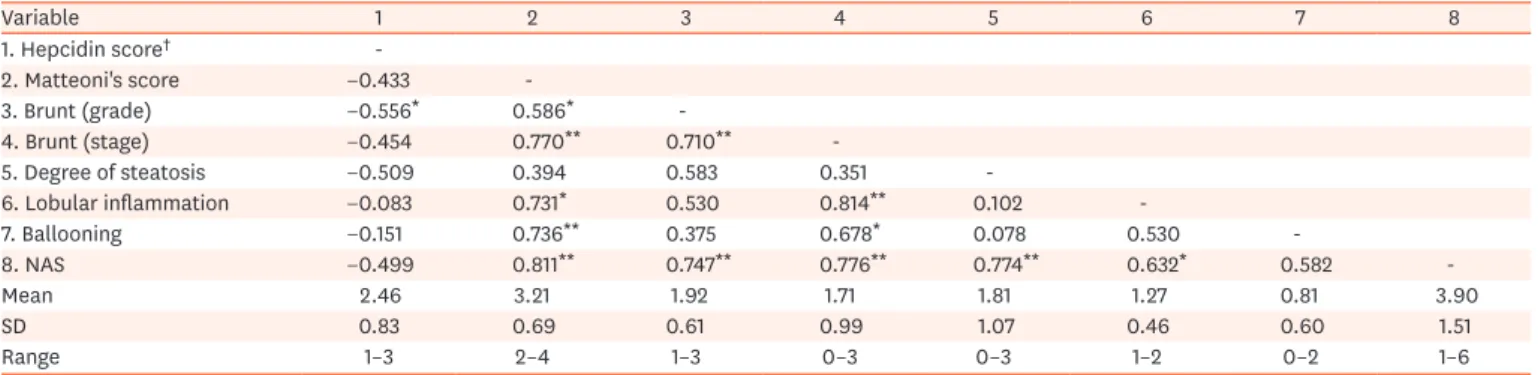
We compared the levels of serum hepcidin and the various characteristics of children with fatty liver and those of controls. No correlation was observed between serum hepcidin levels and age in subjects with or without a hepcidin score of 5. However, a statistically significant inverse correlation was observed between hepcidin and Brunt grade scores. No statistically significant correlation was observed between hepcidin scores and the stage of inflammation or ballooning (Table 2). However, there was an inverse correlation between the hepcidin score on immunostaining with Matteoni's score, the Brunt stage, and steatosis.
We compared hepcidin scores and laboratory data obtained from children with fatty liver. A statistically inverse correlation was observed between hepcidin scores and γGTP, hyaluronic acid, and WBC levels. Conversely, no statistically significant correlation was observed between hepcidin scores and other data. We observed a trend towards an inverse correlation between hepcidin scores and AST, ALT, and TBA, with a correlation coefficient of >0.4. In addition, there was a trend of a positive correlation between hepcidin immunostaining, IgG, and platelet count, with a high correlation coefficient of >0.4. Conversely, no correlation was observed between hepcidin levels and serum iron and ferritin levels (Table 3 and Supplementary Table 1). Table 2. Correlation between hepcidin scores (excluding scores of 5) and other scores and characteristics of fatty liver
Variable 1 2 3 4 5 6 7 8 1. Hepcidin score† -2. Matteoni's score −0.433 -3. Brunt (grade) −0.556* 0.586* -4. Brunt (stage) −0.454 0.770** 0.710** -5. Degree of steatosis −0.509 0.394 0.583 0.351 -6. Lobular inflammation −0.083 0.731* 0.530 0.814** 0.102 -7. Ballooning −0.151 0.736** 0.375 0.678* 0.078 0.530 -8. NAS −0.499 0.811** 0.747** 0.776** 0.774** 0.632* 0.582 -Mean 2.46 3.21 1.92 1.71 1.81 1.27 0.81 3.90 SD 0.83 0.69 0.61 0.99 1.07 0.46 0.60 1.51 Range 1–3 2–4 1–3 0–3 0–3 1–2 0–2 1–6
NAS: NAFLD (nonalcoholic fatty liver disease) Activity Score, SD: standard deviation.
*p<0.05, **p<0.01. †The hepatocytes were classified into five categories according to the degree of cytoplasm and nuclear staining by hepcidin staining as follows: 1, negative; 2, weakly positive; 3, moderately positive; 4, strongly positive; and 5, cytoplasmic and nuclear staining.
Table 3. Correlation between hepcidin scores† (excluding a score of 5) and laboratory data in children with fatty liver
Variable Spearman's rank
correlation coefficient rs (both sides) Mean SD Range
AST (U/L) −0.505 0.055 40.2 19.3 16–90
ALT (U/L) −0.464 0.081 76.7 44.3 11–155
γGTP (U/L) −0.571 0.026* 56 33.1 16–140
T-bil (mg/dL) −0.210 0.452 0.81 0.50 0.37–1.98
D-bil (mg/dL) 0.227 0.415 0.13 0.13 0.01–0.55
Total bile acids (μmol/L) −0.612 0.144 5.7 2.61 2.6–9.9
Type IV collagen (ng/mL) 0.139 0.720 162.8 43.9 96–246 Hyaluronic acid (ng/mL) −0.828 0.042* 18.8 9.2 10–31 Iron (μg/dL) 0.184 0.566 78.2 17.6 51–108 Ferritin (ng/mL) 0.029 0.589 90.6 44.9 42–178.4 IgG (mg/dL) 0.558 0.119 1,002 143.8 872–1,320 UA (mg/dL) −0.177 0.528 6.38 1.35 4.1–8.3 WBC (/μL) −0.607 0.016* 7,593 1,454 5.1–10.1 (×103) Neutrophils (%) 0.006 0.982 54.5 6.82 44.4–69.3 Lymphocytes (%) 0.133 0.637 35.8 6.74 23.8–45.8 Platelets (/μL) 0.485 0.067 28.8 (×104) 5.65 (×104) 18.7–42.5 (×104)
rs: relative score, SD: standard deviation, AST: aspartate aminotransferase, ALT: alanine aminotransferase, γGTP: γ-glutamyltranspeptidase, T-bil: total bilirubin, D-bil: direct bilirubin, IgG: immunoglobulin G, UA: uric acid, WBC: white blood cells.
*p<0.05. †The hepatocytes were classified into five categories according to the degree of cytoplasm and nuclear staining by hepcidin staining as follows: 1, negative; 2, weakly positive; 3, moderately positive; 4, strongly positive; and 5, cytoplasmic and nuclear staining.
DISCUSSION
Hepcidin was first identified as an antimicrobial peptide in urine and plasma [21], and was subsequently discovered to have a profound role in the regulation of iron metabolism. Mice with dietary iron overload showed increased hepcidin mRNA expression and decreased iron absorption [22]. Serum hepcidin levels in children with NAFLD/NASH were significantly higher than those in controls in our study. Serum hepcidin levels in obese patients with NAFLD were reported to be significantly higher than those in obese patients without NAFLD [23]. Cakir et al. [24] reported no significant difference in hepcidin levels in 34 children with chronic liver diseases (12, 4, 6, and 12 cases of Wilson's disease, tyrosinemia, glycogen storage disease, and other chronic diseases, respectively) compared with age- and sex-matched healthy children. However, they observed that hepcidin levels in children with chronic liver diseases were positively correlated with ferritin, pediatric end-stage liver disease score, and total antioxidant status. Transferrin saturation and the hepcidin-to-ferritin ratio were significantly lower in patients with severe fibrosis. Our results further showed that serum hepcidin levels in children with chronic liver diseases reflected both liver function and antioxidant status, and that severe fibrosis was associated with a low hepcidin-to-ferritin ratio in children with chronic liver diseases. The subjects did not have fatty liver diseases, such as NAFLD. Therefore, we can conclude that high serum hepcidin levels are a specific phenomenon observed in fatty liver disease.
NAFLD/NASH is characterized by fatty liver changes, inflammation, fibrosis of the portal area, absence of perisinusoidal fibrosis, and hepatocyte ballooning. Matteoni et al. [18] classified NAFLD into four types, based on pathological findings. Type one, simple fatty liver (only fatty liver); type two, steatohepatitis (fatty liver and lobular inflammation); type three, steatonecrosis as well as ballooning and swelling of hepatocytes; and type four, steatonecrosis and Mallory bodies (liver cell ballooning degeneration) and fibrosis. Progression to liver cirrhosis or liver-associated death was observed in patients with type three and four NAFLD [18]. The grading system of necrosis and inflammation and the staging system of fibrosis defined by Brunt are commonly used [20].
Conversely, NAFLD/NASH can present with different characteristics in adults and children [25]. Patients with strong evidence of fibrosis were classified as having type 2 NAFLD/ NASH. According to Brunt's pathological classification, necrosis and inflammation grades are extremely low, and the fibrosis stage is very high in many children. Schwimmer et al. [26] previously reviewed patients aged 2–18 years with NAFLD, and classified them into two different groups of steatohepatitis by agglomerative hierarchical cluster analysis. Type one was characterized by steatosis, ballooning degeneration, and perisinusoidal fibrosis; type two was characterized by steatosis, portal inflammation, and portal fibrosis. Simple steatosis and advanced fibrosis were observed in 16% and 8% of the subjects, respectively. Type one NASH was observed in 17% of subjects and type two NASH in 51%. NASH type differed significantly according to race and ethnicity, with type one NASH being more common in Caucasian children, while type two NASH was more common in children of Asian ethnicity. In cases of advanced fibrosis, the pattern was generally that of type two NASH. The authors concluded that type one and type two NASH are distinct subtypes of pediatric NAFLD, with type two being the most common presentation in children [26].
Takahashi et al. [25] reported that pediatric steatosis was more severe than adult steatosis after analyzing the clinical and histopathological characteristics of NAFLD in 34 pediatric and
23 adult patients. Perisinusoidal fibrosis, lobular inflammation, and ballooning were milder in pediatric patients than in adult patients. Conversely, portal inflammation was more severe in pediatric patients than in adult patients. Half of the pediatric patients showed overlapping features of types one and two NASH.
Hepatic fibrosis is caused by reactive changes in several liver diseases and is characterized by the accumulation of type I collagen fibers. Liver cirrhosis was the final stage, and extreme fibrosis was observed at this stage. The pattern of fibrosis differs between the lobular and portal regions. Both fibrosis patterns progress and finally lead to liver cirrhosis through bridging necrosis between the portal vein and central vein regions. In our study, hyaluronic acid, a marker of liver fibrosis, was inversely correlated with hepcidin score, suggesting that hepcidin may be associated with liver fibrosis.
When hepatocytes are stimulated with inflammatory cytokines, such as interleukin (IL)-6, hepcidin production is markedly enhanced [27]. A similar increase in hepcidin production was observed when humans were intravenously administered IL-6 and was accompanied by a decrease in iron and transferrin saturation [28]. In normal mice, serum iron rapidly decreases immediately after administration of proinflammatory cytokines; however, this response is not observed in hepcidin-deficient mice or IL-6-deficient mice. These findings demonstrate that in infections and chronic inflammation, hepcidin is produced in response to IL-6, and iron transport by ferroportin is suppressed to reduce the supply of iron to the blood, resulting in functional iron deficiency anemia [29]. Similarly, hepcidin expression by IL-6 is dependent on signal transducer and activator of transcription [30].
The role of hepcidin in NAFLD has recently gained increasing attention. It has been shown that liver necrosis can lead to iron leakage in combination with phagocytosis by liver macrophages and increase hepcidin levels (induced by inflammatory cytokines), resulting in iron accumulation in Kupffer cells. Similarly, leptin can directly regulate hepatic hepcidin expression. Therefore, leptin-induced hepcidin synthesis may favor iron perturbation in patients with NAFLD [31,32]. A highly significant correlation has been reported in patients with dysmetabolic iron overload syndrome between hepcidin levels and tumor necrosis factor-α, indicating that the proinflammatory milieu contributes to hepcidin production in NAFLD patients [33]. From a clinical standpoint, in NAFLD patients, increased hepcidin and proinflammatory cytokines can be derived from adipose tissue and the liver. In NAFLD patients, the expression level of the duodenal iron exporter ferroportin is lower than that in controls and NAFLD patients without liver iron accumulation [34]. These reports indicate that iron and inflammation both contribute to hepcidin synthesis in patients with NAFLD. Two patterns of hepcidin immunostaining were observed in the present study. Decreased hepcidin levels in the cytoplasm of hepatocytes exacerbate liver tissue inflammation. Thus, although the serum hepcidin levels were high, the decrease in hepcidin levels in hepatocytes may have contributed to uncontrolled iron metabolism, inflammation, and fatty changes in the hepatocytes. We excluded samples with an unusual type of hepcidin staining, wherein both the cytoplasm and nucleus were stained. Similarly, there may be other pathophysiological changes in hepcidin expression. One patient was excluded from the study because of Wilson's disease, which was diagnosed by genomic and other analyses showing a relatively high copper level. Therefore, there may be other pathophysiological mechanisms of copper transport underlying pediatric NAFLD.
Mesenchymal iron deposition is more common than hepatocellular iron accumulation; however, both compartments are usually affected by fatty liver disease. Conversely, in tissue iron deposition in hemochromatosis, iron is almost exclusively found in the hepatocellular compartment, and macrophages are iron-deficient [35]. Hepatic stellate cells (HSCs) play a central role in liver fibrosis. In the fibrotic state, type I collagen is produced by HSCs in response to endothelin-1 stimulation. HSCs are activated by various cytokines and changes in the extracellular matrix (ECM). When this transformation occurs, myofibroblasts are formed and collagen synthesis is accelerated [34].
Liver fibrosis is a state in which the balance between the accumulation and decomposition of the hepatic ECM is biased towards accumulation. Protein synthesis, particularly ECM production, is remarkably enhanced and plays a major role in liver fibrosis. Regarding studies on the facilitation of fibrosis by HSC activation upon liver injury, Han et al. [36] showed that hepcidin inhibits HSC activation and ameliorates liver fibrosis. Hepcidin levels are inversely correlated with the exacerbation of fibrosis in patients and animal models. Adenoviral delivery of hepcidin attenuates the liver fibrosis induced by carbon tetrachloride treatment or bile duct ligation in mice [37]. Hepcidin from hepatocytes or exogenous hepcidin suppresses HSC activation by inhibiting transforming growth factor β1-mediated Smad3 phosphorylation, controlled via Akt. In activated HSCs, ferroportin is upregulated, which can also be prevented by hepcidin treatment.
Although our results strongly suggest that hepcidin may suppress liver fibrosis, the limitation of this study is the lack of data on tissues sequentially obtained from the same patient. As fibrosis in subjects with pediatric NASH and NAFLD is mild, other severe types should be investigated to clarify the pathophysiology of fibrosis.
Mitsuyoshi et al. [38] reported that liver hepcidin mRNA levels were lower in patients with NAFLD than in those with simple steatosis, which is similar to the results observed in the pediatric NAFLD/NASH patients in our study. However, serum hepcidin levels in NAFLD patients in our study were higher than those in healthy controls. The source of high serum hepcidin levels in pediatric NAFLD/NASH is considered to be adipocytes and macrophages, in addition to the liver. Reportedly, the increase in hepcidin levels in the adipose tissue of obese subjects is independent of NASH [39]. This represents the second-hit theory of NASH, which postulates that when the ability to produce hepcidin in the liver in response to iron is reduced, iron overload occurs in liver cells, which in turn causes an increase in oxidative stress [14] acting as a second hit, eventually leading to fibrosis.
In conclusion, we observed a statistically significant inverse correlation between hepcidin immunoreactivity and fibrosis in pediatric NAFLD patients, although serum hepcidin levels were significantly higher in these patients than in adult NAFLD patients. These findings suggest a reduced ability of the liver to produce hepcidin in response to iron deficiency in these patients, which is linked to subsequent fibrosis. Therefore, hepcidin levels can be used as a potential marker to identify the progression of fibrosis in patients with NAFLD. Further studies on the role of hepcidin in liver tissue in children with fatty liver disease may provide a better understanding of how NASH differs between children and adults.
SUPPLEMENTARY MATERIAL
Supplementary Table 1
Correlation between hepcidin scores (excluding scores of 5) and laboratory data in children with fatty liver
Click here to view
REFERENCES
1. American Gastroenterological Association. American Gastroenterological Association medical position statement: nonalcoholic fatty liver disease. Gastroenterology 2002;123:1702-4.
PUBMED | CROSSREF
2. Neuschwander-Tetri BA, Caldwell SH. Nonalcoholic steatohepatitis: summary of an AASLD single topic conference. Hepatology 2003;37:1202-19.
PUBMED | CROSSREF
3. Adams LA, Lymp JF, St Sauver J, Sanderson SO, Lindor KD, Feldstein A, et al. The natural history of nonalcoholic fatty liver disease: a population-based cohort study. Gastroenterology 2005;129:113-21.
PUBMED | CROSSREF
4. Eguchi Y, Hyogo H, Ono M, Mizuta T, Ono N, Fujimoto K, et al.JSG-NAFLD. Prevalence and associated metabolic factors of nonalcoholic fatty liver disease in the general population from 2009 to 2010 in Japan: a multicenter large retrospective study. J Gastroenterol 2012;47:586-95.
PUBMED | CROSSREF
5. Moran JR, Ghishan FK, Halter SA, Greene HL. Steatohepatitis in obese children: a cause of chronic liver dysfunction. Am J Gastroenterol 1983;78:374-7.
PUBMED
6. Chalasani N, Younossi Z, Lavine JE, Charlton M, Cusi K, Rinella M, et al. The diagnosis and management of nonalcoholic fatty liver disease: practice guidance from the American Association for the Study of Liver Diseases. Hepatology 2018;67:328-57.
PUBMED | CROSSREF
7. Anderson EL, Howe LD, Jones HE, Higgins JP, Lawlor DA, Fraser A. The prevalence of non-alcoholic fatty liver disease in children and adolescents: a systematic review and meta-analysis. PLoS One 2015;10:e0140908.
PUBMED | CROSSREF
8. Schwimmer JB, Deutsch R, Kahen T, Lavine JE, Stanley C, Behling C. Prevalence of fatty liver in children and adolescents. Pediatrics 2006;118:1388-93.
PUBMED | CROSSREF
9. Tominaga K, Kurata JH, Chen YK, Fujimoto E, Miyagawa S, Abe I, et al. Prevalence of fatty liver in Japanese children and relationship to obesity. An epidemiological ultrasonographic survey. Dig Dis Sci 1995;40:2002-9.
PUBMED | CROSSREF
10. Park HS, Han JH, Choi KM, Kim SM. Relation between elevated serum alanine aminotransferase and metabolic syndrome in Korean adolescents. Am J Clin Nutr 2005;82:1046-51.
PUBMED | CROSSREF
11. Milic S, Mikolasevic I, Orlic L, Devcic E, Starcevic-Cizmarevic N, Stimac D, et al. The role of iron and iron overload in chronic liver disease. Med Sci Monit 2016;22:2144-51.
PUBMED | CROSSREF
12. Zhao N, Zhang AS, Enns CA. Iron regulation by hepcidin. J Clin Invest 2013;123:2337-43.
PUBMED | CROSSREF
13. Ramos E, Ruchala P, Goodnough JB, Kautz L, Preza GC, Nemeth E, et al. Minihepcidins prevent iron overload in a hepcidin-deficient mouse model of severe hemochromatosis. Blood 2012;120:3829-36.
PUBMED | CROSSREF
14. Ahmed U, Latham PS, Oates PS. Interactions between hepatic iron and lipid metabolism with possible relevance to steatohepatitis. World J Gastroenterol 2012;18:4651-8.
15. Messner DJ, Rhieu BH, Kowdley KV. Iron overload causes oxidative stress and impaired insulin signaling in AML-12 hepatocytes. Dig Dis Sci 2013;58:1899-908.
PUBMED | CROSSREF
16. Nelson JE, Wilson L, Brunt EM, Yeh MM, Kleiner DE, Unalp-Arida A, et al. Relationship between the pattern of hepatic iron deposition and histological severity in nonalcoholic fatty liver disease. Hepatology 2011;53:448-57.
PUBMED | CROSSREF
17. Chalasani N, Younossi Z, Lavine JE, Diehl AM, Brunt EM, Cusi K, et al. The diagnosis and management of non-alcoholic fatty liver disease: practice guideline by the American Association for the Study of Liver Diseases, American College of Gastroenterology, and the American Gastroenterological Association. Am J Gastroenterol 2012;107:811-26.
PUBMED | CROSSREF
18. Matteoni CA, Younossi ZM, Gramlich T, Boparai N, Liu YC, McCullough AJ. Nonalcoholic fatty liver disease: a spectrum of clinical and pathological severity. Gastroenterology 1999;116:1413-9.
PUBMED | CROSSREF
19. Kleiner DE, Brunt EM, Van Natta M, Behling C, Contos MJ, Cummings OW, et al. Design and validation of a histological scoring system for nonalcoholic fatty liver disease. Hepatology 2005;41:1313-21.
PUBMED | CROSSREF
20. Brunt EM, Janney CG, Di Bisceglie AM, Neuschwander-Tetri BA, Bacon BR. Nonalcoholic steatohepatitis: a proposal for grading and staging the histological lesions. Am J Gastroenterol 1999;94:2467-74.
PUBMED | CROSSREF
21. Park CH, Valore EV, Waring AJ, Ganz T. Hepcidin, a urinary antimicrobial peptide synthesized in the liver. J Biol Chem 2001;276:7806-10.
PUBMED | CROSSREF
22. Leong WI, Lönnerdal B. Hepcidin, the recently identified peptide that appears to regulate iron absorption. J Nutr 2004;134:1-4.
PUBMED | CROSSREF
23. Vuppalanchi R, Troutt JS, Konrad RJ, Ghabril M, Saxena R, Bell LN, et al. Serum hepcidin levels are associated with obesity but not liver disease. Obesity (Silver Spring) 2014;22:836-41.
PUBMED | CROSSREF
24. Cakir M, Erduran E, Turkmen ES, Aliyazicioglu Y, Reis GP, Cobanoglu U, et al. Hepcidin levels in children with chronic liver disease. Saudi J Gastroenterol 2015;21:300-5.
PUBMED | CROSSREF
25. Takahashi Y, Inui A, Fujisawa T, Takikawa H, Fukusato T. Histopathological characteristics of nonalcoholic fatty liver disease in children: comparison with adult cases. Hepatol Res 2011;41:1066-74.
PUBMED | CROSSREF
26. Schwimmer JB, Behling C, Newbury R, Deutsch R, Nievergelt C, Schork NJ, et al. Histopathology of pediatric nonalcoholic fatty liver disease. Hepatology 2005;42:641-9.
PUBMED | CROSSREF
27. Lee P, Peng H, Gelbart T, Wang L, Beutler E. Regulation of hepcidin transcription by interleukin-1 and interleukin-6. Proc Natl Acad Sci U S A 2005;102:1906-10.
PUBMED | CROSSREF
28. Tomosugi N. [The discovering of hepcidin and following develop]. Nihon Naika Gakkai Zasshi 2010;99:1180-7. Japanese.
CROSSREF
29. Gardenghi S, Renaud TM, Meloni A, Casu C, Crielaard BJ, Bystrom LM, et al. Distinct roles for hepcidin and interleukin-6 in the recovery from anemia in mice injected with heat-killed Brucella abortus. Blood 2014;123:1137-45.
PUBMED | CROSSREF
30. Verga Falzacappa MV, Vujic Spasic M, Kessler R, Stolte J, Hentze MW, Muckenthaler MU. STAT3 mediates hepatic hepcidin expression and its inflammatory stimulation. Blood 2007;109:353-8.
PUBMED | CROSSREF
31. Datz C, Felder TK, Niederseer D, Aigner E. Iron homeostasis in the metabolic syndrome. Eur J Clin Invest 2013;43:215-24.
PUBMED | CROSSREF
32. Chung B, Matak P, McKie AT, Sharp P. Leptin increases the expression of the iron regulatory hormone hepcidin in HuH7 human hepatoma cells. J Nutr 2007;137:2366-70.
PUBMED | CROSSREF
33. Aigner E, Theurl I, Theurl M, Lederer D, Haufe H, Dietze O, et al. Pathways underlying iron accumulation in human nonalcoholic fatty liver disease. Am J Clin Nutr 2008;87:1374-83.
34. Barisani D, Pelucchi S, Mariani R, Galimberti S, Trombini P, Fumagalli D, et al. Hepcidin and iron-related gene expression in subjects with Dysmetabolic Hepatic Iron Overload. J Hepatol 2008;49:123-33.
PUBMED | CROSSREF
35. Aigner E, Weiss G, Datz C. Dysregulation of iron and copper homeostasis in nonalcoholic fatty liver. World J Hepatol 2015;7:177-88.
PUBMED | CROSSREF
36. Rockey DC, Chung JJ. Endothelin antagonism in experimental hepatic fibrosis. Implications for endothelin in the pathogenesis of wound healing. J Clin Invest 1996;98:1381-8.
PUBMED | CROSSREF
37. Han CY, Koo JH, Kim SH, Gardenghi S, Rivella S, Strnad P, et al. Hepcidin inhibits Smad3 phosphorylation in hepatic stellate cells by impeding ferroportin-mediated regulation of Akt. Nat Commun 2016;7:13817.
PUBMED | CROSSREF
38. Mitsuyoshi H, Yasui K, Harano Y, Endo M, Tsuji K, Minami M, et al. Analysis of hepatic genes involved in the metabolism of fatty acids and iron in nonalcoholic fatty liver disease. Hepatol Res 2009;39:366-73.
PUBMED | CROSSREF
39. Bekri S, Gual P, Anty R, Luciani N, Dahman M, Ramesh B, et al. Increased adipose tissue expression of hepcidin in severe obesity is independent from diabetes and NASH. Gastroenterology 2006;131:788-96.