(-)-Epigallocatechin gallate (EGCG)
induces apoptosis in osteoclasts
differentiated from RAW 264.7 cells via
caspase activation
Jeong-Ho Yun
The Graduate School
Yonsei University
(-)-Epigallocatechin gallate (EGCG)
induces apoptosis in osteoclasts
differentiated from RAW 264.7 cells via
caspase activation
A Dissertation Thesis
Submitted to the Department of Dental Science
and the Graduate School of Yonsei University
in partial fulfillment of the
requirements for the degree of
Doctor of Philosophy of Dental Science
Jeong-Ho Yun
This certifies that the dissertation thesis
of Jeong-Ho Yun is approved.
Thesis Supervisor: Seong-Ho Choi
Chong-Kwan Kim
Kyoo-Sung Cho
Yun-Jung Yoo
Yong-Keun Lee
The Graduate School
Yonsei University
감사의 글
이 작은 결실을 맺을 수 있도록 부족한 저를 항상 따뜻한 관심과 지도로 격려해 주시고 이끌어 주신 최성호 교수님께 깊은 감사를 드립니다. 그리고, 많은 조언과 격려를 해주신 김종관 교수님, 채중규 교수님, 조규성 교수님, 유윤정 교수님께 진심으로 감사드립니다. 또한, 본 연구에 많은 관심과 도움을 주신 김창성 교수님께도 감사의 마음을 전합니다. 본 연구 내내 많은 도움을 아끼지 않은 치주과 의국원 여러분께 고마움을 전합니다. 항상 곁에서 든든하게 후원해주시고, 언제나 끝이 없는 사랑으로 저를 감싸주시는 아버지, 어머니, 장인, 장모님께 감사드립니다. 그리고 늘 아낌없는 사랑으로 나를 복 돋아 주고 헌신적인 도움으로 따뜻한 버팀목이 되어준 사랑하는 나의 아내 지현이에게 다시 한번 감사하며 진정으로 고마움을 담아 이 논문을 드립니다. 끝으로, 우리 가족의 소중한 보물인 아들 준우와 함께 이 작은 결실의 기쁨을 함께 하고자 합니다. 모든 분께 진심으로 감사드립니다. 2007년 6월Table of Contents
Abstract(English)
ⅵ
Ⅰ. Introduction
1
Ⅱ. Material and Methods
4
A. Cell culture and cell viability assay
4
B. In vitro osteoclast differentiation
5
C. Tartrate-resistant acid phosphatase (TRAP) staining
5
D. DNA-fragmentation assay
6
E. Caspase activity assay
7
F. Western blot analysis
8
G. Statistical analysis
8
Ⅲ. Results
9
A. Effect of EGCG on the viability of RAW 264.7 cell
9
B. Effect of EGCG and Z-VAD-FMK on the survival of RAW
264.7 cell-derived osteoclasts
10
C.
EGCG-induced
apoptosis
in
RAW
264.7
cell-derived
osteoclasts
12
D.
EGCG-induced
activation
of
caspase-3
in
RAW
264.7
cell-derived osteoclasts
14
Ⅳ. Discussion
17
Ⅴ. Conclusion
21
References
22
v
List of Figures
Figure 1. The effect of EGCG on the viability of RAW 264.7
cells
9
Figure 2. Inhibitory effect of EGCG on the survival of RAW
264.7 cell-derived osteoclasts
10
Figure 3. General caspase inhibitor (Z-VAD-FMK) blocked the
effect of EGCG on the survival of RAW 264.7
cell-derived osteoclasts
11
Figure 4. EGCG-induced nucleosomal DNA-fragmentation in
RAW 264.7 cell-derived osteoclasts
12
Figure 5. The effect of EGCG on the activation of caspase-3 in
RAW 264.7 cell-derived osteoclasts
15
Figure 6. The effect of EGCG on the activtity of caspase-3 in
Abstract
(-)-Epigallocatechin gallate (EGCG) induces
apoptosis in osteoclasts differentiated from
RAW 264.7 cells via caspase activation
Alveolar bone resorption is a characteristic feature of periodontal diseases and involves the removal of both the mineral and the organic constituents of the bone matrix, a process mainly caused by multinucleated osteoclast cells. (-)-Epigallocatechin gallate (EGCG), the main constituent of green tea polyphenols, has been reported to induce the apoptotic cell death of osteoclasts and modulate caspase activation in various tumor cells.In the present study, we investigated the inhibitory effect of EGCG on osteoclast survival and examined if EGCG mediates osteoclast apoptosis via caspase activation.
The effect of EGCG on osteoclast survival was examined by tartrate-resistant acid phosphatase (TRAP) staining in osteoclasts differentiated from RAW 264.7 cells. In addition, we evaluated the apoptosis of osteoclasts by EGCG using a DNA fragmentation assay. Involvement of caspase in EGCG-mediated osteoclast apoptosis was evaluated by treatment with a general caspase inhibitor, Z-VAD-FMK. Moreover, the effect of EGCG on the activation of caspase-3 was assessed by a colorimetric activity assay and western blotting.
EGCG significantly inhibited the survival of osteoclasts differentiated from RAW 264.7 cells in a dose-dependent manner and induced the apoptosis of
vii
osteoclasts. Treatment with EGCG resulted in DNA fragmentation and induced the activation of caspase-3 in RAW 264.7 cell-derived osteoclasts. Additional treatment with Z-VAD-FMK suppressed these effects of EGCG.
From these findings, we could suggest that EGCG might prevent alveolar bone resorption by inhibiting osteoclast survival through the caspase-mediated apoptosis.
1)
(-)-Epigallocatechin gallate (EGCG) induces
apoptosis in osteoclasts differentiated from
RAW 264.7 cells via caspase activation
Jeong-Ho Yun, D.D.S., M.S.D.
Department of Dental Science, Graduate School,
Yonsei University
(Directed by Prof. Seong-Ho Choi, D.D.S., M.S.D., PhD.)
I. Introduction
Bone resorption is clinically the most important issue in bone disease such as periodontitis, because it leads to tooth loss (Schwartz Z et al. 1997). Bone resorption involves the removal of both the mineral and the organic constituents of the bone matrix. Osteoclasts, the cells principally responsible for this process (Baron R 1989), are the only cells that are known to have the capacity to dissolve crystallized hydroxyapatite and degrade the organic bone matrix. Osteoclasts are bone-resorbing, multinucleated cells differentiated from hemopoietic progenitor cells located mainly in bone marrow (Suda T et al. 1992).
Green tea is one of the most popular beverages in the world, and it has received considerable attention because of its many scientifically proven beneficial effects on human health. Several epidemiologic and experimental
2
-observations have confirmed that there is a close relationship between green tea consumption and the prevention of both cancer development and cardiovascular disease (Yang CS et al. 1993). These effects have been largely attributed to the most prevalent polyphenol contained in green tea, namely (-)-epigallocatechin gallate (EGCG). EGCG is known to induce apoptosis in various types of tumor cells, but has little or no effect on normal cells (Yang CS et al. 2002; Bode AM et al. 2004; Chen ZP et al. 1998). Recently, it has been reported that EGCG could induce the apoptotic cell death of osteoclasts (Nakagawa H et al. 2002).
Apoptosis is a pathway of fundamental biochemical cell death, which is essential for normal tissue homeostasis, cellular differentiation, and development within a multicellular organism. Apoptotic cells may be characterized by specific morphological and biochemical changes, including cell shrinkage, chromatin condensation, and internucleosomal cleavage of genomic DNA (Kerr JF et al. 1972). This fragmentation of the genomic DNA is the biochemical hallmark of apoptosis (Wyllie AH 1980). Several recent studies have identified the involvement of multiple caspases in the proteolytic cascade of apoptosis in osteoclasts and different cell types (Grutter MG 2000; Zimmermann KC et al. 2001; Okahashi N et al. 1998). Caspases are part of a growing family of cysteine proteases, and 14 mammalian caspase sequences have been reported up to date. Caspases are synthesized as inactive precursors (zymogens) that are proteolytically processed to generate active enzymes. These activating cleavage events are conducted by other caspases, and are though to represent a major regulatory step in the apoptotic pathway (Grutter MG 2000; Zimmermann KC et al. 2001; Nicholson DW; Stennicke HR et al. 2000). Recently, EGCG has been shown to modulate caspase activation (Islam S et al. 2000; Hayakawa S et al. 2001).
modulates caspase activation remains to be elucidated. In addition, mechanisms of EGCG-mediated inhibition of osteoclast survival are incompletely understood. Such an understanding could be critical for developing EGCG as agents for prevention and therapy of bone disease such as periodontitis.
Therefore, the present study was undertaken to determine the effect of EGCG on osteoclast survival in vitro. We investigated if EGCG mediates osteoclast apoptosis via caspase activation in osteoclastic cells differentiated from the RAW 264.7 cells.
4
-II. Material and Methods
A. Cell culture and cell viability assay
Cell of the murine monocyte/macrophage cell line, Raw 264.7, were cultured to confluence in Dulbecco's modified Eagle's medium (DMEM)* supplemented with 10% fetal bovine serum (FBS)* and antibiotic-antimycotic (100 units/㎖ penicillin G sodium, 100 ㎍/㎖ streptomycin sulfate and 0.25 ㎍/㎖ amphoteicin B)* in 100-㎜ culture dishes at 37℃ in an atmosphere of 5% CO2. The cells
were then detached from the culture dish by treatment with trypsin-EDTA* and collected by centrifugation.
The MTT colorimetric assay was used to measure the viability of cells after treatment with EGCG. The number of viable cells was determined based on the reduction of MTT (3-[4,5-dimethylthiazol-2-yl]-2,5-dipheyltetrazolium bromide) dye† by mitochondrial dehydrogenase in live cells to form blue
formazan crystals (Mosmann T 1983). Raw 264.7 cells (2×10³ cell /well) were seeded in 48-well plates and grown in 400 ㎕ of DMEM containing 10% FBS. Twenty-four hours after cell seeding, the cells were treated with various concentrations of EGCG‡ for an additional 5 days. Subsequently, 100 ㎕ of
MTT solution (5 ㎎/㎖) was added to each well and the cells incubated for 4 h at 37℃. The supernatant was discarded and 400 ㎕ of dimethyl sulfoxide (DMSO) was added to each well to dissolve the formazan crystals. The optical density of the formazan solution was measured at 570 ㎚.
* Gibco BRL, Life Technologies, Carlsbad, CA, USA † Sigma Chemical Co., St. Louis, MO, USA ‡ Calbiochem, La Jolla, CA, USA
B. In vitro osteoclast differentiation
Raw 264.7 cells were seeded in 48-well plates at a density of 2×10³ cell/well [for tartrate-resistant acid phosphatase (TRAP) staining], or in 60-㎜ culture dishes at a density of 6×10³ cell/dish (for DNA-fragmentation assay, caspase activity assay and western blot analysis)in DMEM containing 10% FBS. After 1 day incubation at 37℃ in an atmosphere of 5% CO2, the
differentiation of osteoclasts from RAW 264.7 cells was induced with either 50 ng/㎖ of recombinant mouse soluble receptor activator of nuclear factor-κB ligand (RANKL)§ for TRAP staining, or 50 ng/㎖ of RANKL + 50 ng/㎖ recombinant mouse macrophage-colony stimulating factor (M-CSF)§ for DNA- fragmentation assay, caspase activity assay and western blot analysis.
C. Tartrate-resistant acid phosphatase (TRAP) staining
Concomitant with RANKL, RAW 264.7 cells were treated with various concentrations of EGCG for 5 days. In addition, to examine whether caspase is involved in the action of EGCG, the cell cultures were treated with EGCG and/or the general caspase inhibitor, Z-VAD-FMK**, as follows; (ⅰ) no treatment, (ⅱ) Z-VAD-FMK (10 μM and 50 μM), (ⅲ) EGCG (50 μM) (ⅳ) EGCG (50 μM) + Z-VAD-FMK (10 μM and 50 μM). After treatment, the cells were fixed and stained for TRAP, an enzyme generally accepted as a marker for osteoclasts (Minkin C 1982), using an acid phosphatase kit†. TRAP-positive multinucleated cells showing more than three nuclei were considered to be osteoclasts and were counted as such.
§ Koma Biotech Inc., Seoul, Korea
6 -D. DNA-fragmentation assay
After 4 days of incubation to allow the differentiation of osteoclasts from RAW 264.7 cells by RANKL and M-CSF, the cell cultures were treated in the presence or absence of RANKL for 24 h, as follows; (ⅰ) no treatment, (ⅱ) Z-VAD-FMK (50 μM), (ⅲ) EGCG (50 μM), (ⅳ) EGCG (50 μM) + Z-VAD-FMK (50 μM). After treatment, TACSTM apoptotic DNA laddering kit** was used to assay for apoptosis by detecting internucleosomal DNA fragmentation and displaying DNA laddering. Briefly, after washing with cold phosphate-buffered saline (PBS, pH 7.2), the DNA was isolated by lysing the cells and purified by organic extraction and isopropanol precipitation. Precipitated DNA was pelleted by centrifugation (12,000×g, 10 min, 4℃), washed with 70% ethanol, dried and dissolved in DNase-free water. The purified DNA was quantified using a spectrophotometer and resolved by electrophoresis through 1.5% TreviGel 500 gels included in the kit. The gel was stained with ethidium bromide and observed under ultraviolet illumination.
E. Caspase activity assay
After 4 days of incubation to allow the differentiation of osteoclasts from RAW 264.7 cells by RANKL and M-CSF, the cell cultures were treated in the presence of RANKL for 24 h, as follows; (ⅰ) no treatment, (ⅱ) Z-VAD-FMK (50 μM), (ⅲ) EGCG (50 μM), (ⅳ) EGCG (50 μM) + Z-VAD-FMK (50 μM). Caspase-3 activity in cells was measured using Caspase-3 colorimetric activity assay kit ††, according to the manufacturers' protocol. Briefly, the cells were washed with cold PBS and lysed with cell lysis buffer included in the kit. The cell lysates were centrifuged (10,000×g, 5 min, 4℃), and the supernatants were collected. Equal amounts of protein (100 μg), 30 μg of colorimetric caspase-3 substrate (Ac-DEVD-pNA) and assay buffer were added to each reaction mix, which were then incubated for 2 h at 37℃. Caspase activity was determined by measuring the absorbance at 405 ㎚.
8 -F. Western blot analysis
Cultures of RAW 264.7 cells were treated, as detailed above for the caspase activity assay. Then, the cells were washed with cold PBS and scraped into cell lysis buffer‡‡ after incubation on ice for 5 min. The lysate was cleared by centrifugation at 14,000×g for 10 min at 4℃, and the supernatant was used for western blotting. Protein concentration was determined by the Bradford assay§§. Equal amounts of protein (25-50 μg) for each sample were resolved by sodium dodecyl sulfate-polyacrylamide gel electrophoresis in 4-12 % NuPAGE Bis-Tris gels*** and blotted onto nitrocellulose membrane. After blocking with blocking buffer***, the blotted membrane was incubated with purified anti-caspase-3 immunoglobulin (1:1,000 dilution)‡‡ or anti-actin immunoglobulin (1:100 dilution)† overnight at 4℃. This was followed by incubation with alkaline phosphatase-conjugated secondary anti-rabbit immunoglobulin (1:5,000 dilution)†††, and protein expression was detected by chemiluminescence using a WesternBreeze Chemiluminescent kit***.
G. Statistical analysis
Data were expressed as means ± standard deviation (S.D.). Statistical differences were determined by analysis of variance (ANOVA) using the SAS 8.02 program. Tukey's test was used for the post hoc comparison of specific groups. Statistical significance was determined at the P<0.05 level.
‡‡ Cell Signaling Technology, Inc., Danvers, MA, USA §§ Bio-Rad, Richmond, CA, USA
*** Invitrogen, Life Technologies, Carlsbad, CA, USA
III. Results
A. Effect of EGCG on the viability of RAW 264.7 cell
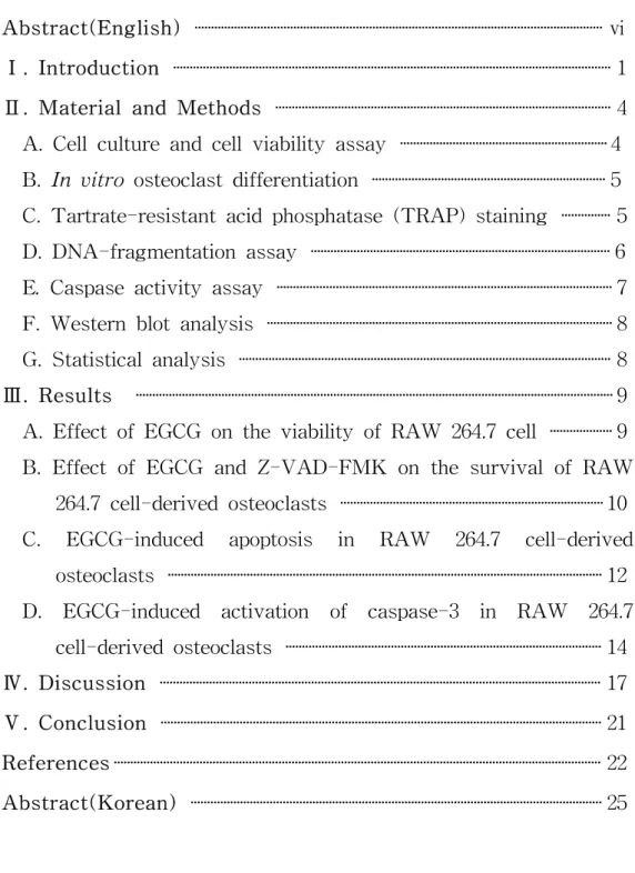
The MTT assay was performed to assess the effect of EGCG on the viability of RAW 264.7 cells and to determine the appropriate concentration for treating cell cultures. Compared with the non-treated cells, concentrations of up to 50 μM EGCG showed no effect, after 5 days of treatment, on the viability of RAW 264.7 cells. However, inhibition of cell growth was observed at the higher concentrations of EGCG (100 μM) (Fig. 1). Therefore, EGCG was used at a concentration of less than 50 μM in the subsequent studies.
* 0 0.3 0.6 0.9 1.2 1.5 0 5 10 20 50 100 Concentration of EGCG (µM) O .D at 57 0 n m
Figure 1. The effect of EGCG on the viability of RAW 264.7 cells. RAW 264.7 cells were treated with indicated concentrations of EGCG for 5 days. The cellular activity was then estimated by MTT assay, and the results are expressed as the mean ± S.D. of six cultures. The data are representative of three separate experiments. *P<0.05; significantly different from the non-treated group.
10
-B. Effect of EGCG and Z-VAD-FMK on the survival of RAW 264.7 cell-derived osteoclasts
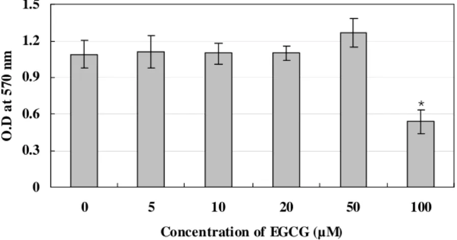
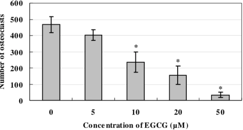
To examine the effect of EGCG and on osteoclast survival, osteoclasts were differentiated from RAW 264.7 cell by RANKL and treated with various concentrations of EGCG. Treatment with EGCG resulted in a significant decrease in the number of TRAP-positive osteoclasts in a dose-dependent manner (Fig. 2). In addition, to examine whether caspase is involved in the effect of EGCG, the cell cultures were treated with EGCG and/or Z-VAD-FMK. Z-VAD-FMK prevented an EGCG-induced decrease in the number of osteoclasts. This effect was more prominent at 50 μM Z-VAD-FMK than at 20 μM. However, Z-VAD-FMK itself had no effect on the number of TRAP-positive osteoclasts, compared with the non-treated cells (Fig. 3). * * * 0 10 0 20 0 30 0 40 0 50 0 60 0 0 5 10 20 5 0
Co nce ntration o f EGCG (µM )
N u m b er of o st eoc la st s
Figure 2. Inhibitory effect of EGCG on the survival of RAW 264.7 cell-derived osteoclasts. Osteoclasts were differentiated from RAW 264.7 cell by RANKL and treated with the indicated concentrations of EGCG. TRAP-positive
multinucleated cells with more than three nuclei were counted as osteoclasts. The results are expressed as the mean ± S.D. of five cultures. The data are representative of three separate experiments. *P<0.05; significantly different from the non-treated group.
* * *† 0 100 200 300 400 500 600 700 Co n tr o l Z-VAD -F M K (1 0 µ M ) Z-VA D-F M K (5 0 µ M ) EG CG ( 5 0 µ M ) EG CG ( 5 0 µ M ) + Z -V AD-F M K (1 0 µ M ) EG C G ( 5 0 µ M ) + Z-VAD -F M K (5 0 µ M ) N u m b er of os te oc la st s
Figure 3. General caspase inhibitor (Z-VAD-FMK) blocked the effect of EGCG on the survival of RAW 264.7 cell-derived osteoclasts. The cell cultures were treated with EGCG and/or Z-VAD-FMK to examine whether caspase is involved in the action of EGCG. The TRAP-positive multinucleated cells with more than 3 nuclei were counted as osteoclasts. The results are expressed as the mean ± S.D. of six cultures. The data are representative of three separate experiments. *P<0.05; significantly different from the non-treated control group. †P<0.05; significantly different from the EGCG-treated group.
12
-C. EGCG-induced apoptosis in RAW 264.7 cell-derived osteoclasts
To investigate whether treatment with EGCG induced apoptosis in RAW 264.7 cell-derived osteoclasts, cells were treated with EGCG (50 μM) in the presence or absence of RANKL (50 ng/㎖) for 24 h. In each case, nucleosomal DNA fragmentation, which is typical of apoptosis, was visible and the fragmentation pattern was most prominent in cells treated with EGCG in the absence of RANKL. This result shows that the presence of RANKL in culture medium slightly sustains the survival of osteoclasts. In addition, 50 μM Z-VAD-FMK reduced the extent of DNA fragmentation and significantly blocked EGCG from inducing apoptosis (Fig. 4).
RANKL (50 ng/㎖) - - - - + + + + EGCG (50 μM) - - + + - - + + Z-VAD-FMK (50 μM) - + - + - + - +
Figure 4. EGCG-induced nucleosomal DNA fragmentation in RAW 264.7 cell-derived osteoclasts. Cells were incubated with EGCG or Z-VAD-FMK for 24 h in the presence or absence of RANKL. The data are representative of duplicate experiments.
14
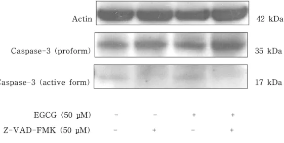
-D. EGCG-induced activation of caspase-3 in RAW 264.7 cell-derived osteoclasts
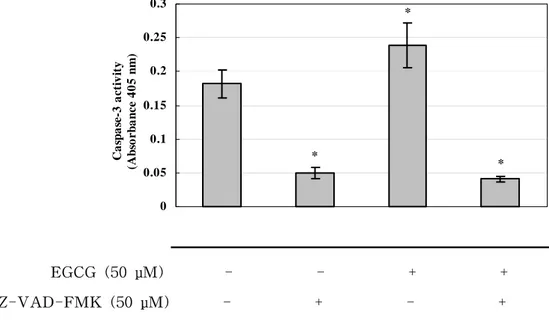
Another feature of apoptotic cell death, the activation of caspase-3, was evaluated by western blotting and colorimetric activity assay of caspase-3. RAW 264.7 cell-derived osteoclasts were treated with EGCG (50 μM) in the presence of RANKL (50 ng/㎖) for 24 h. In western blotting, the antibody used recognizes the intact 35-kDa proform and the cleaved 17-kDa active form of caspase-3. The treatment with EGCG (50 μM) increased the amount of the cleaved 17-kDa fragment of caspase-3 and decreased the amount of the 35-kDa pro-caspase-3, indicating the activation of caspase-3, compared with untreated control cells (Fig. 5). In addition, 50 μM Z-VAD-FMK reduced the intensity of the active form of caspase-3 and blocked the activation of caspase-3 by EGCG (Fig. 5). The activity of caspase-3 in EGCG-treated and untreated RAW 264.7 cell-derived osteoclasts was also measured. The cells treated with EGCG showed a significantly higher activity of caspase-3, which was blocked by Z-VAD-FMK (Fig. 6).
These results indicate that EGCG induces the activation of caspase-3, which is associated with reduced cell survival and apoptosis of EGCG-treated osteoclasts, as shown in Fig. 3.
Actin 42 kDa
Caspase-3 (proform) 35 kDa
Caspase-3 (active form) 17 kDa
EGCG (50 μM) - - + + Z-VAD-FMK (50 μM) - + - +
Figure 5. The effect of EGCG on the activation of caspase-3 in RAW 264.7 cell-derived osteoclasts. Cells were treated with EGCG or Z-VAD-FMK for 24 h. Western blotting analysis was performed using anti-caspase-3 immunoglobulin that recognizes the intact proform (32 kDa) and cleaved active form (17 kDa) of caspase-3. Actin served as an endocontrol. The data are representative of duplicate experiments.
16 * * * 0 0.05 0.1 0.15 0.2 0.25 0.3 C a sp as e -3 ac ti vi ty (A bs o r b a nc e 4 0 5 nm ) EGCG (50 μM) - - + + Z-VAD-FMK (50 μM) - + - +
Figure 6. The effect of EGCG on the activtity of caspase-3 in RAW 264.7 cell-derived osteoclasts. Cells were treated with EGCG or Z-VAD-FMK for 24 h. The results are expressed as the mean ± S.D. of four cultures. The data are representative of duplicate experiments. *P<0.05; significantly different from the non-treated group.
IV. Discussion
Recently, green tea has been focused on as a result of numerous biological effects of its constituents (Yang et al. 1993). Green tea consists mainly of polyphenols (catechins), which constitute up to 30% of the dry weight. Among these polyphenols, EGCG is the most abundant catechin and the one that has been the most extensively studied (Graham HN 1992). On the other hand, the bone resorbing activity of osteoclasts, which is principally responsible for this process, is significantly increased in the pathogenesis of bone disease such as periodontitis.
In our recent study, we reported that 20 μM EGCG significantly inhibited osteoclast formation in the co-culture system of bone marrow cells and primary osteoblastic cells. However, the precise mechanism of this effect was not established (Yun et al. 2004). Some studies have shown that apoptosis is the mechanism of action of EGCG for inhibiting cell growth in various type of tumor cells (Yang CS et al. 2002; Bode AM et al. 2004; Chen ZP et al. 1998). Thus, in the present study, we re-evaluated the effects of EGCG on osteoclasts differentiated from RAW 264.7 cells and investigated if caspase-mediated apoptosis is involved in the effect of EGCG on osteoclasts. Our present findings clearly show that EGCG was highly effective in inhibiting the survival of RAW 264.7 cell-derived osteoclasts in vitro (Fig. 2) and demonstrate that this inhibition might be mediated in part through the apoptosis of osteoclasts by the formation of a DNA fragmentation pattern, the most significant feature of apoptosis (Fig. 4).
In addition, these inhibitory effects of EGCG on osteoclasts were mildly suppressed by Z-VAD-FMK, suggesting the partial involvement of caspase in the EGCG-induced apoptosis of RAW 264.7 cells-derived osteoclasts (Fig. 3).
18
-This finding is supported by the present results that EGCG stimulates the activation of caspase-3: the elevation of caspase-3 activity and the cleavage of pro-caspase-3 (Fig. 5 and Fig. 6). It is known that pro-caspase-3 (35 kDa) is cleaved to yield 17-kDa and 12-kDa fragments when activated. In addition, caspase-3, defined as a key executioner of apoptosis, is the most prevalent caspase in the cell. Moreover, it is known that caspase-3 is the caspase ultimately responsible for the majority of the apoptotic effect, although it is supported by caspase-6 and -7 (Zimmermann KC et al. 2001). We also found that this EGCG-induced activation of caspase-3 was suppressed by Z-VAD-FMK (Fig. 5 and Fig. 6). Therefore, our results suggest that the induction of apoptosis in RAW-264.7 cell-derived osteoclasts involves the activation of caspase, in particular, caspase-3.
However, although Z-VAD-FMK strongly suppressed the EGCG-induced activation of the caspase-3, it only partially suppressed the EGCG-induced apoptosis (Fig.4). In addition, Z-VAD-FMK could not completely restore the decrease in the number of osteoclasts by EGCG (Fig. 3). This suggests that mechanisms using different signaling pathways could be involved in the EGCG-induced apoptosis of osteoclasts as well as the caspase pathway. In this respect, numerous studies have shown that the transcription factor, nuclear factor-kappa B (NF-kB), regulates susceptibility of cells to apoptosis through the transcriptional control of genes. NF-kB plays a critical role in the regulation of genes related to cell survival, proliferation and apoptosis (Gupta S et al. 2004; Bours V et al. 2000; Karin M et al. 2002). In a previous study, it has been reported that EGCG is capable of inducing apoptosis by inhibiting NF-kB activation in cancer cells (Gupta S et al. 2004; Ahmad N et al. 2000). However, it remains to be determined which pathway is more important in the apoptosis of osteocalsts by EGCG.
apoptosisis has not yet fully established. It was suggested that the binding of EGCG to Fas, presumably on the cell surface, triggers the Fas-mediated apoptosis in tumor cells (Hayakawa S et al. 2001). Moreover, it was reported that Fas-mediated caspase activation is involved in apoptosis in osteoclasts (Wu X et al. 2003).
In previous studies, osteoclasts are reported to have a short half-life. They undergo apoptosis in vitro within a few days (Jimi E et al. 1995). It was also reported that the absolute number of osteoclasts undergoing apoptosis, at any given time, ranges from 10-80% (Parfitt AM et al. 1996), and more than 80% of the purified osteoclasts were found to undergo apoptotic cell death by 48 h during the culture (Okahashi N et al. 1998). Similarly, in our experiment, untreated control cells showed a relatively high level of the apoptosis and caspase-3 activity (Fig. 4 - Fig. 6). In addition, the number of osteoclasts is dependent upon the relative rates of osteoclastogenesis and apoptosis. Although we showed that EGCG induced the apoptosis of osteoclasts, we cannot exclude the possibility that EGCG plays a role in the regulation of osteoclast formation. On the other hand, EGCG had no effect on RAW 264.7 cells themselves. This result suggests that EGCG has an effect on differentiated osteoclasts but not the undifferentiated cells. Several studies have indicated that tumor cells are much more sensitive to apoptosis induction by EGCG than the normal counterparts (Yang CS et al. 2002; Bode AM et al. 2004; Chen ZP et al. 1998). In our previous study, we also showed that EGCG had no inhibitory effect on the cell viability of either co-culture system or primary osteoblastic cells at concentrations of up to 20 μM. Taken together, these results show that EGCG, at an appropriate dose, inhibits osteoclasts but not other osteogenic cells. Therefore, EGCG could have an advantage as a chemopreventive agent specific for osteoclasts.
20
-osteoclasts with EGCG significantly lowered the number of -osteoclasts and there was evidence of the induction of apoptosis, as shown by DNA fragmentation and partial involvement of caspase-3. In addition, the ability of Z-VAD-FMK to block apoptosis shows the caspase-dependent property of EGCG-induced apoptosis. Our results also show that the treatment of osteoclasts with EGCG significantly activated the caspase-3, compared with the controls. Thus, our data revealed a novel finding that the EGCG-induced apoptosis in RAW 264.7 cell-derived osteoclasts was mediated, in part, through the activation of caspase-3.
From these findings, we suggest that EGCG might prevent alveolar bone resorption, which occurs in periodontal diseases, by inhibiting osteoclast survival through caspase-mediated apoptosis. Therefore, our results indicate that the inhibitory activity of EGCG could be utilized in the development of a therapeutic agent for the treatment of bone diseases, such as periodontitis. However, because we examined only the in vitro effects of EGCG in the present study, it remains to be determined whether EGCG also exerts these effects in vivo.
V. Conclusion
The purpose of the present study was to examine the effect of EGCG on osteoclast survival in vitro. For this purpose, we investigated if EGCG mediates osteoclast apoptosis via caspase activation in osteoclastic cells differentiated from the RAW 264.7 cells.
The effect of EGCG on osteoclast survival was examined by TRAP staining in osteoclasts differentiated from RAW 264.7 cells. In addition, we evaluated the apoptosis of osteoclasts by EGCG using DNA-fragmentation assay. Involvement of caspase in EGCG-mediated osteoclast apoptosis was evaluated by treatment with a general caspase inhibitor, Z-VAD-FMK. Moreover, the effect of EGCG on the activation of caspase-3 was assessed by colorimetric activity assay and western blotting. The results were follows.
1. Treatment with EGCG significantly lowered the number of osteoclasts differentiated from RAW 264.7 cells in a dose-dependent manner.
2. Treatment with EGCG resulted in DNA fragmentation and induced the activation of caspase-3 in RAW 264.7 cell-derived osteoclasts. Additional treatment with Z-VAD-FMK suppressed these effects of EGCG.
The present study shows that EGCG induces apoptosis in osteoclasts differentiated from RAW 264.7 through the activation of caspase-3. From these findings, we suggest that EGCG might prevent alveolar bone resorption by inhibiting osteoclast survival through the caspase-mediated apoptosis.
22
-VI. Reference
Ahmad N, Gupta S, Mukhtar H. Green tea polyphenol epigallocatechin-3-gallate differentially modulates nuclear factor kB in cancer cells versus normal cells. Arch Biochem Biophys 376:338-346, 2000.
Baron R. Molecular mechanisms of bone resorption by the osteoclast. Anat Rec 224:317-324, 1989.
Bode AM, Dong Z. Targeting signal transduction pathways by chemopreventive agents. Mutat Res 555:33-51, 2004.
Bours V, Bentires-Alj M, Hellin AC, Viatour P, Robe P, Delhalle S, Benoit V, Merville MP. Nuclear factor-kappa B, cancer, and apoptosis. Biochem
Pharmacol 60:1085-1090, 2000.
Chen ZP, Schell JB, Ho CT, Chen KY. Green tea epigallocatechin gallate shows a pronounced growth inhibitory effect on cancerous cells but not on their normal counterparts. Cancer Lett ;129:173-179, 1998.
Graham HN. Green tea composition, consumption, and polyphenol chemistry.
Prev Med 21:334-350, 1992.
Grutter MG. Caspases: key players in programmed cell death. Curr Opin Stuct
Biol 10:649-655, 2000.
Gupta S, Hastak K, Afaq F, Ahmad N, Mukhtar H. Essential role of caspases in epigallocatechin-3-gallate-mediated inhibition of nuclear factor kappa B and induction of apoptosis. Oncogene 23:2507-2522, 2004.
Hayakawa S, Saeki K, Sazuka M, Suzuki Y, Shoji Y, Ohta T, Kaji K, Yuo A, Isemura M. Apoptosis induction by epigallocatechin gallate involves its binding to Fas. Biochem Biophys Res Commun 285:1102-1106, 2001.
Islam S, Islam N, Kermode T, Johnstone B, Mukhtar H, Moskowitz RW, Goldberg VM, Malemud CJ, Haqqi TM. Involvement of caspase-3 in
epigallocatechin-3-gallate-mediated apoptosis of human chondrosarcoma cells. Biochem Biophys Res Commun 270:793-797, 2000.
Jimi E, Shuto T, Koga T. Macrophage colony-stimulating factor and interleukin-1 alpha maintain the survival of osteoclast-like cells.
Endocrinology 136:808-811, 1995.
Karin M, Lin A. NF-kB at the crossroads of life and death. Nat Immunol 3:221-227, 2002.
Kerr JF, Wyllie AH, Currie AR. Apoptosis: a basic biological phenomenon with wide-ranging implications in tissue kinetics. Br J Cancer 26:239-257, 1972. Minkin C. Bone acid phosphatase: tartrate-resistant acid phosphatase as a
marker of osteoclast function. Calcif Tissue Int 34:285-290, 1982.
Mosmann T. Rapid colorimetric assay for cellular growth and survival: application to proliferation and cytotoxicity assays. J Immunol Methods 65:55-63, 1983.
Nakagawa H, Wachi M, Woo JT, Kato M, Kasai S, Fuminori T, Lee IS, Nagai K. Fenton reaction is primarily involved in a mechanism of (-)-epigallocatechin-3-gallate to induce osteoclastic cell death. Biochem
Biophys Res Commun 292:94-101, 2002.
Nicholson DW. Caspase structure, proteolytic substrates, and function during apoptotic cell death. Cell Death Differ 6:1028-1042, 1999.
Okahashi N, Koide M, Jimi E, Suda T, Nishihara T. Caspases (interleukin-1-converting enzyme family proteases) are involved in the regulation of the survival of osteoclasts. Bone 23:33-41, 1998.
Parfitt AM, Mundy GR, Roodman GD, Hughes DE, Boyce BF. A new model for the regulation of bone resorption, with particular reference to the effects of bisphosphonates. J Bone Miner Res 11:150-159, 1996.
Schwartz Z, Goultschin J, Dean DD, Boyan BD. Mechanisms of alveolar bone destruction in periodontitis. Periodontol 2000 141:158172, 1997.
24
-Stennicke HR, Salvesen GS. Caspases - controlling intracellular signals by protease zymogen activation. Biochim Biophys Acta 1477:299-306, 2000. Suda T, Takahashi N, Martin TJ. Modulation of osteoclast differentiation.
Endocrine Rev 13:66-80, 1992.
Wu X, McKenna MA, Feng X, Nagy TR, McDonald JM. Osteoclast apoptosis: the role of Fas in vivo and in vitro. Endocrinology 144:5545-5555, 2003. Wyllie AH. Glucocorticoid-induced thymocyte apoptosis is associated with
endogenous endonuclease activation. Nature 284:555-556, 1980. Yang CS, Wang ZY. Tea and cancer. J Natl Cancer 85:1038-1049, 1993.
Yang CS, Maliakal P, Meng X. Inhibition of carcinogenesis by tea. Annu Rev
Pharmacol Toxicol 42:25-54, 2002.
Yun JH, Pang EK, Kim CS, Yoo YJ, Cho KS, Chai JK, Kim CK, Choi SH. Inhibitory effects of green tea polyphenol (-)-epigallocatechin gallate on the expression of matrix metalloproteinase-9 and on the formation of osteoclasts. J Periodontal Res 39:300-307, 2004.
Zimmermann KC, Bonzon C, Green DR. The machinery of programmed cell death. Pharmacol Ther 92:57-70, 2001.
국문 요약
RAW 264.7 세포로부터 분화된 파골세포에서
caspase 활성화에 의한 (-)-epigallocatechin
gallate (EGCG)의 apoptosis 유도
연세대학교 대학원 치의학과 (지도 최성호 교수)
윤 정 호
치조골 흡수는 치주질환의 특징적인 양상이며, 골의 무기질과 유기질의 제거가 일어나는데, 이러한 과정에는 파골세포가 주로 관여한다. (-)-epigallocatechin gallate (EGCG)는 녹차의 주된 성분으로서, 파골세포의 apoptosis를 유도하고 다양한 형태의 종양 세포에서 caspase 활성화를 조절하는 것으로 보고 되었다.
본 연구에서는 파골세포의 생존에 미치는 EGCG의 억제 효과를 조사하였다. 또한, EGCG가 caspase 활성화를 통해 파골세포의 apoptosis를 유도하는지를 조사하였다.
EGCG가 파골세포의 생존에 미치는 효과는 RAW 264.7 세포로부터 분화된 파골세포에서 tartrate-resistant acid phosphatase (TRAP) 염색에 의해 조사하였으며, DNA fragmentation 분석을 사용하여 EGCG에 의해 파골세포의 apoptosis가 일어나는지 평가하였다. EGCG에 의한 파골세포의 apoptosis에 caspase가 관여되는지의 여부는 통상적인 caspase 억제제인 Z-VAD-FMK를 처리함으로써 평가하였다. 또한, caspase-3의 활성화에 미치는 EGCG의 효과는 colorimetric activity assay와 western blotting을 시행하여 조사하였다.
26
-EGCG의 처리는 RAW 264.7 세포로부터 분화된 파골세포의 생존을 농도가 증가함에 따라 더욱 억제하였다. 또한, EGCG의 처리는 RAW 264.7 세포로부터 분화된 파골세포에서 DNA Fragmentation을 초래하고, caspase-3의 활성화를 유도하여, EGCG가 파골세포의 apoptosis를 유도하는 것으로 보여 졌다. 더하여, 통상적인 caspase 억제제인 Z-VAD-FMK의 부가적인 처리는 이러한 EGCG의 효과들을 감소시켰다.
이상의 결과에서 볼 때, EGCG가 caspase 활성화에 의한 apoptosis를 통해 파골세포 생존을 억제하므로, 치조골 흡수를 억제할 수 있을 것으로 사료되며, 이러한 EGCG의 작용은 치주염 같은 골 질환의 치료를 위한 치료제 개발에 유용하게 적용될 수 있을 것이다.
‡‡‡
핵심되는말 : 파골세포, (-)-epigallocatechin gallate (EGCG), apoptosis, caspase