Association between
single nucleotide polymorphisms in
deoxycytidine kinase and lamivudine
monotherapy response among patients
with chronic hepatitis B
Hyun Woong Lee
Department of Medicine
Association between
single nucleotide polymorphisms in
deoxycytidine kinase and lamivudine
monotherapy response among patients
with chronic hepatitis B
Directed by Professor Kwang-Hyub Han
The Doctoral Dissertation
submitted to the Department of Medicine,
the Graduate School of Yonsei University
in partial fulfillment of the requirements for the degree
of Doctor of Philosophy
Hyun Woong Lee
This certifies
that the Doctoral Dissertation
of Hyun Woong Lee is approved.
---
Thesis Supervisor: Kwang-Hyub Han
---
Min Goo Lee: Thesis Committee Member#1
---
Jin Sung Lee: Thesis Committee Member#2
---
Byong Ro Kim: Thesis Committee Member#3
---
Sang Hoon Ahn: Thesis Committee Member#4
The Graduate School
Yonsei University
ACKNOWLEDGEMENTS
I am deeply grateful to Prof. Kwang-Hyub Han who has guided me throughout several years in my learning and research. I especially acknowledge him for giving me a good run in Hepatology division. He has been my role model whom I have learned from and who shaped my perspective career as a doctor.
I want to express my thanks to Prof. Min Goo Lee, Prof. Jin Sung Lee, Prof. Byong Ro Kim, and Prof. Sang Hoon Ahn who have provided valuable insights as well as perspectives to my study.
In addition, I am so grateful to Prof. Min Goo Lee, who gave me the first insights about pharmacogenetics; Prof. Jin Sung Lee, who gave professional advices about single nucleotide polymorphisms; Prof. Byong Ro Kim, who gave me courage and wisdom; and Prof. Sang Hoon Ahn, whose excellence and integrity inspire me.
I want to express my gratitude to Dr. Sung Hee Lee who has provided key ideas and helped me in countless ways. Without his help, this outcome could not be made.
Special thanks are owed to my colleagues in Gastroenterology: Hye Young Chang, Yong Kwang Park, and Prof. Ki Jun Han who have helped in laboratory steps and clinical analysis.
Heartfelt thanks to my parents and parents-in-law who love me, pray for me, and always encourage me. I want to thank my mentor, Minister Young Ho Jung who has poured spiritual wisdom into my life.
First of all, I owe a debt of gratitude to my wife Sook Yi Han who has always been beside me understanding and encouraging me. Without her patience and supports, writing this thesis would not have been possible.
At last, I want to give my love to my daughter, Ye Jin Lee, and my new baby who will be born soon. God has richly blessed me through each of you.
For my patients, my research, efforts and courage can not, should not and will not be stopped. I would like to dedicate this paper to all of you with all of my heart.
Thanks God.
<TABLE OF CONTENTS>
Abstract ……… 1
I. INTRODUCTION ……… 4
II. PATIENTS AND METHODS………. 8
1.
Study population ..……… 8
2.
Definitions ………... 9
3.
Assay methodology ………... 10
4.
Sequence analysis of deoxycytidine kinase(dCK) gene. 10
5.
Statistical analysis ………. 15
III. RESULTS………... 16
1.
Identification of SNPs in dCK gene in the Korean
population ……….……….. 16
2.
Correlation between dCK SNP genotypes and the
treatment responses with lamivudine in 125 chronic
hepatitis B patients ………... 23
3.
Cumulative virologic breakthrough rates between the
groups according to dCK SNP genotypes ………. 31
4.
Cumulative HBeAg loss rates between the groups
according to dCK SNP haplotypes ……….. 33
5.
Haplotype structure between control and patients with
chronic hepatitis B ……….. 36
6.
Haplotype structure and association with lamivudine
response among patients with chronic hepatitis B …… 39
IV. DISCUSSION ... 44
V. CONCLUSION ... 49
VI. REFERENCES ……… 50
LIST OF FIGURES
Figure 1. Human deoxycytidine kinase (dCK) gene structure
and single nucleotide polymorphism (SNP) sites for
the haplotype SNP sets studied ... ... ... ... ... ... ...22
Figure 2. Linkage disequilibrium (LD) plot of dCK in patients
with chronic hepatitis B and healthy control ……. 29
Figure 3. Cumulative virologic breakthrough rates between the
g r o u p o f g . - 3 6 0 G / g . - 2 0 1 T h a p l o t y p e a n d
g.-360C/g.-201C haplotype by using Kaplan-Meier
method ... 32
Figure 4. Cumulative HBeAg loss rates between the group of
g.-360G/g.-201T haplotype and g.-360C/g.-201C
haplotype by using Kaplan-Meier method ……. 35
LIST OF TABLES
Table 1. PCR primers and amplification conditions used for
dCK ……….. 12
Table 2. Positions, sequences and frequencies of dCK
variations …..……… 18
Table 3. Clinical characteristics of 125 patients with chronic
hepatitis B ... 25
Table 4. Genotype distribution and allele frequency of
g.-1329C>T, g.-360C>G, g.-201C>T, c.364C>T,
IVS6+41T>A, and c.948T>C in two groups of
chronic hepatitis B ………. 26
Table 5. Multivariate logistic regression analysis of predictive
factors associated with good response …………. 30
Table 6. Genotype distribution and allele frequency of g.-360
C>G and g.-201C>T in two groups according to loss
of HBeAg during lamivudine treatment ………. 34
Table 7. Haplotype clusters and frequencies of 3 single
nucleotide polymorphism sets at the dCK gene in
patients with chronic hepatitis B
and control
samples………. 37
Table 8. Haplotype clusters and frequencies of 3 single
nucleotide polymorphism sets at the dCK gene in
patients with good response and poor response ….. 40
Table 9. Multivariate logistic regression analysis of predictive
factors associated with good response …………. 43
<ABSTRACT>
Association between single nucleotide polymorphisms in
deoxycytidine kinase and lamivudine monotherapy response
among patients with chronic hepatitis B
Hyun Woong Lee
Department of Medicine
The Graduate School, Yonsei University
(Directed by Professor Kwang-Hyub Han)
Background and Aims: Deoxycytidine kinase (dCK) is the important rate-limiting enzyme of the intracellular phosphorylation of lamivudine to its active triphosphates. This study was tried to confirm the known dCK polymorphisms in Chinese and Caucasian, to discover new dCK polymorphisms in Korean and to evaluate whether the discovered single nucleotide polymorphisms (SNPs) of dCK gene is associated with the treatment outcomes of lamivudine monotherapy in patients with chronic hepatitis B (CHB).
patients with CHB for discovering the SNPs. A total of 125 CHB patients were enrolled to compare the treatment outcomes of lamivudine according to genetic variants in dCK gene. They all treated with lamivudine 100mg once daily for at least 48 weeks. At week 48, those who achieved virological and biochemical response with undetectable HBV DNA and ALT normalization were classified as good response. Those who were occurred primary non-response or virologic breakthrough at 48 weeks were considered as poor response.
Results: In Korean, 15 SNPs of dCK gene were detected. Among them, 7 SNPs, had been previous reported in public SNP database. Eight novel dCK SNPs were found (g.-2052C>A, g.-360C>G, c.364C>T (P122S), IVS3-46G>del., IVS4+40G>T, IVS5+39T>C, IVS5-72A>T, and c.966~975T10>T11). 2 SNPs within the promoter region (g.-360C>G and
g.-201C>T) and 3 SNPs (c.364C>T, IVS6+41T>A, and c.948T>C) were in strong linkage disequilibrium, respectively (r2>0.9). Especially, the allele frequencies of two SNPs (g.-360C>G and g.-201C>T) were more frequent than what has been reported from Caucasians and Chinese (26% vs. 2% and 15.6%). At week 48, those with g.-360CG/g.-201CT and g.-360GG/g.-201TT compound genotype displayed more favourable virological response to lamivudine monotherapy than those with g.-360CC/g.-201CC (70.6% vs. 48.2%, P = 0.027). The allele frequency of g.-360G/g.-201T was significantly higher in good response group (20.0% vs. 9.3%, P = 0.021). In multivariate
analysis, the genotype containing g.-360G/g.-201T haplotype was independent factor for virological response [odds ratio (OR), 2.846; 95% confidence interval (CI), 1.121 – 7.227, P = 0.028). In addition, specific haplotype cluster 5-SNP-B (GTCTT), which was g.-360 C>G, g.-201C>T, c.-364C>T (P122S), IVS6+41T>A, and c.948T>C (3'UTR), was highly associated with good response in patients with CHB, while haplotype cluster 5-SNP-C (CCTAC) was associated with poor response (OR, 2.830; 95% CI, 1.035 – 7.737, P = 0.043).
Conclusions: These results anticipate that SNP haplotypes of dCK gene may play an important role as a genetic marker for predicting lamivudine responsiveness in patients with CHB.
---
Key words : deoxycytidine kinase, single nucleotide polymorphisms, chronic hepatitis B, lamivudine
Association between single nucleotide polymorphisms in
deoxycytidine kinase and lamivudine monotherapy response
among patients with chronic hepatitis B
Hyun Woong Lee
Department of Medicine
The Graduate School, Yonsei University
(Directed by Professor Kwang-Hyub Han)
I. INTRODUCTION
The recent development of new and potent anti-viral agents may offer many therapeutic options against chronic hepatitis B virus (HBV) infection. Coincidently, antiviral resistance has become an increasingly common problem during long-term treatment with anti-viral agent. Recently, multi-drug resistant HBV has been reported in patients who received sequential treatment with nucleoside/tide analog (NA) monotherapy.1-3 Therefore, an understanding of the mechanisms of drug resistance is important for preventing the emergence of resistance and designing new drugs.4
NAs replace natural nucleosides during the synthesis of the first or second strand (or both) of HBV DNA. They function as competitive inhibitors of the viral reverse transcriptase and DNA polymerase.5 Because NAs partially and reversibly suppress viral replication, they must be given for more than 1 year in most cases to achieve maximal efficacy. Unfortunately, a long duration of NA treatment is associated with an increasing risk of emergence of drug resistance.6
Up to recently, lamivudine has been considered as first-line therapy for individuals with non-cirrhotic and cirrhotic liver disease.Nevertheless, the efficacy of lamivudine is limited by the development of drug-resistant HBV mutants, restricting its utility as a long-term therapy for chronic hepatitis B (CHB).7 Numerous studies have shown that resistance to antiviral agent such as lamivudine is the major cause of treatment failure. Although viral factors and poor compliance are the most important factors in treatment failure, another important factor to be considered is cellular factor affecting anti-viral efficacy. However, it remains unclear if any of these mechanisms play a role in the response to anti-HBV drugs.8,9
The cellular factors of drug resistance include host genetic factors and ability to efficiently convert the agent to its active metabolite.Over the past decade, the relationship between the genetic composition and drug responses has become an important focus of medical practice, especially with the discovery of single nucleotide polymorphisms (SNPs) in genes encoding
receptors and target enzymes mediating drug effects or enzymes directly implicated in drug metabolism.10-14
NAs are prodrugs that must be metabolized intracellularly to exert their activity. The nucleoside-based reverse transcriptase inhibitors (NRTIs) approved for anti-viral agents include compounds that mimic endogenous pyrimidine or purine nucleosides. They need to be activated for their anti-viral activity via a phosphorylation process to their nucleoside triphosphates or nucleoside diphosphate that functions as the inhibitor of polymerase.15-18One or more steps in sequential phosphorylation of NRTIs from initial monophosphate to diphosphate to triphosphate formation can be rate limiting in formation of active drug. Usually, the initial phosphorylation step is the rate-limiting step in the activation process and may elucidate some of differences in potency among the various NAs.19
To date, three different kinases are known to be involved in mediating DNA synthetic process leading to lamivudine phosphorylation. Among them, deoxycytidine kinase (dCK) is considered as the important enzyme because phosphorylation into its monophosphate form is initiating step for the activation of lamivudine.20,21The dCK gene is located on chromosome 4q13.3, spanning over 37 kb and containing seven exons. With an open reading frame of 783 bp, its protein product functions as a homodimer of 60 kDa, consisting of two subunits of 30.5 kDa.22-24 Recently, inactivated dCK transcripts due to alternative splicing were reported in acute myeloid leukemia (AML) patients
resistant to chemotherapy.25 Another report suggested that specific SNPs in dCK gene affecting variations in gene expression would be considered as a new mechanism contributing to the resistance to 1-β-arabinofuranosylcytosine (Ara-C) in AML patients.26 However, there was no analysis demonstrated that the dCK gene contributes to the development of lamivudine resistance.
Therefore, the potential influence of a genetic variation such as SNPs of the dCK gene on clinical sensitivity to lamivudine is worthy of investigation. The objectives of this study are to confirm the known dCK polymorphisms in Chinese and Caucasian, to discover new dCK polymorphisms in Korean and to evaluate whether the discovered SNPs of dCK gene is associated with the treatment outcomes of lamivudine therapy in patients with CHB.
II. PATIENTS AND METHODS
1.
Study population
A total of 125 patients with CHB, who visited Severance hospital between Jan. 2002 and Dec. 2003 and were treated with lamivudine 100mg for at least 48 weeks, were enrolled in the current study. Patients enrolled in the study meet the following entry criteria: they were 18–75 years of age; the presence of serum HBsAg was observed for at least 6 months; they had elevated serum alanine aminotransferase (ALT) on two occasions, at least 1 month apart, with an average value of ≥ 2 times the upper limit of normal (ULN); the
presence of serum
hepatitis B e antigen
(HBeAg) and HBV DNA had been documented on two occasions, at least 1 month apart. Additional requirements included: a hemoglobin value of ≥ 10 g/dl, a platelet count of ≥ 70,000 mm3, a white cell count of ≥ 3,000 mm3, a polymorphonuclear count of ≥ 1,500 mm3 and normal renal function with normal serum creatinine levels. Candidates were required to have compensated liver disease with a prothrombin time of less than 4 sec, prolonged over control values, a serum albumin of ≥ 3.0 g/dl, a total bilirubin of ≤ 4 mg/dl, and no history ofhepatic encephalopathy or bleeding esophageal varices and imaging features suggestive of cirrhosis on ultrasonography.
within 6 months of entry; previous therapy with IFN; the presence of antibody to human immunodeficiency virus (HIV), hepatitis C virus (HCV) or hepatitis D virus (HDV); a history of malignancy, evidence of other forms of liver disease; or a history of intravenous drug abuse. Patients with other significant medical or psychiatric problems were also excluded.
Serum HBeAg, anti-HBe, HBV DNA and ALT were tested every 3 or 6 months, and whenever necessary during medication and after drug cessation. In addition, peripheral blood samples from 112 healthy donors were also used as control. Patients and controls were compared for genotype and allele frequencies of the SNPs of the dCK gene.
2.
Definitions
Patients without decline in serum HBV DNA by ≥ 1 log10 copies/mL
after the first 6 months of therapy were classified as “primary non-response (NR).” Virologic breakthrough (VB) was defined as patients with increase in serum HBV DNA by > 1 log10 copies/mL (10-fold) above nadir after
achieving virological response, during continued treatment.27 Those who achieved virological and biochemical response with undetectable HBV DNA and ALT normalization at least 48 weeks were classified as good response. By contrast, those who were classified as NR and were occurred VB at 48 weeks were considered as poor response.
3.
Assay methodology
Commercially available enzyme-linked immunoadsorbent assay (ELISA) was used for the detection of serum HBsAg, anti-HBs, anti-HBc, HBeAg, and anti-HBe (Abbott Laboratories, North Chicage, IL, USA). The HBV DNA was detected by Digene Hybrid Capture II Assay (Digene Diagnostics Beltsville, MD, USA; detection limit, 0.5 pg/ml = 1.4 x 10⁵ copies/mL).
4.
Sequence analysis of dCK gene
24 healthy volunteers and 24 patients with CHB were screened for discovering the SNPs of the dCK gene. A 5-ml aliquot of EDTA blood was collected from each individual. Genomic DNA from peripheral blood stored samples will be isolated using a NucleoGen Genomic DNA isolation Kit (NucleoGen, Seoul, Korea) according to the manufacturer's instructions and subsequently stored at 4℃ until analysis. 13 primer sets were used to amplify all seven dCK exons and the core promoter region (Table 1). Several base pair fragments of the dCK gene were amplified with forward primers and reverse primers in a final volume of 25㎕ containing 50~100 ng of genomic DNA, 20 pmol of each primer, 0.2 mM dNTPs, 2 U of Taq polymerase (iNtRON biotechnology, Seoul, Korea) and manufacturer's standard polymerase chain reaction (PCR) buffer. Amplification was performed in a GeneAmp PCR
system 9700 (Perkin Elmer Corp., Norwalk, CT, USA). PCR conditions were 94℃ for 5 min, followed by 35 cycles of 94℃ for 30 sec, 55℃ for 30 sec and 72℃ for 50 sec, with final extension at 72℃ for 7 min. PCR products were identified by electrophoresis, then PCR products were purified with PCR purification kit (iNtRON biotechnology, Seoul, Korea) and analyzed by direct sequencing (bionex Co. Ltd, Seoul, Korea).
Table 1. PCR primers and amplification conditions used for dCK
Region Strand PCR-product forward primer Reverse primer
Promoter1 Forward
5' - CAG GAA ACA GCT ATG ACC CAG CCA GAA ACT GGT AGA AC - 3'
5' - TGT AAA ACG ACG GCC AGT TGA CTC CAT CCT CAG AAA TAA - 3'
Promoter2 Forward
5' - CAG GAA ACA GCT ATG ACC TGA TTC TTG CTG TTT AAT CCT - 3'
5' - TGT AAA ACG ACG GCC AGT GAC CTA ATG AGG CAA GAG AA - 3'
Promoter3 Forward
5' - CAG GAA ACA GCT ATG ACC CTG CCT AGT CTT GCC TAG AA - 3'
5' - TGT AAA ACG ACG GCC AGT CCA GAA ATA CCG TCA TTA GC - 3'
Exon1 Reverse
5' - CAG GAA ACA GCT ATG ACC CTA GAG AGG CGG GTT TTC - 3'
5' - TGT AAA ACG ACG GCC AGT GTA AGG GAA GGA TGC TCT G - 3'
Exon2 Forward
5' - CAG GAA ACA GCT ATG ACC GGT GGC CAT TAG GAG TAT TA - 3'
5' - TGT AAA ACG ACG GCC AGT GAA ACC CAT TGA TAT GGA GA - 3'
Exon3 Forward
5' - CAG GAA ACA GCT ATG ACC ATC ATC TTG GTT TTG CTG AT - 3'
5' - TGT AAA ACG ACG GCC AGT CAC CAT ATT CCC AAC AGT TT - 3'
Exon4 Reverse
5' - CAG GAA ACA GCT ATG ACC AAG TAA TCT GGC CTC TCA CA - 3'
5' - TGT AAA ACG ACG GCC AGT TAA TTT AGA GGT GGG GAG TG - 3'
Exon5 Forward
5' - CAG GAA ACA GCT ATG ACC TGT GGA TGG ATA CCA AAA AT - 3'
5' - TGT AAA ACG ACG GCC AGT TCA TTG CAA TCA AGA GAA TG - 3'
Exon6 Forward
5' - CAG GAA ACA GCT ATG ACC CCC AGC TGT ATA TAT TTT GTA CC - 3'
5' - TGT AAA ACG ACG GCC AGT CCG AAG GAT CTT TAT TTT AGC - 3'
Exon7-A Forward and Reverse
5' - CAG GAA ACA GCT ATG ACC AGC TGG GGT CTT CAA CTA TT - 3'
5' - TGT AAA ACG ACG GCC AGT TAA GAA ACT GGT CAC CAA CG - 3'
Exon7-B Forward
5' - CAG GAA ACA GCT ATG ACC TTT CTC ATA GCA GGA AAT GTA G - 3'
5' - TGT AAA ACG ACG GCC AGT TTA ATG GAT GCT TTC TAG CC - 3'
Exon7-C Forward
5' - CAG GAA ACA GCT ATG ACC TAT CCT GAA AGC ATT ATT TTT - 3'
5' - TGT AAA ACG ACG GCC AGT TGA TTA TTA CAT CTT TTT AGA ACT G - 3'
Exon7-D Forward
5' - CAG GAA ACA GCT ATG ACC TTT TTA GTT TGT TTT TGT TTG G - 3'
5' - TGT AAA ACG ACG GCC AGT ACC TTT CTA GGA GAG CAA ACT - 3'
5.
Statistical analysis
All data were expressed as means ± standard deviation (SD). Student’s t test compared mean age, pretreatment ALT levels and HBV DNA levels. The chi-square test or Fisher’s exact test compared the sex ratio, and frequencies of novel and known dCK genotype. Based on the gene frequencies, predicted phenotype frequencies were calculated according to the Hardy-Weinberg equation and compared with the observed frequencies using the chi-square test. We assessed all variables using the logistic regression model. Cumulative virologic breakthrough rates and HBeAg loss rates were estimated using Kaplan-Meier method and compared by log-rank test. All statistical analysis was performed using Statistical Package for Social Science (SPSS Inc. Chicago, Illinois.) version 12.0. A P value of less than 0.05 was considered significant.
III. RESULTS
1.
Identification of SNPs in dCK gene in the Korean population
All the 7 coding exon of dCK and 1.5kb of the proximal promoter were analyzed in a group of 24 healthy volunteers and 24 patients with CHB. A total of 15 variants were identified in 48 Korean. Among them, four SNPs were present in the 5' regulatory region (the first nucleotide upstream of the translation initiation site being defined as -1 position), which were g.-2052C>A, g.-1329C>T, g.-360C>G, and g.-201C>T with allele frequencies of 0.979:0.021, 0.979:0.021, 0.74:0.26, and 0.734:0.266, respectively. Three SNPs were detected in exons which were c.364C>T (P122S), c.948T>C, and c.966-975T10>T11 with allele frequencies of 0.969:0.031, 0.979:0.021, and
0.979:0.021, respectively. In addition, we detected 7 SNPs in introns, and 1 SNP in 3' UTR, respectively. To detect population structure, an additional 14 SNPs were chosen from the National Center for Biotechnology Information (NCBI) public database. However, they were not detected in Korean. Among 15 SNPs, which were detected in 48 Korean, 7 SNPs had been previous reported in public SNP database (NCBI reference number). Interestingly, eight novel dCK SNPs were found (g.-2052C>A, g.-360C>G, c.364C>T (P122S), IVS3-46G>del., IVS4+40G>T, IVS5+39T>C, IVS5-72A>T, and c.966~975T10>T11) (Table 2).
healthy volunteers and 24 patients with CHB in 15 SNPs. Especially, the two promoter SNPs g.-360C>G and g.-201C>T were previously confirmed in Caucasians and Chinese. They were in perfect linkage disequilibrium and found at an allele frequency of 26%. The allele frequencies of two SNPs were more frequent than what has been reported from Caucasians and Chinese (26% vs. 2% and 15.6%). Linkage analysis revealed significant disequilibrium between g.-360C>G and g.-201C>T (r-squre 1.0, D' 1.0). One SNP is non-synonymous and results in the substitution of proline at amino acid position 122 by serine (c.364C>T in exon 3, P122S).
6 SNPs were selected to compare for genotype and allele frequencies between lamivudine response groups. Three promoter SNPs and one intronic SNP, which were g.-1329C>T, g.-360C>G, g.-201C>T and IVS6+41T>A were selected according to high frequencies of alleles. Two SNPs located in exon 3 and 3’UTR were selected (c.364C>T, and c.948T>C) (Fig. 1)
Table 2. Positions, sequences and frequencies of dCK variations (N=48*)
Position SNP name†††† Nucleotide sequence Allele frequency‡‡‡‡ OH EH
HWE p
Promoter g.-2052C>A§
5' – TCC TCT CGG TAG CTG CCA GTA CT[C/A] TTA CCA TTT CAT GAG TTT TCC CC - 3'
C:A=0.979:0.021 0 0.041 0.0211
Promoter g.-1431A>G
5' – AAC CAA GTG CTT CAA GAG TCC CA[A/G] TTG CAG TGT TCT TAA GGG TTA CT - 3'
A:G=1:0 0 0 1
Promoter g.-1359A>G
5' – TAG GTG CCC TTT ACA GTC GTC CA[A/G] TTG GTT GCA CCC TAT GAA GGA TT - 3'
A:G=1:0 0 0 1
Promoter g.-1329C>T
5' – GCA CCC TAT GAA GGA TTG GCC TG[C/T] GACC AAT CAG AGG CTG AAA TGG A - 3'
C:T=0.979:0.021 0.042 0.041 1 Promoter g.-360C>G§ 5' – CCT CTT CCC GCG CCC TGC CCG GG[C/G] GCC TGG CTG CTT GGG GTA GAG GC - 3' C:G=0.74:0.26 0.271 0.385 0.0824 Promoter g.-201C>T 5' – CGG CTT GAG GAG GGC GGG GCC GC[C/T] CCG CAG GCC CGC CAG TGT CCT CA - 3' C:T=0.734:0.266 0.277 0.39 0.092 Intron 1 IVS1+37G>C
5' – GGA AAT GTG GGA CGC AAG GCT GG[G/C] GTG TCG CGG CAG TGG CTG AAG CT - 3'
G:C=1:0 0 0 1
GGA GCC TTT TCA TTT TCT TC - 3'
Intron 1 IVS1-110T>G
5' – AAA GTT TAG AAA GTT GAA TGT TT[T/G] GGG CTT TTT ATG TTG CTT GCT AT - 3'
T:G=1:0 0 0 1
Intron 2 IVS2+114G>A
5' – CTT TCA AAT CTC GCT TTA GGT AT[G/A] TAT CTT CAT CTA GGT GGT GGT TA - 3'
G:A=0.969:0.031 0.062 0.061 1
Exon 3 A100A
5' – TCT TTT ACC TTC CAA ACA TAT GC[C/T] TGT CTC AGT CGA ATA AGA GCT CA - 3'
C:T=1:0 0 0 1
Exon 3 P122S§
5' – GCA AGC TCA AAG ATG CAG AGA AA[C/T] CTG TAT TAT TTT TTG AAC GAT CT - 3'
C:T=0.969:0.031 0.062 0.061 1
Intron 3 IVS3-46G>del.§
5' – GTT CTC TTT TTT ATT TCT TTG TT[G/-] TTT TTT TTT GAA ATG ATA CAT GT - 3'
G:del.=0.978:0.022 0.043 0.043 1
Intron 3 IVS3-45T>del.
5' – TTC TCT TTT TTA TTT CTT TGT TG[T/-] TTT TTT TTG AAA TGA TAC ATG TG - 3'
T:del.=1:0 0 0 1
Intron 4 IVS4+40G>T§
5' – ATG TGT TTC ACT GAA AAT TTA AA[G/T] AAA TAT TTA GAA CTC TTT TCA GT - 3'
G:T=0.989:0.011 0.022 0.022 1
Intron 5 IVS5+39T>C§
5' – ATT TAC TAT TCA TTT TAA ATA CC[T/C] TTG TTA CCT TTG TTA AAT TTT AA - 3'
T:C=0.99:0.01 0.021 0.021 1
Intron 5 IVS5-72A>T§
5' – TTT GAT TTT CCA AGG ACA TAC GA[A/T] TGA ATT TTT AAT GTT TCT CTC TT - 3'
Intron 7 IVS6+41T>A
5' – ACA AAC AAA TAT TTG TTT TTT CT[A/T] AAA AAG TGT ACT GAG TGG TGA GA - 3'
T:A=0.969:0.031 0.062 0.061 1
Exon 7 (3' UTR)
c.948T>C
5' – GAA TCT TAT GCA AAA CTT TTT GA[T/C] CAG TTT CTT TTC TTT TGT TTT TT - 3'
T:C=0.979:0.021 0.042 0.041 1
Exon 7 (3' UTR)
c.966~975T10>T11
§ 5' – TTT GAY CAG TTT CTT TTC TTT TG[T10/T11] AAA AAA GAC ATT TAA AGA CAA AG - 3'
T10/T11=0.979:0.021 0.042 0.041 1
Exon 7 (3' UTR)
c.1796A>G
5' – TTT AGA AAA TTT TAT GTA TTT TA[A/G] AAT AAG GGG AAG AGT CAT TTT CA - 3'
A:G=1:0 0 0 1
Exon 7 (3' UTR)
c.1797A>G
5' – TTA GAA AAT TTT ATG TAT TTT AA[A/G] ATA AGG GGA AGA GTC ATT TTC AC - 3'
A:G=1:0 0 0 1
Exon 7 (3' UTR)
c.1827del.>A
5' – GAA GAG TCA TTT TCA CTT TTA AA[-/A] CTA CTA TTT TTC TTT CCA AGT CA - 3'
del.:A=1:0 0 0 1
Exon 7 (3' UTR)
c.1904del.>C
5' – TAA TTT AGT GGA TTA ACC AGT CC[-/C] AGA CGC ACT GAT CTT TGC AAA GG - 3'
del.:C=1:0 0 0 1
Exon 7 (3' UTR)
c.1952A>C
5' – GAC TTA ATT TCA AAT CTG TAA TT[A/C ]CCA TAC ATA AAC TGT CTC ATT AT - 3'
A:C=1:0 0 0 1
Exon 7 (3' UTR)
c.2031A>G
5' – GTA TAA ATT AAT TTG TTA ATT AA[A/G] TAT TTC TTA AGT ATA AAC CTT AT - 3'
A:G=1:0 0 0 1
(3' UTR) AAT CAT TTT AGA ATT AA - 3' Exon 7
(3' UTR)
c.2254TT>del.
5' – AAC CTT TAA AAA TGT ATA ATG AC[TT/-] TTA AAA TTT GTA GAA TTG AAA AG - 3'
TT:del..=1:0 0 0 1
3' UTR c.2419A>T
5' – TCA TGT CTT CCA TTA GAA TTT AT[A/T] TAT TGC TCT TTA TAG TTT GCT CT - 3'
A:T=0.978:0.022 0.043 0.043 1
*
24 healthy volunteers and 24 patients with CHB were screened for the SNP of the dCK gene †
Base pair position numbered from translation initiation site ‡
Frequency for the variant genotype
§
Eight novel dCK SNPs were found (g.-2052C>A, g.-360C>G, P121S, IVS3-46G>del., IVS4+40G>T, IVS5+39T>C, IVS5-72A>T, and 966~975T10>T11)
Abbreviation: SNP, Single nucleotide polymorphism; EH, Expected heterozygous; OH, Observed heterozygous; HWE p, Hardy-Weinberg equilibrium p-value
5’ UTR Exon 7 3’ UTR Exon 1 Exon 2 Exon 3 Exon 4 Exon 5 Exon 6 g.-360C>G g.-201C>T (rs2306744) g.-1329C>T (rs1906021) promoter c.364C>T (P122S) IVS6+41T>A (rs1486271) c.948T>C (rs4643786) 2-SNP Set (159 b) 5-SNP Set (36 kb) 3-SNP Set (7 kb) 5’ UTR Exon 7 3’ UTR Exon 1 Exon 2 Exon 3 Exon 4 Exon 5 Exon 6 g.-360C>G g.-201C>T (rs2306744) g.-1329C>T (rs1906021) promoter c.364C>T (P122S) IVS6+41T>A (rs1486271) c.948T>C (rs4643786) 2-SNP Set (159 b) 5-SNP Set (36 kb) 3-SNP Set (7 kb) Fig. Fig. Fig.
Fig. 1 1 1.... 1 Human deoxycytidine kinase (dCK) gene structure and single nucleotide polymorphism (SNP) sites for the haplotype SNP sets studied.
The size of the gene is over 37 kilobases(kb). 6 SNPs were selected to compare for genotype and allele frequencies between lamivudine response groups. Three promoter SNPs and one intronic SNP, which were g.-1329C>T, g.-360C>G, g.-201C>T and IVS6+41T>A were selected according to high frequencies of alleles. Two SNPs located in exon 3 and 3’UTR were selected (c.364C>T, and c.948T>C).
2.
Correlation between dCK SNP genotypes and the treatment responses
with lamivudine in 125 chronic hepatitis B patients
The 125 CHB patients were divided into two groups according to their responses to lamivudine monotherapy. 69 patients achieved good responses which are virological response with undetectable HBV DNA and ALT normalization at least 48 weeks. On the contrary, 56 patients showed poor responses, including 9 patients who did not have decline in serum HBV DNA by ≥ 1 log10 copies/mL after the first 6 months of therapy, 47 patients who had
increase in serum HBV DNA by > 1 log10 copies/mL (10-fold) above nadir after
achieving virological response at 48 weeks.
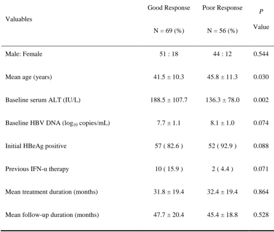
The univariate analysis of clinical and virological factors showed that younger age (P = 0.030) and higher serum ALT levels (P = 0.002) were associated with a higher probability of good response. There were no obvious differences in clinical data such as sex, baseline serum HBV DNA, and so on, before treatment between the two groups (Table 3).
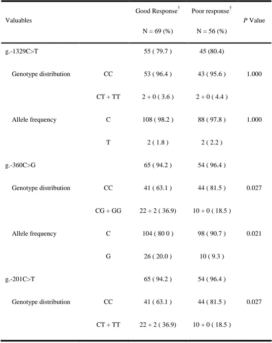
The relationship between six dCK SNP genotypes and the treatment responses was analyzed (Table 4). Interestingly, there was statistically significant difference between the two groups in terms of the distribution of genotype and allele frequency of the g.-360C>G, g.-201C>T. Allele frequency analysis of the g.-201C/T and g.-360C/G in 125 CHB patients, as well as 112 peripheral blood samples from normal individuals, is in agreement with the
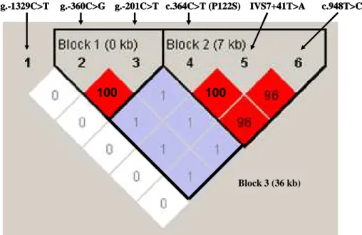
Hardy-Weinberg equilibrium (HWp = 0.699). Sequence analysis showed that g.-360C/g.-201C and g.-360G/g.-201T were in perfect linkage disequilibrium (Fig. 2.)
Multivariate logistic regression analysis was performed using one categorized value according to dCK SNP genotype and three continuous values that were previous known as predictive factors of good response. Age, baseline serum ALT level, HBV DNA level and the genotypes containing g.-360G/g.-201T haplotype were independent predictive factors for good response (Table 5).
Table 3. Clinical characteristics of 125 patients with chronic hepatitis B Valuables Good Response N = 69 (%) Poor Response N = 56 (%) P Value Male: Female 51 : 18 44 : 12 0.544
Mean age (years) 41.5 ± 10.3 45.8 ± 11.3 0.030 Baseline serum ALT (IU/L) 188.5 ± 107.7 136.3 ± 78.0 0.002 Baseline HBV DNA (log10 copies/mL) 7.7 ± 1.1 8.1 ± 1.0 0.074 Initial HBeAg positive 57 ( 82.6 ) 52 ( 92.9 ) 0.088 Previous IFN-α therapy 10 ( 15.9 ) 2 ( 4.4 ) 0.071 Mean treatment duration (months) 31.8 ± 19.4 32.4 ± 19.4 0.864 Mean follow-up duration (months) 47.7 ± 20.4 45.4 ± 18.8 0.528
NOTE. Values are given as mean ± standard error of the mean
Abbreviation: ALT, alanine aminotransferase; HBV, hepatitis B virus; IFN-α, interferon alpha
Table 4. Genotype distribution and allele frequency of g.-1329C>T, g.-360C>G, g.-201C>T, c.364C>T, IVS6+41T>A, and c.948T>C in two groups of chronic hepatitis B* Valuables Good Response† N = 69 (%) Poor response† N = 56 (%) P Value g.-1329C>T 55 ( 79.7 ) 45 (80.4) Genotype distribution CC CT + TT 53 ( 96.4 ) 2 + 0 ( 3.6 ) 43 ( 95.6 ) 2 + 0 ( 4.4 ) 1.000 Allele frequency C T 108 ( 98.2 ) 2 ( 1.8 ) 88 ( 97.8 ) 2 ( 2.2 ) 1.000 g.-360C>G 65 ( 94.2 ) 54 ( 96.4 ) Genotype distribution CC CG + GG 41 ( 63.1 ) 22 + 2 ( 36.9) 44 ( 81.5 ) 10 + 0 ( 18.5 ) 0.027 Allele frequency C G 104 ( 80 0 ) 26 ( 20.0 ) 98 ( 90.7 ) 10 ( 9.3 ) 0.021 g.-201C>T 65 ( 94.2 ) 54 ( 96.4 ) Genotype distribution CC CT + TT 41 ( 63.1 ) 22 + 2 ( 36.9) 44 ( 81.5 ) 10 + 0 ( 18.5 ) 0.027
Allele frequency C T 104 ( 80 0 ) 26 ( 20.0 ) 98 ( 90.7 ) 10 ( 9.3 ) 0.021 c.364C>T (P122S) 64 ( 92.8 ) 54 ( 96.4 ) Genotype distribution CC CT + TT 58 ( 90.6 ) 6 + 0 ( 9.4 ) 44 ( 81.5 ) 8 + 2 ( 18.5 ) 0.148 Allele frequency C T 122 ( 95.3 ) 6 ( 4.7 ) 96 ( 88.9 ) 12 ( 11.1 ) 0.064 IVS6+41T>A 64 ( 92.8 ) 54 ( 96.4 ) Genotype distribution TT TA + AA 58 ( 90.6 ) 6 + 0 ( 9.4 ) 44 ( 81.5 ) 8 + 2 ( 18.5 ) 0.148 Allele frequency T A 122 ( 95.3 ) 6 ( 4.7 ) 96 ( 88.9 ) 12 ( 11.1 ) 0.064 c.948T>C 55 ( 79.7 ) 44 (78.6 ) Genotype distribution TT TC + CC 50 ( 90.9 ) 5 + 0 ( 9.1 ) 37 ( 84.1 ) 6 + 1 ( 15.9 ) 0.302 Allele frequency T C 105 ( 95.5 ) 5 ( 4.5 ) 80 ( 90.9 ) 8 ( 9.1 ) 0.199
*
125 patients with CHB who were treated with lamivudine were retrospectively analyzed.
†
Good response: virological response with undetectable HBV DNA and ALT normalization at 48 weeks. Poor response: virological response without decline in serum HBV DNA by ≥ 1 log10 copies/mL after the first 6 months of
therapy and with increase in serum HBV DNA by > 1 log10 copies/mL (10-fold)
g.-360C>G g.-201C>T g.-1329C>T c.364C>T (P122S) IVS7+41T>A c.948T>C Block 3 (36 kb) 100 100 g.-360C>G g.-201C>T g.-1329C>T c.364C>T (P122S) IVS7+41T>A c.948T>C Block 3 (36 kb) g.-360C>G g.-201C>T g.-1329C>T c.364C>T (P122S) IVS7+41T>A c.948T>C Block 3 (36 kb) 100 100
Fig. 2. Linkage disequilibrium (LD) plot of dCK in samples with chronic hepatitis B patients and healthy control
LD plots were generated in Haploview using genotype data from 125 patients with chronic hepatitis B and 112 healthy control samples. The scheme is: black when r2 > 0.9; shades of grey light 0 < r2 < 0.9. SNPs that are linked r2 > 0.9 are categorized in the same haplotype block. We used 2-SNP (g.-360C>G and g.-201C>T), 3-SNP (c.364C>T, IVS6+41T>A, and c.948T>C), and 5-SNP (g.-360C>G, g.-201C>T, c.364C>T, IVS6+41T>A, and c.948T>C) sets to create the windows for performing each of 3 separate analyses
Table 5. Multivariate logistic regression analysis of predictive factors associated with good response
Valuables
Odds ratio
95% CI P Value
Genotypes containing g.-360G/g.-201T haplotype 2.846 1.121 – 7.227 0.028 Age (years) 0.962 0.925 – 1.000 0.047 Baseline serum ALT (IU/L) 1.007 1.002 – 1.013 0.005 Baseline HBV DNA (log10 copies/mL) 0.564 0.371 – 0.857 0.007
Abbreviation: CI, confidence interval; ALT, alanine aminotransferase; HBV, hepatitis B virus
3.
Cumulative virologic breakthrough rates between the groups
according to dCK SNP genotypes
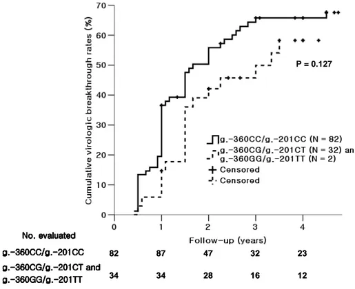
Because 9 patients who were classified as NR were excluded, the genotype of two SNPs (g.-360C>G and g.-201C>T) was available in only 116 CHB patients. With a median follow-up of 48 (12 – 90) months, the 3-year cumulative virologic breakthrough rates for the group of g.-360CG/g.-201CT (N = 32) and g.-360GG/g.-201TT (N = 2) were 49.9%, whereas that for the g.-360CC/g.-201CC (N = 82) was 65.8%. Cumulative virologic breakthrough rates of the two groups were presented in Fig. 3 using Kaplan-Meier method. Although those of the genotypes containing g.-360G/g.-201T haplotype were slightly lower, there was no statistically significant difference between two groups (P = 0.127).
82 87 47 32 23 34 34 28 16 12 P = 0.127 g. g. g. g.---360CC/g.-360CC/g.360CC/g.360CC/g.----201CC201CC201CC201CC g. g. g.
g.---360CG/g.-360CG/g.360CG/g.360CG/g.----201CT and201CT and201CT and201CT and g. g. g. g.---360GG/g.-360GG/g.360GG/g.360GG/g.---201TT-201TT201TT201TT No. evaluated No. evaluated No. evaluated No. evaluated 82 87 47 32 23 34 34 28 16 12 P = 0.127 g. g. g. g.---360CC/g.-360CC/g.360CC/g.360CC/g.----201CC201CC201CC201CC g. g. g.
g.---360CG/g.-360CG/g.360CG/g.360CG/g.----201CT and201CT and201CT and201CT and g. g. g. g.---360GG/g.-360GG/g.360GG/g.360GG/g.---201TT-201TT201TT201TT No. evaluated No. evaluated No. evaluated No. evaluated
Fig. 3. Cumulative virologic breakthrough rates between the group of g.-360G/g.-201T haplotype and g.-360C/g.-201C haplotype by using Kaplan-Meier method.
The genotype of two SNPs was available in only 116 CHB patients. With a median follow-up of 48 (12 – 90) months, the 3-year cumulative virologic breakthrough rates for the group of g.-360CG/g.-201CT (N = 32) and g.-360GG/g.-201TT (N = 2) were 49.9%, whereas that for the g.-360CC/g.-201CC (N = 82) was 65.8%. Although those of the genotypes containing g.-360G/g.-201T haplotype were slightly lower, there was no statistically significant difference between two groups (P = 0.127).
4
Cumulative HBeAg loss rates between the groups according to dCK
SNP haplotypes
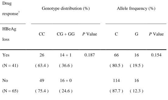
Among 125 patients, 19 patients with HBeAg negative CHB were excluded. Allele frequency analysis of the g.-201C/T and g.-360C/G in 106 HBeAg positive CHB patients, as well as 112 peripheral blood samples from normal individuals, is also in agreement with the Hardy-Weinberg equilibrium (HWp = 0.533). The 106 HBeAg positive CHB patients were divided into two groups according to loss of HBeAg during lamivudine monotherapy. 41 patients achieved HBeAg loss during treatment period, whereas another 65 patients did not achieve HBeAg loss. Unfortunately, there was no significant difference between the two groups in terms of the distribution of SNP genotype and allele frequency (P = 0.187, P = 0.154) (Table 6).
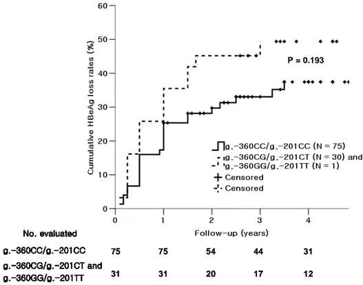
The follow-up data of cumulative HBeAg loss rates were available for 106 HBeAg positive CHB patients. With a median follow-up of 51 (12 – 90) months, the 3-year cumulative HBeAg loss rate for the group of g.-360CG/g.-201CT (N = 30) and g.-360GG/g.-201TT (N = 1) was 49.4%, whereas that for the g.-360CC/g.-201CC (N = 75) was 33.0%. Cumulative HBeAg loss rates of the two groups were presented in Fig. 4 using Kaplan-Meier method. Although those of the genotypes containing g.-360G/g.-201T haplotype were slightly higher, there was no statistically significant difference between two groups (P = 0.193).
Table 6. Genotype distribution and allele frequency of g.-360C/G and g.-201C/T in two groups according to loss of HBeAg during lamivudine treatment*
Drug response†
Genotype distribution (%) Allele frequency (%)
HBeAg loss CC CG + GG P Value C G P Value Yes (N = 41) 26 ( 63.4 ) 14 + 1 ( 36.6 ) 0.187 66 ( 80.5 ) 16 ( 19.5 ) 0.154 No (N = 65) 49 ( 75.4 ) 16 + 0 ( 24.6 ) 114 ( 87.7 ) 16 ( 12.3 ) *
106 patients with HBeAg positive CHB who were treated with lamivudine were analyzed.
†
75 75 54 44 31 31 31 20 17 12 P = 0.193 g. g.g. g.----360CC/g.360CC/g.360CC/g.360CC/g.----201CC201CC201CC201CC g. g.g.
g.----360CG/g.360CG/g.360CG/g.360CG/g.----201CT and201CT and201CT and201CT and g.
g.g.
g.----360GG/g.360GG/g.360GG/g.360GG/g.---201TT-201TT201TT201TT No. evaluated No. evaluatedNo. evaluated No. evaluated 75 75 54 44 31 31 31 20 17 12 P = 0.193 g. g.g. g.----360CC/g.360CC/g.360CC/g.360CC/g.----201CC201CC201CC201CC g. g.g.
g.----360CG/g.360CG/g.360CG/g.360CG/g.----201CT and201CT and201CT and201CT and g.
g.g.
g.----360GG/g.360GG/g.360GG/g.360GG/g.---201TT-201TT201TT201TT No. evaluated No. evaluatedNo. evaluated No. evaluated
Fig. 4. Cumulative HBeAg loss rates between the group of g.-360G/g.-201T haplotype and g.-360C/g.-201C haplotype by using Kaplan-Meier method.
The follow-up data of cumulative HBeAg loss rates were available for 106 HBeAg positive CHB patients. With a median follow-up of 51 (12 – 90) months, the 3-year cumulative HBeAg loss rate for the group of g.-360CG/g.-201CT (N = 30) and g.-360GG/g.-201TT (N = 1) was 49.4%, whereas that for the g.-360CC/g.-201CC (N = 75) was 33.0%. Although those of the genotypes containing g.-360G/g.-201T haplotype were slightly higher, there was no statistically significant difference between two groups (P = 0.193).
5.
Haplotype structure between control and patients with chronic
hepatitis B
We used three SNP sets for haplotype-based association analyses, grouping SNPs on the basis of the level of LD strength. We used 2-SNP (g.-360C>G and g.-201C>T), 3-SNP (c.364C>T, IVS6+41T>A, and c.948T>C), and 5-SNP (g.-360C>G, g.-201C>T, c.364C>T, IVS6+41T>A, and c.948T>C) sets to create the windows for performing each of 3 separate analyses (Fig. 1, 2). In 2-SNP haplotype block, there were 2 configuration (A: CC; and B: GT) that accounted for 100% of all chromosomes in patients with CHB. The haplotype pattern did not differ significantly between control and patients. In addition, specific haplotypes in the 3-SNP and 5-SNP block did not differ significantly between two groups (Table 7).
Table 7. Haplotype clusters and frequencies of 3 single nucleotide polymorphism sets at the dCK gene in patients with
chronic hepatitis B* and control samples†
SNP‡ Frequency
Haplotype Cluster
g.-360C>G g.-201C>T c.364C>T IVS6+41T>A c.948T>C Control Patients Chi Square P value
2-SNP Cluster A C C 0.862 0.835 0.594 0.441 B G T 0.138 0.165 0.594 0.441 Total 1.0 1.0 3-SNP Cluster A C T T 0.929 0.924 0.036 0.850
B T A C 0.071 0.071 0.0 0.990 Total 1.0 0.995 5-SNP Cluster A C C C T T 0.790 0.759 0.612 0.434 B G T C T T 0.138 0.165 0.598 0.439 C C C T A C 0.071 0.071 0.0 0.990 Total 0.999 0.995 *
125 patients with CHB who were treated with lamivudine were analyzed. †
Peripheral blood samples from 112 healthy donors were also used as control
‡
Two promoter, two exon and one intronic SNPs, which were g.-360C>G, g.-201C>T, c.364C>T, c.948T>C and IVS6+41T>A were selected according to high frequencies of alleles.
6.
Haplotype structure and association with lamivudine response among
patients with chronic hepatitis B
Using all 5 available SNPs, we simultaneously evaluated. The specific haplotype in the 5-SNP block differed significantly between good response and poor response. One of the clusters, 5-SNP-B (GTCTT) corresponding to 2-SNP-B (GT), was higher frequency in good response than poor response (0.203 vs 0.093; P = 0.019; OR, 2.830; CI, 1.035 – 7.737). In contrast, cluster 5-SNP-C (CCTAC) corresponding to 3-SNP-B (TAC), was slightly more abundant in poor response (0.039 vs 0.100; P = 0.063). In lamivudine monotherapy, these data suggested that haplotype cluster 5-SNP-B (GTCTT) was highly associated with good response in patients with CHB, while haplotype cluster 5-SNP-C (CCTAC) was associated with poor response (Table 8, 9).
Table 8. Haplotype clusters and frequencies of 3 single nucleotide polymorphism sets at the dCK gene in patients with good
response and poor response*
SNP† Frequency Haplotype Cluster g.-360C>G g.-201C>T c.364C>T IVS6+41T>A c.948T>C Good Response‡ Poor Response‡ Chi Square P value Odds Ratio 95% CI 2-SNP Cluster A C C 0.797 0.907 5.536 0.019 0.351 0.138 – 0.892 B G T 0.203 0.093 5.536 0.019 2.846 1.121 – 7.227 Total 1.0 1.0
3-SNP Cluster A C T T 0.953 0.898 2.649 0.104 B T A C 0.039 0.100 3.470 0.063 Total 0.989 0.998 5-SNP Cluster A C C C T T 0.750 0.806 1.038 0.308 B G T C T T 0.203 0.093 5.536 0.019 2.830 1.035 – 7.737 C C C T A C 0.039 0.100 3.470 0.063 Total 0.992 0.999 *
†
Two promoter, two exon and one intronic SNPs, which were g.-360C>G, g.-201C>T, c.364C>T, c.948T>C and IVS6+41T>A were selected according to high frequencies of alleles.
‡
Good response: virological response with undetectable HBV DNA and ALT normalization at 48 weeks. Poor response: virological response without decline in serum HBV DNA by ≥ 1 log10 copies/mL after the first 6 months of therapy and with increase in serum HBV DNA by > 1 log10 copies/mL (10-fold) above nadir after achieving virological response, during continued treatment.
Table 9. Multivariate logistic regression analysis of predictive factors associated with good response
Valuables Odds ratio 95% CI P Value
Specific haplotype cluster (GTCTT)* 2.830 1.035 – 7.737 0.043 Age (years) 0.955 0.916 – 0.997 0.035 Baseline serum ALT (IU/L) 1.007 1.001 – 1.012 0.012 Baseline HBV DNA (log10 copies/mL) 0.548 0.344 – 0.873 0.011 *
Two promoter, two exon and one intronic SNPs, which were g.-360C>G, g.-201C>T, c.364C>T, c.948T>C and IVS6+41T>A were selected according to high frequencies of alleles.
Abbreviation: CI, confidence interval; ALT, alanine aminotransferase; HBV, hepatitis B virus
IV. DISCUSSION
Novel eight dCK SNPs were found (g.-2052C>A, g.-360C>G, c.364C>T (P122S), IVS3-46G>del., IVS4+40G>T, IVS5+39T>C, IVS5-72A>T, and c.966~975T10>T11). Especially, the two promoter SNPs g.-360C>G and
g.-201C>T, which were more confirmed in Caucasians and Chinese, were more frequent in Korean (26% vs. 2% and 15.6%). In addition, we found one SNP (P122S) in exon 3, which leads to change the amino acid sequence. Although Joerger M et al. found these SNPs in Caucasian healthy volunteers, Shi et al. did not find any exon 3 polymorphisms in Chinese study cohort.26,28
Deoxycytidine kinase is responsible for the phosphorylation of several deoxyribonucleosides and their analogs. It is the rate limiting enzyme in the activation of many important antiviral drugs.29 Deficiency of this enzyme activity is associated with resistance to antiviral and anticancer chemotherapeutic agents, whereas increased enzyme activity is associated with increased activation of these compounds to antiviral nucleoside triphosphate derivatives. It is clinically important because of its relationship to drug resistance and sensitivity.30-32
According to Shi et al, acute myelogenous leukaemia patients carrying the g.−360CG/g.−201CT or g.−360GG/g.−201TT genotype expressed significantly higher levels of dCK mRNA than patients carrying the g.−360CC/g.−201CC
genotype, and the former also displayed a favourable response to 1-β-arabinofuranosylcytosine (AraC)-containing induction chemotherapy.26 These results confirm the functionality of the g.−360CG/g.−201CT promoter polymorphism. On the basis of these results, CHB patients carrying the g.−360CG/−201CT or g.−360GG/−201TT genotype may have active lamivudine triphosphate derivatives and show a favorable response to drug sensitivity.
In present study, we explored the potential relationship between the clinical response to lamivudine and the SNPs of the dCK gene, a key enzyme in the metabolism of lamivudine. When CHB patients were divided into two groups according to clinical outcomes (virological and biochemical response at 48 weeks) to lamivudine, there was statistically significant difference between the two groups in terms of the distribution of SNP genotype and allele frequency (P = 0.027, P = 0.021). Also, the genotypes containing g.-360G/g.-201T haplotype were independent predictive factors for good drug response (P = 0.028; OR, 2.846; 95% CI, 1.121 – 7.227). In addition, our data suggested that haplotype cluster 5-SNP-B (GTCTT) was highly associated with good response in patients with CHB (0.203 vs 0.093; P = 0.019; OR, 2.830; CI, 1.035 – 7.737). The cumulative HBeAg loss rates showed a tendency to increase for long-term follow-up in CHB patients carrying the g.−360CG/g.−201CT or g.−360GG/g.−201TT genotype. However, there was no statistically significant difference between two groups (P = 0.193).
Shi et al. suggested that g.-360G/g.-201T haplotype may provide the cellular transcriptional machinery with more efficient promoter/enhancer elements.26 According to this hypothesis, a possible explanation of our results could be that the g.-360C/g.-201C homozygous genotype may be considered as new factor contributing to the resistance to lamivudine.
Recently, Lamba JK et al. reported that the homozygous genotype (c.364CC) of c.364C>T (P122S) had a significantly greater ratio dCK activity to dCK mRNA expression than other subjects (c.364CT or c.364TT). In our data, the frequency of good response was slightly higher in CHB patients with c.364CC genotype than those with c.364CT or c.364TT genotype (Table 4).33
Lamivudine is one of effective antiviral agents used in CHB treatment. However, resistance to lamivudine remains a main drawback in the treatment of CHB. Therefore, the main concern with long-term NA therapies is the emergence of drug resistance. Long-term treatment of CHB patients with NA agents may result in failure of therapy, due to the rapid emergence of resistant virus mutants with decreased susceptibility to therapeutic agents. Besides, cellular factors as well as viral resistance may contribute to the weaken efficiency of antiviral agents. This led to the assumption that altered drug metabolism in host cells may contribute to inefficient activation of lamivudine in CHB patients. Thus, intracellular sub-therapeutic levels of the active compounds may develop. In this intracellular environment, selection of resistant virus populations may be promoted.34,35
Our study may give an answer to the question, whether cellular factors, in addition to viral mutations, may account for the success or failure of lamivudine monotherapy. It may suggest that enzymatic activation of lamivudine probably may have an effect on antiviral activity in CHB patients.
As other factors affecting antiviral efficacy, the pharmacokinetics properties including, intestinal absorption, distribution into the infected liver, and cellular uptake using nucleoside transporters remain unclear if any of these properties play an important role in the response to antiviral agents. In the clinical setting, host determinants such as compliance, severity of the liver disease and associated disorders may also influence antiviral efficacy.36,37
The limitations of this study were due to its small size. Thus the results of the exploratory analysis in CHB patients need to be confirmed in a larger patient population. If the correlation could be confirm in a larger number of patients, the SNPs of dCK gene would be a genetic marker predicting treatment response for antiviral agents. Another perspective is that the data generated in this study must be used to elucidate the functionality of dCK candidate SNPs in patients receiving antiviral agents. Experimental research must be explored to find the exact mechanism of how SNPs regulate dCK gene expression. Lastly, the functionality of exon and intronic SNPs and the relationship between SNPs have to be studied. Although CHB is a very heterogeneous disease with different genotype and viral mutation that are of prognostic significance, the pharmacogenetics of lamivudine could help in better understanding of drug
responsiveness and guide us to develop individualized antiviral therapy in CHB patients receiving NA.
In summary, 15 dCK SNPs were found. Among them, eight dCK SNPs were newly discovered. Especillay, the two promoter SNPs g.-360C/G and g.-201C/T were more frequent in Korean than other ethnic group. We found one SNP (P122S) in exon 3, which leads to change the amino acid sequence. The genotype and the allele frequency of g.-360G/g.-201T were significantly higher in good response. Also, the genotypes containing g.-360G/g.-201T haplotype were independent predictive factors for good drug response. In addition, specific haplotype cluster 5-SNP-B (GTCTT) was highly associated with good response in patients with CHB. These results anticipate that SNP haplotypes of dCK gene may play an important role as a genetic marker for predicting lamivudine responsiveness in patients with CHB.
We have shown in a pilot study of the clinical implication of dCK polymorphisms in CHB patients undergoing treatment with lamivudine. In future, further studies will be tried whether these SNP haplotypes are associated with the levels of transcriptional expression of dCK gene, as well as the level of active NAs triphosphates. Our data will provide important evidence of a relationship between dCK gene and antiviral agents
V. CONCLUSION
We hypothesize that a genetic variation such as SNPs of the dCK gene may influence on the clinical responsiveness to lamivudine monotherapy.
1. Eight dCK SNPs were newly discovered (g.-2052C>A, g.-360C>G, c.364C>T (P122S), IVS3-46G>del., IVS4+40G>T, IVS5+39T>C, IVS5-72A>T, and c.966~975T10>T11).
2. Especillay, the two promoter SNPs g.-360C/G and g.-201C/T were more frequent in Korean than other ethnic group.
3. We found one SNP (P122S) in exon 3, which leads to change the amino acid sequence.
4. The genotype and the allele frequency of g.-360G/g.-201T were significantly higher in good response.
5. The genotypes containing g.-360G/g.-201T haplotype were independent predictive factors for good response.
6. The specific haplotype cluster 5-SNP-B (GTCTT) was highly associated with good response in patients with CHB.
7. These results anticipate that SNP haplotypes of dCK gene may play an important role as a genetic marker for predicting lamivudine responsiveness in patients with CHB.
VI. REFERENCES
1. Yim HJ, Hussain M, Liu Y, Wong SN, Fung SK, Lok AS. Evolution of
multi-drug resistant hepatitis B virus during sequential therapy. Hepatology 2006; 44:703-12.
2. Mutimer D, Pillay D, Cook P, Ratcliffe D, O'Donnell K, Dowling D, et al. Selection of multiresistant hepatitis B virus during sequential nucleoside-analogue therapy. J Infect Dis 2000; 181:713-6.
3. Villet S, Pichoud C, Villeneuve JP, Trepo C, Zoulim F. Selection of a multiple drug-resistant hepatitis B virus strain in a liver-transplanted patient. Gastroenterology 2006; 131:1253-61.
4. Ghany M, Liang TJ. Drug targets and molecular mechanisms of drug resistance in chronic hepatitis B. Gastroenterology 2007; 132:1574-85. 5. Nair S, Perrillo RP. In: Zakim D, Boyer T, Eds.; Hepatitis B and D
Hepatology: A Textbook of Liver Disease. Saunders Philadelphia: 2003. p. 959-1016.
6. Perrillo RP. Overview of Treatment of Hepatitis B: Key Approaches and Clinical Challenges. Semin Liver Dis 2004; 24(suppl 1): 23-9.
7. Locarnini SA. Molecular virology of hepatitis B. Semin Liver Dis 2004; 24(suppl 1): 3-10.
8. Locarnini S, Mason WS. Cellular and virological mechanisms of HBV drug resistance. J Hepatol 2006; 44:422-31.
9. Locarnini SA, Hatzakis A, Heathcote J. Management of antiviral resistance in patients with chronic hepatitis B. Antivir Ther 2004; 9: 679-93.
10. Evans WE, Relling MV. Pharmacogenomics: translating functional genomics into rational therapeutics. Science 1999; 286:487–91.
11. Hoehe MR, Timmermann B, Lehrach H. Human inter-individual DNA sequence variation in candidate genes, drug targets, the importance of haplotypes and pharmacogenomics. Curr Pharm Biotechnol 2003; 4:351–78.
12. Ahn SH, Kim DY, Chang HY, Hong SP, Shin JS, Kim YS, et al. Association of genetic variations in CCR5 and its ligand, RANTES with clearance of hepatitis B virus in Korea. J Med Virol 2006; 78:1564-71. 13. Eriksson S, Wang L. The role of cellular deoxynucleoside kinases in the
activation of nucleoside analogs used in chemotherapy. Recent Advances in Nucleosides: Chemistry and Chemotherapy, Chu C.K.ed. Amsterdam: Elsevier, 2002: 455-75.
14. Ahn SH, Han KH, Park JY, Lee CK, Kang SW, Chon CY, et al. Association between hepatitis B virus infection and HLA-DR type in Korea. Hepatology 2000; 31:1371-3.
15. Waqar MA, Evans MJ, Manly KF, Hughes RG, Huberman JA. Effects of 29, 39-dideoxynucleosides on mammalian cells and viruses. J Cell Physiol 1984; 121:402-8.
Rapid evolution of RNA genomes. Science 1982; 215:1577-85.
17. Deeks SG, Barditch-Crovo P, Lietman PS, Hwang F, Cundy KC, Rooney JF, et al. Safety, pharmacokinetics, and antiretroviral activity of intravenous 9-[2-(R)-phosphonomethoxy)propyl]adenine, a novel anti-human immunodeficiency virus (HIV) therapy, in HIV-infected adults. Antimicrob Agents Chemother 1998; 42:2380-4.
18. Barditch-Crovo P, Toole J, Hendrix CW, Cundy KC, Ebeling D, Jaffe HS, et al. Anti-human immunodeficiency virus (HIV) activity, safety, and pharmacokinetics of adefovir dipivoxil (9-[2-(bispivaloyloxy-methyl) phosphonylmethoxyethyl] adenine) in HIV-infected patients. J Infect Dis 1996; 176:406-13.
19. De Clercq E. Strategies in the design of antiviral drugs. Nat Rev Drug Discov 2002; 1:13-25.
20. Kim MY, Ives DH. Human deoxycytidine kinase: kinetic mechanism and end product regulation. Biochemistry 1989; 28:9043–7.
21. Shewach DS, Reynolds KK, Hertel L. Nucleotide specificity of human deoxycytidine kinase. Mol Pharmacol 1992; 42:518–24.
22. Hengstschlager M, Denk C, Wawra E. Cell cycle regulation of deoxycytidine kinase: evidence for post-transcriptional control. FEBS Lett 1993; 321:237–40.
23. Chen EH, Johnson EE, Vetter SM, Mitchell BS. Characterization of the deoxycytidine kinase promoter in human lymphoblast cell lines. J Clin
Invest 1995; 95:1660–8.
24. Johansson M, Norda A, Karlsson A. Conserved gene structure and transcription factor sites in the human and mouse deoxycytidine kinase genes. FEBS Lett 2000; 487:209–12.
25. Veuger MJ, Honders MW, Landegent JE, Willemze R, Barge RM. High incidence of alternatively spliced forms of deoxycytidine kinase in patients with resistant acute myeloid leukemia. Blood 2000; 96:1517–24.
26. Shi JY, Shi ZZ, Zhang SJ, Zhu YM, Gu BW, Li G, et al. Association between single nucleotide polymorphisms in deoxycytidine kinase and treatment response among acute myeloid leukaemia patients. Pharmacogenetics 2004; 14:759–68.
27. Lok AS, Zoulim F, Locarnini S, Bartholomeusz A, Ghany MG, Pawlotsky JM, et al. Antiviral drug-resistant HBV: standardization of nomenclature and assays and recommendations for management. Hepatology 2007; 46:254-65.
28. Joerger M, Bosch TM, Doodeman VD, Beijnen JH, Smits PHM, Schellens JHM. Novel deoxycytidine kinase gene polymorphisms: a population screening study in Caucasian healthy volunteers. Eur J Clin Pharmacol 2006; 62:681-4.
29. Stein DS, Moore KHP. Phosphorylation of Nucleoside Analog Antiretrovirals: A Review for Clinicians. Pharmacotherapy 2001; 21:11-34. 30. Groschel B, Cinatl J, Cinatl J Jr. Viral and cellular factors for resistance
against antiretroviral agents. Intervirology 1997; 40:400-7.
31. Jacobsson B, Britton S, He Q, Karlsson A, Eriksson S. Decreased thymidine kinase levels in peripheral blood cells from HIV-seropositive individuals: implications for zidovudine metabolism. AIDS Res Hum Retroviruses 1995; 11:805-11.
32. Antonelli G, Turriziani O, Verri A, Narciso P, Ferri F, D'Offizi G, et al. Long-term exposure to zidovudine affects in vitro and in vivo the efficiency of phosphorylation of thymidine kinase. AIDS Res Hum Retroviruses 1996; 12:223-8.
33. Lamba JK, Crews K, Pounds S, Schuetz E, Gresham J, Gandhi V, et al. Pharmacogenetics of Deoxycytidine kinase: identification and characterization of novel genetic variants. J Pharmacol Exp Ther 2007; 12:`935-45.
34. Shaw T, Bartholomeusz A, Locarnini S. HBV drug resistance: mechanisms, detection and interpretation. J Hepatol 2006; 44:593-606.
35. Zoulim F. Mechanism of viral persistence and resistance to nucleoside and nucleotide analogs in chronic hepatitis B virus infection. Antiviral Res 2004; 64:1-15.
36. Lee K, Chu CK. Molecular modeling approach to understanding the mode of action of L-nucleosides as antiviral agents. Antimicrob Agents Chemother 2001; 45:138-44.
a hepatitis B virus polymerase may be a primer-unblocking mechanism. Proc Natl Acad Sci U S A. 2001; 98:4984-9.
< ABSTRACT (IN KOREAN)>
만성 B형 간염 환자에서 Deoxycytidine kinase 단일 염기
다형성과 라미부딘 치료반응과의 상관성 연구
<지도교수 한 광 협>
연세대학교 대학원 의학과
이 현 웅
연 연 연 연구배경구배경구배경구배경 및및및및 목적목적목적목적: : : Deoxycytidine kinase (dCK)는 간세포내에서 라미: 부딘이 약리작용을 할 수 있는 형태로 인산화하는데 가장 중요한 효 소로 알려져 있다. 본 연구의 목적은 서양인과 중국인에서 이미 알려 진 dCK 단일 염기 다형성을 확인하며, 한국인에서 새로운 단일 염기 다형성을 발견하고, 발견된 단일 염기 다형성이 만성 B형 간염 환자 에서 라미부딘 치료효과와 관련이 있는지를 알아보고자 하였다. 대상 대상 대상 대상 및및및및 방법방법방법: 방법: : : 24명의 건강한 사람과 24명의 만성 B형 간염 환자로부 터 dCK 유전자를 모두 분석하여 단일 염기 다형성을 평가하였다. 또한, 라미부딘으로 48주 이상 치료를 받은 125명의 만성 B형 간염 환 자를 대상으로 임상자료 분석과 발견된 단일 염기 다형성과의 상관관 계를 분석하였다. 라미부딘 치료 48주에 혈청 HBV DNA 가 음전되고 ALT 정상을 획득한 군을 “약물 반응 군”으로, 초기 치료 반응이 없 고 치료 48주 이내에 바이러스 돌파가 발생한 군을 “약물 비반응 군” 으로 정의하였다. 결과 결과 결과 결과:::: 한국인의 dCK 유전자에서 확인된 15개의 단일 염기 다형성 중 7개의 단일 염기 다형성은 기존 데이터 베이스에 알려져 있으나 8개 의 단일 염기 다형성은 새롭게 발견되었다 (g.-2052C>A, g.-360C>G, c.364C>T (P122S), IVS3-46G>del., IVS4+40G>T, IVS5+39T>C, IVS5-72A>T, and c.966~975T10>T11). 특히,
promoter 위치에서 발견된 g.-360C/G 와 g.-201C/T 단일 염기 다 형성은 강한 연관 불균형 관계를 보이고 있으며 대립유전자형 (allele)의 빈도는 서양인이나 중국인에 비해 높았다 (26% vs. 2% and 15.6%). 라미부딘 치료 48주에 g.-360CG/g.-201CT 와 g.-360GG/g.-201TT 유전형을 가진 환자 군이 g.-360CC/ g.-201CC 유전형을 가진 환자 군에 비해 효과적인 치료 반응을 보였 다 (70.6% vs. 48.2%, P = 0.027). 또한, g.-360G/g.-201T 대립유전 자형의 빈도가 약물반응 군에서 의미있게 높았다 (20.0% vs. 9.3%, P


