저작자표시-비영리-변경금지 2.0 대한민국 이용자는 아래의 조건을 따르는 경우에 한하여 자유롭게 l 이 저작물을 복제, 배포, 전송, 전시, 공연 및 방송할 수 있습니다. 다음과 같은 조건을 따라야 합니다: l 귀하는, 이 저작물의 재이용이나 배포의 경우, 이 저작물에 적용된 이용허락조건 을 명확하게 나타내어야 합니다. l 저작권자로부터 별도의 허가를 받으면 이러한 조건들은 적용되지 않습니다. 저작권법에 따른 이용자의 권리는 위의 내용에 의하여 영향을 받지 않습니다. 이것은 이용허락규약(Legal Code)을 이해하기 쉽게 요약한 것입니다. Disclaimer 저작자표시. 귀하는 원저작자를 표시하여야 합니다. 비영리. 귀하는 이 저작물을 영리 목적으로 이용할 수 없습니다. 변경금지. 귀하는 이 저작물을 개작, 변형 또는 가공할 수 없습니다.
A Doctoral Dissertation
Cytoprotective effects of Baicalein against
oxidative stress-induced cell damage
Ki-Cheon Kim
Department of Medicine
GRADUATE SCHOOL
JEJU NATIONAL UNIVERSITY
산화적 스트레스에 대한 Baicalein
화합물의 세포 보호 작용 기전 연구
지도교수 현 진 원, 고 영 상
김 기 천
이 논문을 의학 박사학위 논문으로 제출함
2014년 2월
김기천의 의학 박사학위 논문을 인준함
심사위원장 印
위 원 印
위 원 印
위 원 印
위 원 印
제주대학교 대학원
2014년 2월
Cytoprotective effects of Baicalein against
oxidative stress-induced cell damage
Ki-Cheon Kim
(Supervised by Professor Young-Sang Koh and Professor Jin-Won
Hyun)
A thesis submitted in partial fulfillment of the requirement
for Doctor of Philosophy degree in Medicine
2014. 02.
This thesis has been examined and approved.
Thesis director, Yung-Hyun Choi, Professor of Dongeui University Young-Sang Koh, Professor of Jeju National University
Hye-Kyoung Kang, Professor of Jeju National University
Gi-Young Kim, Professor of Jeju National University
Jin-Won Hyun, Professor of Jeju National University
2014. 02
.
Date
Department of Medicine
GRADUATE SCHOOL
JEJU NATIONAL UNIVERSITY
CONTENTS
CONTENT OF PART I... i
CONTENT OF PART II... ii
LIST OF FIGURES... iii
BACK GROUND... 1
PART I... 2
PART II... 21
i
CONTENT OF PART I
ABSTRACT ... 3
INTRODUCTION... 4
MATERIALS AND METHODS... 6
1. Cell culture... 6
2. Reagents... 6
3. Reverse transcription-polymerase chain reaction... 7
4. Western blot analysis... 7
5. Determination of MMP-1 activity... 8
6. Transient transfection and AP-1 luciferase assay... 8
7. Statistical analysis... 9
RESULTS... 10
1. Baicalein reduces the H2O2-induced MMP-1 expression and activity... 10
2. Baicalein attenuates the H2O2-induced activation of AP-1... 14
3. Baicalein reduces the H2O2-induced phosphorylation of MEK-ERK and SEK-JNK... 16
ii
CONTENT OF PART II
ABSTRACT ... 22
INTRODUCTION... 23
MATERIALS AND METHODS... 26
1. Reagents... 26
2. Cell culture... 26
3. Measurement of intracellular ROS... 26
4. Comet assay... 27
5. Western blot analysis... 28
6. Detection of 8-OxoG... 28
7. Transient transfection and OGG1 promoter luciferase assay... 29
8. Reverse transcriptase polymerase chain reaction... 29
9. Cell viability... 30
10. TUNEL assay... 30
11. Statistical analysis... 31
RESULTS... 32
1. Baicalein shows intracellular ROS scavenging activity... 32
2. Baicalein reduces the induction of DNA strand breaks by oxidative stress... 34
3. Baicalein inhibits oxidative stress-induced DNA base modification... 38
4. Baicalein prevents the inhibition of OGG1 protein and mRNA expression by H2O2 treatment... 40
5. Baicalein activates the Akt signaling pathway... 42
6. Baicalein inhibits apoptotic DNA fragmentation induced by oxidative stress.. 44
iii
LIST OF FIGURES
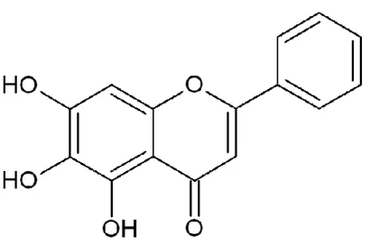
FIGURE 1. Chemical structure of baicalein (5,6,7-trihydroxyflavone)... 6
FIGURE 2. Effects of baicalein in a dose dependent manner on H2O2-induced MMP-1 activity... 11
FIGURE 3. Effects of baicalein on H2O2-induced MMP-1 mRNA level... 12
FIGURE 4. Effects of baicalein on H2O2-induced MMP-1 protein expression... 13
FIGURE 5. Effects of baicalein and NAC on H2O2-induced MMP-1 activity... 14
FIGURE 6. Effects of baicalein on H2O2-induced AP-1 expression... 15
FIGURE 7. Effects of baicalein on H2O2-induced activity of AP-1 transcription factor... 16
FIGURE 8. Effects of baicalein on H2O2-induced ERK signal transduction... 17
FIGURE 9. Effects of baicalein on H2O2-induced JNK signal transduction... 18
FIGURE 10. Effects of baicalein in a dose dependent manner on the scavenging activity against H2O2 treatment... 33
FIGURE 11. Effects of baicalein on H2O2-induced intracellular ROS... 34
FIGURE 12. Effects of baicalein on H2O2-induced cellular DNA damage... 35
FIGURE 13. Effects of baicalein on H2O2-induced phospho-histone H2A.X... 36
FIGURE 14. Effects of baicalein on H2O2-reduced Ku70 and phospho-DNA-PKcs... 37
FIGURE 15. Effect of baicalein on H2O2-induced 8-OxoG levels... 38
FIGURE 16. Effect of baicalein on H2O2-induced 8-OxoG formation in cells... 39
FIGURE 17. Effects of baicalein on H2O2-reduced transcriptional activity of OGG1. 40
FIGURE 18. Effects of baicalein on H2O2-reduced mRNA level of OGG1... 41
FIGURE 19. Effects of baicalein on H2O2-reduced protein expression of OGG1... 42
FIGURE 20. Effects of baicalein on H2O2-reduced Akt phosphorylation... 43 FIGURE 21. Effects of baicalein on H2O2-reduced cell viability via akt signal
iv
pathway... 44
1
BACK GROUND
This dissertation consisted of two sections, one is the effects of baicalein against the senescence of skin keratinocytes induced by oxidative stresses and the other is the effects of baicalein against the DNA damage of lung fibroblasts induced by oxidative stresses. Lung and skin is typical tissue of human should be exposed to oxygen from the natural environments. According to the scientific development of civilization has been progressed, the natural environment has been destroyed. Human body has the various immune systems against the pollutions of environment, and the immune responses more progressed in tissues which contacted with outer environment. The immune responses generates the ROS and RNS, called free radicals, and they has harmful effects to the normal tissue when the redox balancing was broken.
Baicalein (5,6,7-trihydroxyflavone) is a flavonoid derived from the roots of Scutellaria
baicalensis. Baicalein attenuates oxidative stress and protects cardiomyocytes from lethal
oxidant damage in an ischemia-reperfusion model [13,14]. In addition, our recent work showed that baicalein ameliorated mitochondrial oxidative stress by activating nuclear factor (erythroid-derived 2)-like 2-mediated induction of manganese superoxide dismutase [15] and protected cellular components against oxidative damage by scavenging ROS and inhibiting apoptosis [16]. On the other hand, the protective effect of baicalein against ROS-associated stimulation of MMP-1 expression and depletion of DNA damage response against DNA double strands breaks by non-homologous end joing repair. These current reseaches focused on the ability of baicalein to safeguard cultured human keratinocytes and Chinese hamster lung fibroblasts agianst H2O2-mediated senescence and destructions of DNA repair
2
PART I
Baicalein attenuates oxidative stress-induced expression of matrix
metalloproteinase-1 by regulating via ERK and JNK mediated AP-1 pathway in
human keratinocytes
3 ABSTRACT
The matrix metalloproteinase (MMP) family is involved in the breakdown of the extracellular matrix during normal physiological processes such as embryonic development, reproduction, and tissue remodeling, as well as in disease processes such as pathological aging, arthritis, and metastasis. Oxidative conditions generate reactive oxygen species (ROS) (e.g., hydrogen peroxide [H2O2]) in cells, which subsequently induce the synthesis of matrix
metalloproteinase-1 (MMP-1). MMP-1, an interstitial collagenase, in turn stimulates an aging phenomenon. In this study, baicalein (5,6,7-trihydroxyflavone) was investigated for its in vitro activity against H2O2-induced damage using a human skin keratinocyte model.
Baicalein pretreatment signifi cantly inhibited H2O2-induced up-regulation of MMP-1
mRNA, MMP-1 protein expression and MMP-1 activity in cultured HaCaT keratinocytes. In addition, baicalein decreased the transcriptional activity of activator protein-1 (AP-1) and the expression of c-Fos and c-Jun, both components of the heterodimeric AP-1 transcription factor. Furthermore, baicalein reduced phosphorylation of extracellular signal-regulated kinase (ERK) and c-Jun-N-terminal kinase (JNK), which are upstream of the AP-1 transcription factor. The results of this study suggest that baicalein is involved in the inhibition of oxidative stress-induced expression of MMP-1 via inactivation of the ERK/JNK/AP-1 signaling pathway.
Key Words: Baicalein, Matrix metalloproteinase, Oxidative stress, Reactive oxygen species, Hydrogen peroxide, Signal transduction
4
INTRODUCTION
The degradation of the extracellular matrix (ECM) is essential for embryonic development, morphogenesis, reproduction, and tissue remodeling. The family of matrix metalloproteinases (MMPs) in general, and matrix metalloproteinase-1 (MMP-1) in particular, play a central role in these processes. MMP-1, or interstitial collagenase, is a secreted protein that contributes to the etiology of many age-related degenerative diseases [1,2]. MMP-1 is a prominently involved in the proteolytic release and activation of growth factors, cytokines, and signaling peptides, which also have the potential to modulate the senescent microenvironment [3].
Reactive oxygen species (ROS) such as hydrogen peroxide (H2O2) readily undergo
reactions with thiol groups and may, thus, participate in a common mechanisms underlying the activation of several different MMPs, including MMP-1 [4]. H2O2 regulates the activity
of critical signaling molecules, leading to augmented MMP-1 expression in human skin cells [5]. Furthermore, the redox activation of c-Jun-N-terminal kinase (JNK) controls the activity of the activator protein-1 (AP-1) transcription factor, resulting in an age-dependent increase in MMP-1 expression [3]. Moreover, oxidative stress stimulates the activity of extracellular signal-regulated kinase (ERK), which are also important for the regulation of MMP-1 expression. Blockade of the ERK pathway was found to abrogate the Ras- and serum-induced stimulation of the MMP-1 promoter, indicating a role for ERK in the transcriptional regulation of MMP-1 [6]. These studies suggest that the ERK/JNK/AP-1 pathway may be the major activator of MMP-1 gene and protein expression.
A number of studies demonstrate inhibition of MMP-1 up-regulation by antioxidants [7,8], including N-acetylcysteine (NAC), a precursor of glutathione [9-11]. Previous work from our group demonstrated that triphlorethol-A, an antioxidant, participates in the modulation of MMP-1 level in cultured cells [12]. These data provide further support for the
5
ability of ROS to initiate signaling pathways that lead to MMP-1 induction.
Baicalein (5,6,7-trihydroxyflavone) is a flavonoid derived from the roots of Scutellaria
baicalensis. Baicalein attenuates oxidative stress and protects cardiomyocytes from lethal
oxidant damage in an ischemia-reperfusion model [13,14]. In addition, our recent work showed that baicalein ameliorated mitochondrial oxidative stress by activating nuclear factor (erythroid-derived 2)-like 2-mediated induction of manganese superoxide dismutase [15] and protected cellular components against oxidative damage by scavenging ROS and inhibiting apoptosis [16]. On the other hand, the protective effect of baicalein against ROS-associated stimulation of MMP-1 expression has not been investigated. Therefore, the current study focused on the ability of baicalein to safeguard cultured human keratinocytes against H2O2
6
MATERIALS AND METHODS
1. Cell culture
Human keratinocytes (HaCaT cells) were cultured in Dulbecco’s modified Eagle’s medium containing 10% heat-inactivated fetal calf serum, streptomycin (100 μg/ml) and penicillin (100 U/ml). The cells were maintained at 37°C in a humidified atmosphere containing 5% CO2.
2. Reagents
Baicalein (Figure 1) was purchased from Sigma-Aldrich, Inc. (St. Louis, MO, USA). The primary MMP-1 antibody was purchased from Epitomics, Inc. (Burlingame, CA, USA). Primary antibodies against phospho MEK1 (mitogen-activated protein (MAP) kinase kinase1), MEK1, phospho ERK1/2, ERK2, phospho SEK1 (stress-activated protein kinase (SAPK)/ERK kinase1), SEK1, phospho JNK1/2, JNK1/2, c-Fos, and phospho c-Jun were purchased from Cell Signaling Technology (Beverly, MA, USA).
7
3. Reverse transcription-polymerase chain reaction (RT-PCR)
Cells were seeded in 100φ dish with 10 ml at a density of 1.5×10⁵ cells/ml. Sixteen hours
after plating, the cells were treated with baicalein at a concentration of 5 μg/ml. After 30 min, H2O2 (1 mM) was added to the plate. The cells were incubated for an additional 48 h at 37°C.
Total RNA was isolated from the cells using an easy-BLUE RNA extraction kit obtained from iNtRON Biotechnology Inc. (Daegeon, Korea). The PCR conditions for MMP-1 and the housekeeping gene, glyceraldehyde 3-phosphate dehydrogenase (GAPDH), were as follows: 35 cycles of denaturing at 94°C for 45 sec, annealing at 52°C for 1 min, and elongation at 72°C for 1 min; followed by a final extension at 72°C for 5 min. The forward and reverse primer pairs for MMP-1 and GADPH purchased from Bionics (Seoul, Republic of Korea) were as follows: human MMP-1, GAGGAAATCTTGCTCAT-3’ and 5’-CTCAGAAAGAGCAGCATC-3’; and human GAPDH, 5’-AAGGTCGGAGTCAACGGATTT-3’ and 5’-GCAGTGAGGGTCTCTCTCCT-3’. The amplified products were resolved by electrophoresis in a 1% agarose gel, stained with ethidium bromide, and photographed under ultraviolet light.
4. Western blot analysis
Cells were seeded in 100φ dish with 10 ml at a density of 1.5×10⁵ cells/ml. Sixteen hours
after plating, the cells were treated with baicalein at a concentration of 5 μg/ml. After 30 min, H2O2 (1 mM) was added to the plate. The cells were incubated for an additional 48 h at 37°C.
Cells were then lysed in lysis buffer [100 μl; 120 mM NaCl, 40 mM Tris (pH 8.0), 0.1% NP-40]. Aliquots of the lysates (20 μg protein) were boiled for 5 min and electrophoresed in a 10% SDS-polyacrylamide gel. The proteins in the gels were transferred onto nitrocellulose membranes purchased from Bio-Rad Laboratories Inc. (Hercules, CA, USA), and the membranes were subsequently incubated with the primary antibodies. The membranes were further incubated with secondary anti-IgG-horseradish peroxidase conjugates purchased
8
from Thermo Fisher Scientific Inc. (Rockford, IL, USA) followed by exposure to X-ray film. The protein bands were detected using an enhanced chemiluminescence Western blotting detection kit purchased from GE Healthcare Ltd. (Buckinghamshire, UK).
5. Determination of MMP-1 activity
Cells were seeded in a 96-well plate at a density of 1.5×10⁵ cells/well. Sixteen hours after
plating, the cells were treated with baicalein at indicated concentrations or pretreated with 1 mM of N-acetyl cysteine (NAC) for 1 h. After 30 min, H2O2 (1 mM) was added to the plate.
The cells were incubated for an additional 48 h at 37°C. MMP-1 activity was determined using a Fluorokine® E human active MMP-1 fluorescent assay purchased from R&D
Systems Inc. (Minneapolis, MN, USA) according to the manufacturer's instructions, which uses a quenched fluorogenic substrate. Production of the fluorescent cleavage product was determined using a fluorescence plate reader set purchased from BMG Labtech (Ortenberg, Germany) with an excitation wavelength of 320 nm and emission wavelength of 405 nm.
6. Transient transfection and AP-1 luciferase assay
Cells were transiently transfected with a plasmid harboring the AP-1 promoter using LipofectamineTM 2000 purchased from Invitrogen (Carlsbad, CA, USA), according to the
manufacturer's instructions. Following an overnight transfection, cells were treated with baicalein (5 μg/ml). After an additional incubation for 1 h, cells were treated with H2O2 (1
mM). After 6 h, the cells were washed twice with PBS and then lysed with a passive lysis buffer purchased from Promega (Madison, WI, USA). Following vortex-mixing and centrifugation at 12,000×g for 30 sec at 4°C, the supernatant was stored −70°C until use in the luciferase assay. After mixing the cell extract (20 μl) with the luciferase assay substrate reagent (100 μl) at room temperature, the mixture was placed in an illuminometer to measure the light produced. The amount of light produced provided a measure of AP-1 luciferase
9
activity, and hence, AP-1 transcriptional activity.
7. Statistical analysis
All the measurements were made in triplicate and all values are represented as mean ± standard error of the mean (SEM). The results were subjected to an analysis of the variance (ANOVA) using the Tukey test to analyze the difference. A value of p<0.05 was considered significant.
10 RESULTS
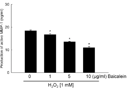
1. Baicalein reduces the H2O2-induced MMP-1 expression and activity
H2O2 and other ROS induce oxidative stress in many types of cells, including human
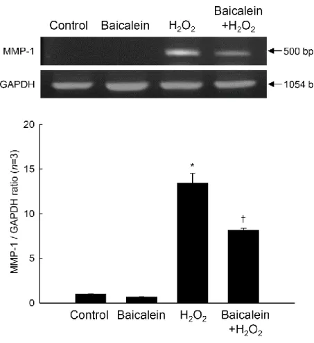
keratinocytes [17,18]. In particular, excessive amounts of ROS can stimulate mRNA and protein expression of MMP-1, a hallmark of oxidative stress [5]. Baicalein treatment at 1, 5, and 10 μg/ml significantly prevented the production of active MMP-1 in a concentration-dependent manner (Figure 2), and 5 μg/ml of baicalein was determined as the optimal concentration for further study. In the current study, H2O2 treatment markedly increased the
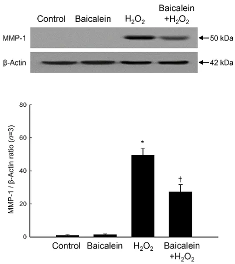
levels of MMP-1 mRNA and protein in cultured HaCaT cells compared with those in control cells (no treatment), as evidenced by RT-PCR (Figure 3) and Western blot (Figure 4) analyses. However, baicalein pretreatment inhibited the increased transcription of MMP-1 mRNA in H2O2-treated cells (Figure 3). Consistent with the RT-PCR results, baicalein also
partially inhibited the H2O2-induced increase in MMP-1 protein expression (Figure 4).
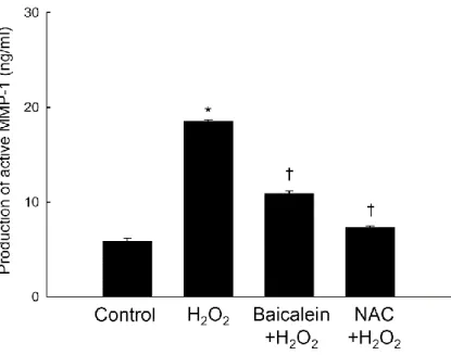
Moreover, H2O2 treatment increased the amount of active MMP-1 in HaCaT cells, whereas
pretreatment with baicalein or an antioxidant NAC significantly prevented this increase (Figure 5). These results suggest that baicalein blocked H2O2-induced MMP-1 expression
11
Figure 2. Effects of baicalein in a dose dependent manner on H2O2-induced MMP-1 activity.
Cells were treated with baicalein at indicated concentrations. After 30 min, H2O2 (1 mM)
was added to the plate. The cells were incubated for an additional 48 h at 37°C. MMP-1 activity was measured using a fluorimetric ELISA assay, as described in Materials and Methods. Each bar represents the mean ± the standard error from triplicate experiments. *Significantly different from H2O2 treatment (p<0.05).
12
Figure 3. Effects of baicalein on H2O2-induced MMP-1 mRNA level. Total RNA was
extracted, and MMP-1 mRNA expression was analyzed by RT-PCR. The GAPDH band is shown to confirm the integrity and equal loading of RNA. Each bar represents the mean ± the standard error from triplicate experiments. *Significantly different from untreated control
13
Figure 4. Effects of baicalein on H2O2-induced MMP-1 protein expression. Cell lysates were
electrophoresed in SDS-polyacrylamide gels and transferred onto nitrocellulose membranes. The expression of the MMP-1 protein was detected with a primary antibody specific for MMP-1. The actin band is shown to confirm equal loading of protein. Each bar represents the mean ± the standard error from triplicate experiments. *Significantly different from
14
Figure 5. Effects of baicalein and NAC on H2O2-induced MMP-1 activity. Cells were treated
with baicalein or NAC and after 30 min, H2O2 (1 mM) was added to the plate. The cells were
incubated for an additional 48 h at 37°C. MMP-1 activity was measured using a fluorimetric ELISA assay, as described in Materials and Methods. Each bar represents the mean ± the standard error from triplicate experiments. *Significantly different from untreated control
(p<0.05). †Significantly different from H2O2 treatment (p<0.05).
2. Baicalein attenuates the H2O2-induced activation of AP-1
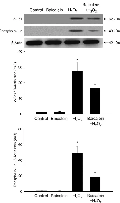
The AP-1 transcription factor exists as either a c-Jun/c-Jun homo-dimer or a c-Jun/c-Fos hetero-dimer and is involved in the activation of MMP genes [19,20]. H2O2 treatment
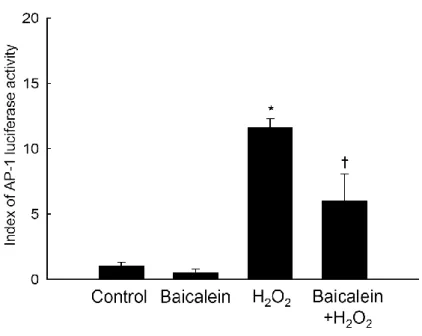
stimulated the expression of both c-Fos and phospho c-Jun, while baicalein pretreatment decreased the extent of this stimulation (Figure 6). Baicalein pretreatment also significantly reduced H2O2-induced AP-1 transcriptional activity, as assessed by the AP-1 luciferase assay
(Figure 7). These results suggest that baicalein blocked H2O2-induced the c-Fos and
15
Figure 6. Effects of baicalein on H2O2-induced AP-1 expression. Cell lysates were
electrophoresed in SDS-polyacrylamide gels, transferred onto nitrocellulose membranes, and immunoblotted using antibodies specific for c-Fos and phospho c-Jun. The actin band is shown as a loading control. Each bar represents the mean ± the standard error from triplicate experiments. *Significantly different from untreated control (p<0.05). †Significantly different
16
Figure 7. Effects of baicalein on H2O2-induced activity of AP-1transcription factor. The
transcriptional activity of AP-1 was assessed using an AP-1 luciferase assay as described in Materials and Methods. Each bar represents the mean ± the standard error in triplicate experiments. *Significantly different from control (p<0.05). †Significantly different from
H2O2 treatment (p<0.05).
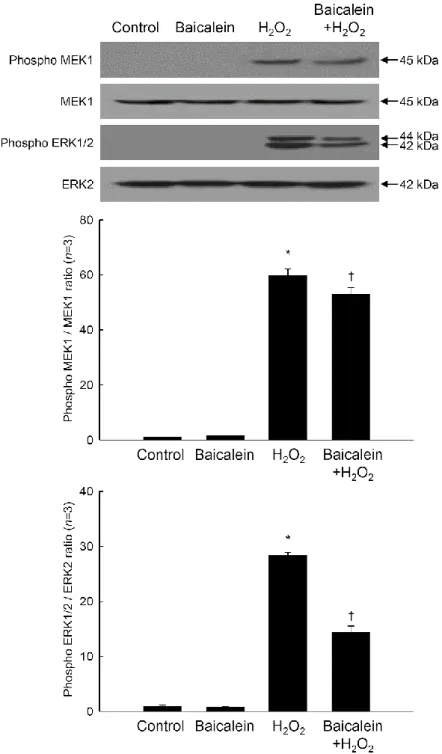
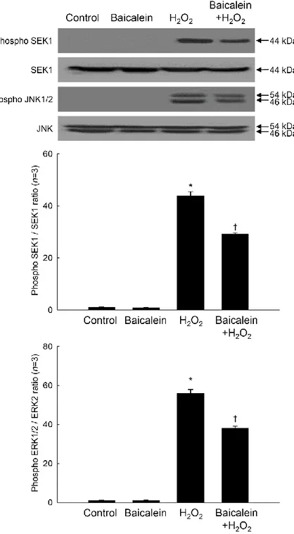
3. Baicalein reduces the H2O2-induced phosphorylation of MEK-ERK and SEK-JNK
Members of the Fos and Jun families of transcription factors are regulated by members of the MAP kinase family, particularly ERK and JNK [21-23]. As shown Figure 8, phospho ERK1/2 and phospho MEK1, which is upstream of ERK1/2, were markedly increased by H2O2 treatment. Baicalein reduced the amount of phospho ERK1/2 and phospho MEK1
induced by H2O2 (Figure 8). Furthermore, baicalein pretreatment attenuated the H2O2
-mediated up-regulation of phospho JNK1/2 and phospho SEK1, which is upstream of JNK1/2 (Figure 9). Taken together, the results of this study suggest that baicalein inhibits oxidative stress-induced expression and activity of MMP-1 by inactivating AP-1 and its associated signaling kinases.
17
Figure 8. Effects of baicalein on H2O2-induced ERK signal transduction. Cell lysates were
electrophoresed in SDS-polyacrylamide gels, transferred onto nitrocellulose membranes, and immunoblotted using primary antibodies specific for phospho MEK1, MEK1, phospho ERK1/2, and ERK2. Each bar represents the mean ± the standard error in triplicate experiments. *Significantly different from control (p<0.05). †Significantly different from
18
Figure 9. Effects of baicalein on H2O2-induced JNK signal transduction. Cell lysates were
electrophoresed in SDS-polyacrylamide gels, transferred onto nitrocellulose membranes, and immunoblotted using primary antibodies specific for phospho SEK1, SEK1, phospho JNK1/2, and JNK1/2. Each bar represents the mean ± the standard error in triplicate experiments. *Significantly different from control (p<0.05). †Significantly different from
19 DISCUSSION
This study suggested that baicalein should reduce the senescences induced by treatment of H2O2. We hypothesized the oxidative stress could induce the senescence identified by the
expressions and activations of MMP-1.
ROS, including H2O2, hasten aging of the skin and contribute to processes such as
ultraviolet B-induced photoaging, loss of elasticity, and wrinkling. These processes result, in large part, from the deterioration of the connective tissue matrix of the skin and, in particular, its collagen component. ROS produced in skin cells may contribute to the biological changes that are observed in the aging organ by accelerating the MMP-related ECM degradation system [24]. Furthermore, ROS are important intermediates in downstream signaling pathways that culminate in the induction of increased steady state levels of MMP-1 [5], an interstitial collagen-degrading enzyme.
In this study, H2O2 caused a pronounced increase in MMP-1 mRNA and protein
expression in cultured human HaCaT keratinocytes. Importantly, H2O2 also increased the
amount of active MMP-1. Pretreatment with baicalein, a flavonoid component of Scutellaria
baicalensis, partially suppressed the actions of H2O2. Furthermore, H2O2 augmented the
activity of the AP-1 transcription factor, which is formed by a c-Fos/c-Jun heterodimer, as well as the expression of c-Fos and phospho c-Jun. AP-1 binds to the 5’ residues of the MMP-1 promoter, stimulating the expression of the MMP-1 gene [25,26]. Notably, baicalein pretreatment also reduced H2O2-induced c-Fos/phospho c-Jun expression and AP-1
transcriptional activity.
c-Fos and c-Jun, the key components of AP-1, are primarily regulated at the transcriptional level by phosphorylated MEK/ERK and SEK/JNK, respectively [27-30]. H2O2 enhanced the phosphorylation of MEK1/ERK1/2 and SEK1/JNK1/2, in keeping with
its activation of AP-1. However, baicalein impaired the H2O2-induced phosphorylation of
20
In conclusion, the results of the present study suggest that baicalein can safeguard HaCaT cells against oxidative stress-induced senescence-associated MMP-1 expression and activation via inhibition of the ERK/JNK/AP-1 pathway. These results have important implications for therapies designed to protect against premature aging of the skin and age-related skin disorders.
21
PART II
Baicalein (5,6,7-trihydroxyflavone) reduces oxidative stress-induced DNA
damage by upregulating the DNA repair system
22 ABSTRACT
Oxidative stress caused by reactive oxygen species (ROS) induces DNA base modifications and DNA strand breaks. In this study, the protective effect of baicalein against H2O2-induced DNA damage was investigated in V79-4 Chinese hamster fibroblast cells.
H2O2 treatment increased the levels of intracellular ROS and DNA double-strand breaks
(DSBs) and decreased the level of Ku70 protein and the phosphorylation (activation) of DNA-dependent protein kinase catalytic subunit (DNA-PKcs), which are involved in the repair of DSBs by nonhomologous end joining. Baicalein effectively scavenged intracellular ROS induced by H2O2, reduced DSBs, and rescued Ku70 protein level and phosphorylation
of DNA-PKcs. In cellular response to DNA base damage, 8-oxoguanine DNA glycosylase 1 (OGG1) plays a vital role in the removal of 8-oxoguanine (8-OxoG), which is formed mainly by oxidative stress. Baicalein significantly decreased the levels of 8-OxoG induced by H2O2,
and this correlated with increases in OGG1 promoter activity and OGG1 mRNA and protein expression. The phosphorylated form of Akt kinase, which is a regulator of OGG1, was sharply decreased by H2O2, but was prevented by baicalein. A specific Akt inhibitor
abolished the cytoprotective effects of baicalein, suggesting that OGG1 induction by baicalein involves the Akt pathway. In conclusion, baicalein exerted protective effects against DNA damage induced by oxidative stress by activating DNA repair systems and scavenging ROS.
Key Words: Baicalein, DNA damage response, Nonhomologous end joining, Base excision repair, 8-Oxoguanine DNA glycosylase, Akt kinase
23
INTRODUCTION
DNA is sensitive to many endogenous and exogenous factors, which may damage it at rates of up to 1×10⁶ lesions per cell per day. Reactive oxygen species (ROS) can lead to
single-stranded and double-stranded breaks in DNA, as well as modification of purine and pyrimidine bases and oxidation of 2’-deoxyribose [31]. Oxidative DNA modifications and mutagenic lesions contribute to the pathogenesis of many diseases, including cancer [32].
H2O2 originates during partial reduction of O2 by light, radiation, ferrous iron, or
enzymes. This nonradical ROS under physiological conditions is more stable than its radical relatives with a half-life of 1 ms, which is 1,000 times longer than that of O2•- [33]. This
extended stability allows H2O2 to engage in the Fenton reaction yielding OH•. Thus, H2O2
damages molecules via the more potent OH•. OH• interacts with all biological molecules and causes subsequent cellular damages such as DNA damage, lipid peroxidation, protein damage, and membrane destruction [34].
Deficiency in the repair of DNA double-strand breaks (DSBs) may cause the accumulation of genomic rearrangements or mutations that ultimately lead to tumorigenesis [35]. Proteins of the nonhomologous end joining (NHEJ) repair pathway assemble at the ends of broken DNA to form a nuclear serine/threonine kinase complex [36]. This complex consists of a heterodimer of Ku70 and Ku80 and a DNA-dependent protein kinase catalytic subunit (DNA-PKcs). After the initial loading of the Ku70/Ku80 heterodimer onto the DNA ends, DNA-PKcs is recruited, forming a synapse which protects the DNA fromexonuclease activity and ensures the juxtaposition of the DNA ends [37]. The presence of Ku70/Ku80 and DNA-PKcs at the DNA ends is not fixed, but rather constitutes a dynamic equilibrium of DNA-bound and DNA-free proteins [38]. The Ku70/Ku80 heterodimer binds to DSBs without DNA sequence specificity [39]. The association of the DNA end-bound Ku70/Ku80 heterodimer with DNA-PKcs activates its kinase activity, which suggests that one major role
24
for Ku is to function as the DNA-binding subunit of this enzyme [40]. Further, DNA-PKcs can phosphorylate various substrates in vitro and may serve as a sensor of DSBs in the context of cellular DSB repair [39].
8-Oxoguanine (8-OxoG) is a major base lesion formed through oxidative damage to DNA [41]. Large amounts of 8-OxoG are produced in mammalian cells, either as a by-product of normal oxidative metabolism or as a result of exogenous ROS [42]. During replication, 8- OxoG can mispair with adenine, giving rise to G:C to T:A transversion mutations, which may compromise essential genes and result in cell death [43,44]. Oxidative base damage in DNA is corrected mainly by base excision repair (BER), although certain types of lesions can also be repaired by the nucleotide excision repair and mismatch repair pathways [45]. The BER process involves the recognition of damaged bases by specific DNA glycosylases, the hydrolysis of the glycosidic bonds between bases and deoxyribose, and the excision of the affected DNA strand by an apurine/apyrimidine endonuclease at the resulting abasic site, thereby creating a DNA single-strand break [46]. In mammals, 8-oxoguanine DNA glycosylase 1 (OGG1) initiates the removal of 8-OxoG via the BER pathway, thus preserving genomic integrity and genetic function. Recently, antioxidant compounds such as luteolin, quercetin, rosmarinic acid, butin, and 7,8-dihydroxyflavone have been shown to protect DNA against oxidative damage and to increase the repair of damaged DNA, suggesting the possibility that these compounds could be used therapeutically to protect against oxidative stress-induced DNA damage, which is associated with many pathological conditions [42,47,48].
Baicalein (5,6,7-trihydroxyflavone) is a flavonoid compound derived from the roots of
Scutellaria baicalensis. Our recent work showed that baicalein ameliorated mitochondrial
oxidative stress by induction of manganese superoxide dismutase [15]. Baicalein also protected cellular components against oxidative damage by scavenging ROS and inhibiting apoptosis [16] and attenuated oxidative stress-induced expression of matrix
25
metalloproteinase-1 by regulating the mitogen-activated protein kinase pathway in human keratinocytes [49]. In the present study, we investigated whether baicalein could prevent oxidative DNA damage by activating NHEJ and BER repair and examined its possible protective mechanisms.
26
MATERIALS AND METHODS
1. Reagents
Baicalein, 2,7-dichlorodihydrofluorescein diacetate (DCF-DA), and [3-(4,5-dimethylthiazol-2-yl)-2,5-diphenyltetrazolium] bromide (MTT) were obtained from Sigma-Aldrich (St. Louis, MO, USA). The OGG1 promoter–luciferase construct was a generous gift from Dr. Ho Jin You, professor of Chosun University (Gwangju, Republic of Korea). The OGG1 antibody was purchased from A.G. Scientific Incorporation (San Diego, CA, USA), and the Ku70, phospho-DNA-PKcs, phospho-Akt, total Akt, and β-actin antibodies were purchased from Santa Cruz Biotechnology (Santa Cruz, CA, USA). The phospho-histone H2A.X antibody was purchased from Upstate Biotechnology (Lake Placid, NY, USA), and the Akt inhibitor IV was purchased from Calbiochem (San Diego, CA, USA).
2. Cell culture
It has reported that many genes were induced by oxidative stress in lung fibroblast cells, which are sensitive to oxidative stress [50]. Therefore, we used the Chinese hamster lung fibroblast (V79-4) in this study. V79-4 cells were obtained from the American Type Culture Collection (Rockville, MD, USA) and cultured in Dulbecco’s modified Eagle’s medium containing 10 % heat-inactivated fetal calf serum, streptomycin (100 μg/ml), and penicillin (100 U/ml). Cells were maintained at 37°C in an incubator with a humidified atmosphere of 5 % CO2. Cell doubling time of V79-4 is approximately 12 h and 60–70 % confluent cells
after seeding were used in this study.
3. Measurement of intracellular ROS
The DCF-DA method was used to measure the levels of intracellular ROS [51]. Previously, we reported that baicalein induced manganese superoxide dismutase activity in a
27
time-dependent manner, showing maximum induction at 24 h of baicalein treatment [15]. So, we detected intracellular ROS at 24 h after H2O2 treatment. Cells were seeded in a 96-well
plate at 1.0×10⁴ cells per well and, 16 h after plating, were treated with baicalein at the
concentration of 1, 5, and 10 μg/ml for 1 h. H2O2 was then added to the medium to a final
concentration of 1 mM, and cells were incubated for an additional 24 h at 37°C. After the addition of 25 μM DCF-DA for 10 min, the fluorescence of 2,7-dichlorofluorescein was detected using a Perkin-Elmer LS-5B spectrofluorometer. The scavenging effect of ROS generation (in percent) was calculated as [(fluorescence value of H2O2-treated cells
alone)−(fluorescence value of H2O2-treated cells with baicalein treatment)/(fluorescence
value of H2O2-treated cells alone)]×100. For detection of intracellular ROS using a flow
cytometer, cells were treated with DCF-DA for 10 min and trypsinized. The fluorescent cells were measured using flow cytometry produced by BD Biosciences (San Jose, CA, USA).
4. Comet assay
A neutral comet assay was performed to assess DNA double-strand breakage [52]. Cells were seeded in a six-well plate at 1.0×10⁵ cells per well and treated with baicalein and H
2O2,
as described above. The cell pellet (1×10⁴ cells) was mixed with 100 μl of 1 % low-melting
agarose at 39°C and the mixture was pipetted onto a slide precoated with 1 % normalmelting agarose, covered with a cover glass, and allowed to gel for 20 min. After solidification of the agarose, the slide was immersed in lysis solution (30 mM ethylenediaminetetraacetic acid [EDTA], 0.5 % sodium dodecyl sulfate [SDS], pH 8.3) for 4 h at 43°C. After removing the cover glass, the slides were rinsed three times in TBE buffer (90 mM Tris, 90 mM boric acid, 2 mM EDTA, pH 8.5) and stored in TBE buffer overnight at 4°C. The slides were then placed in a gel electrophoresis apparatus containing TBE buffer. An electrical field was applied (250 mA, 20 V) for 25 min at room temperature to draw negatively charged DNA toward the anode. After electrophoresis, the slides were washed three times for 5 min at 4°C
28
in distilled and deionized water and then stained with 75 μl of ethidium bromide (20 μg/ml). The slides were then observed under a fluorescence microscope and analyzed using a Komet 5.5 image analysis software produced by Andor Technology plc. (Belfast, UK). The percentage of total fluorescence in the tail and the tail length of 50 cells per slide were recorded.
5. Western blot analysis
Cells were seeded in 1.5×10⁶ cells per 100φ dish and treated with baicalein and H 2O2, as
described above. Cells were harvested and then lysed on ice for 30 min in 100 μl of lysis buffer [120 mM NaCl, 40 mM Tris (pH 8), 0.1 % NP-40] and centrifuged at 13,000×g for 15 min. Lysate supernatants were collected and the protein concentrations were determined. Aliquots of the lysates (20 μg of protein) were boiled for 5 min and electrophoresed on 10 % SDS polyacrylamide gels. Gels were transferred onto nitrocellulose membranes for blotting purchased from Bio-Rad Laboratories Inc. (Hercules, CA, USA), and the membranes were incubated with the appropriate primary antibodies. The membranes were further incubated with secondary immunoglobulin G–horseradish peroxidase conjugates purchased from Thermo Fisher Scientific Inc. (Rockford, IL, USA) and then exposed to X-ray film. Protein bands were detected using an enhanced chemiluminescence Western blotting detection kit purchased from GE Healthcare Ltd. (Buckinghamshire, UK).
6. Detection of 8-OxoG
Cellular DNA was isolated using DNAzol reagent purchased from Life Technologies (Grand Island, NY, USA) and quantified using a spectrophotometer. The amount of 8-hydroxy-2-deoxyguanosine (8-OhdG; a nucleoside of 8-OxoG) in the DNA was determined using the Bioxytech 8-OHdG enzyme-linked immunosorbent assay (ELISA) kit from OXIS Health Products (Portland, OR, USA) according to the manufacturer’s instructions. The
29
amount of 8-OhdG was also estimated in a fluorescent binding assay [53]. Avidin binds with high specificity to 8-OhdG. Cells were fixed and permeabilized with ice-cold methanol for 15 min and avidin-conjugated tetramethylrhodamine isothiocyanate (TRITC, fluorescent dye) were incubated for 1 h at room temperature. 8-OhdG was visualized under a fluorescence microscope. The detected 8-OhdG level was considered as the 8-OxoG level.
7. Transient transfection and OGG1 promoter luciferase assay
Cells were transiently transfected with a plasmid harboring the OGG1 promoter using LipofectamineTM 2000 purchased from Invitrogen (Carlsbad, CA, USA), according to the
manufacturer's instructions. After overnight transfection, cells were treated with baicalein for 1 h, 1mM H2O2 was then added to the medium for 24 h, and then lysed with reporter lysis
buffer purchased from Promega (Madison, WI, USA). The lysate supernatant was then mixed with the luciferase assay reagent and the mixture was placed in a luminometer to measure the light produced.
8. Reverse transcriptase polymerase chain reaction
Total RNA was isolated from cells using an easy-BLUE RNA extraction kit obtained from iNtRON Biotechnology Inc. (Daegeon, Korea). Complementary DNA was synthesized by the reverse transcription process. One microliter of oligo-dT primer (5’-TTTTTTTTTTTTTTTTTT-3’) was added to 5 μl of total RNA, which was the fixed quantity. These products were incubated at 72°C for 5 min, and then cooled down in ice. Reverse transcription was performed using the following protocol: 42°C for 60 min and 72°C for 15 min, after adding the reverse transcriptase, dNTP, RNase inhibitor, and reaction buffer. Polymerase chain reaction of OGG1 and β-actin was conducted using the following protocol: 94°C for 2 min; followed by 35 cycles of 35 cycles of Denaturing at 94°C for 20 se; Annealing at 52°C for 30 sec; and Elongation at 72°C for 1 min; followed by a final
30
extension at 72°C for 5 min. The primer pairs purchased from Bionics (Seoul, Republic of Korea) were as follows: mouse OGG1 sense (5’-GCAGAGCCCTGCTCACTGGA-3’) and mouse OGG1 antisense CGAGGATGGCTTTGGCACTG-3’); mouse β-actin sense (5’-GTGGGCCGCCCTAGGCACCAGG-3’) and mouse β-actin antisense (5’- GGAGGAAGAGGA TGCGGCAGTG-3’). Amplified products were resolved by 1 % agarose gel electrophoresis, stained with ethidium bromide, and photographed under ultraviolet light.
9. Cell viability
Cells were seeded in a 96-well plate at a concentration of 1.0×10⁴ cells per well and were pretreated with 1 μM of Akt inhibitor IV (a specific inhibitor of Akt phosphorylation) for 1 h, followed by treatment with baicalein (10 μg/ml) for 1 h, and then 1 mM H2O2 for 24 h. MTT
(50 μl of a 2 mg/ml stock solution) was then added to each well to yield a total reaction volume of 200 μl. After incubating for 4 h, the plate was centrifuged at 800×g for 5 min and the supernatants were aspirated. The formazan crystals in each well were dissolved in 150 μl dimethyl sulfoxide and the A540 was read on a scanning multi-well spectrophotometer [54].
10. TUNEL assay
The terminal deoxynucleotidyl transferase-mediated digoxigenin-dUTP nick end labeling (TUNEL) assay was performed using an APO-BrdU™ TUNEL Assay Kit purchased from Invitrogen (Carlsbad, CA, USA) according to the manufacturer’s instructions [55]. Cells were seeded on a 24-well culture plate at 5×10⁴ cells per well and treated with baicalein and
H2O2 as described above. After 24 h of treatment, the plated cells were fixed with 1 %
paraformaldehyde for 15 min at 4°C and then with 70 % ethanol for 18 h at −20°C. After fixation, cells were washed twice with wash buffer and incubated with the 5-bromo-2’-deoxyuridine-5-triphosphate DNA labeling reagent mixture for 1 h at 37°C. Cells were then
31
washed twice with rinse buffer and incubated with an Alexa Fluor 488-labeled anti-5-bromo-2’-deoxyuridine (BrdU) antibody for 30 min at room temperature in the dark. After fluorescent BrdU labeling, cells were incubated with propidium iodide for 30 min at room temperature in the dark. The stained cells were then observed under an Olympus IX 70 fluorescence microscope produced by Olympus Optical Co., Ltd. (Tokyo, Japan). The percentage of apoptotic cells was assessed by counting three random fields in three wells per condition.
11. Statistical analysis
All measurements were made in triplicate (n=3), and all values are represented as the mean±standard error. The results were subjected to an analysis of the variance using Tukey’s test for analysis of the differences. Statistical significance was set at p<0.05.
32 RESULTS
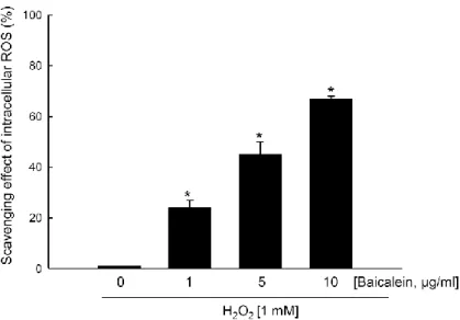
1. Baicalein shows intracellular ROS scavenging activity
Previously, our data showed that 1 mM H2O2 was determined as the concentration of 50 %
growth inhibition, which was assessed at 24 h after H2O2 treatment using the MTT test [15],
and 1 mM H2O2 was determined as the optimal dose in this study. The intracellular ROS
scavenging activity of baicalein after H2O2 treatment was determined using a DCF-DA assay.
Fluorescence spectrometry data revealed that the intracellular ROS scavenging activity of baicalein in H2O2-treated cells was 24 % at 1 μg/ml, 45 % at 5 μg/ml, and 67 % at 10 μg/ml
compared with 0 % of scavenging activity in only H2O2-treated cells (Figure 10). In our cell
culture system, concentrations of baicalein higher than 10 μg/ml were toxic to the cells (data not shown). The intracellular ROS scavenging activity of baicalein determined by this assay was consistent with our previous results [16]. Therefore, 10 μg/ml was chosen as the optimal concentration of baicalein for further cell culture studies. The fluorescence intensity of DCF was also measured using a flow cytometer. The H2O2-treated cells showed two cell
populations in Figure 11. These two populations indicate a high level of ROS and a low level of ROS populations, suggesting that the cells are differently affected by H2O2 treatment.
Two densities of ROS cell populations are not a specific finding in V79-4 cells only. Several papers reported that two densities of ROS cell populations were also found in various cells such as cancer stem cells, neuronal cells, immune cells, or fibroblast cells [56-60]. The DCF fluorescence intensity in cells treated with H2O2 and 10 μg/ml baicalein was decreased more
than three folds compared with that in cells treated with H2O2 alone (Figure 11). These
results suggested that the intracellular ROS induced by H2O2 treatment should be reduced by
33
Figure 10. Effects of baicalein in a dose dependent manner on the scavenging activity
against H2O2 treatment. Cells were treated with baicalein at the concentration of 1, 5, and 10
μg/ml for 1 h and then incubated with 1 mM H2O2 for an additional 24 h. After the addition
of 25 μM DCF-DA for 10min, the intracellular ROS generated were detected by spectrofluorometry. Each bar represents the mean ± the standard error in triplicate experiments. *Significantly different from H
2O2-treated cells with no baicalein treatment
34
Figure 11. Effects of baicalein on H2O2-induced intracellular ROS. The intracellular ROS
were detected by flow cytometry and quantified. FI indicates the fluorescence intensity of DCF. Each bar represents the mean ± the standard error from triplicate experiments.
*Significantly different from untreated control (p<0.05). †Significantly different from H 2O2
treatment (p<0.05).
2. Baicalein reduces the induction of DNA strand breaks by oxidative stress
The interaction between ROS and DNA can lead to several types of oxidative DNA damage, including strand breaks and base modifications [61]. The protective effect of baicalein against the formation of DNA strand breaks induced by H2O2 treatment was
determined using the neutral comet assay [52]. H2O2 treatment induced greater DNA tail
lengths and increased the mean percentage of DNA in the tails to 47 %. Baicalein treatment decreased the percentage of DNA in the tails to 32 % (Figure 12). H2O2 strongly induced the
35
phosphorylation of the histone variant H2A.X (a marker of DSBs), whereas baicalein significantly decreased H2O2-induced phosphohistone H2A.X, as shown in Figure 13. The
results in Figure 12, 13 are consistent with our previous results [16]. H2O2 treatment also
sharply decreased intracellular levels Ku70 protein and phosphorylated DNA-PKcs (components of the NHEJ repair complex); however, baicalein significantly restored their levels, as shown in Figure 14. According to these results, baicalein treatment should suppress the DNA strands breakage induced by H2O2 treatment and increase the levels of
Ku70 and phosphorylated DNA-PKcs decreased by H2O2 treatment.
Figure 12. Effects of baicalein on H2O2-induced cellular DNA damage. The percentage of
cellular DNA damage was detected by comet assay. Representative images are shown (magnification, ×400). Each bar represents the mean ± the standard error from triplicate experiments. *Significantly different from untreated control (p<0.05). †Significantly different
36
Figure 13. Effects of baicalein on H2O2-induced phospho-histone H2A.X. Cell lysates were
electrophoresed, and phospho-histone H2A.X was detected using specific antibodies. Each bar represents the mean ± the standard error from triplicate experiments. *Significantly
different from untreated control (p<0.05). †Significantly different from H2O2 treatment
37
Figure 14. Effects of baicalein on H2O2-reduced Ku70 and phospho-DNA-PKcs. Cell lysates
were electrophoresed, and Ku70 and phospho-DNA-PKcs was detected using specific antibodies. Each bar represents the mean ± the standard error from triplicate experiments.
38
*Significantly different from untreated control (p<0.05). †Significantly different from H 2O2
treatment (p<0.05).
3. Baicalein inhibits oxidative stress-induced DNA base modification
The levels of 8-OxoG incorporated into DNA, a hallmark of oxidative stress and DNA base damage, were assessed by an ELISA assay using specific antibodies against 8-OhdG (a nucleoside of 8-OxoG). Levels of 8-OhdG were significantly higher in H2O2-treated cells
than in control cells. Baicalein decreased the levels of 8-OhdG detected in H2O2-treated cells
by nearly half, though not completely to baseline levels (Figure 15). 8-OhdG in cellular DNA was also visualized using avidin, which binds 8-OhdG with high specificity [53], conjugated to a TRITC dye. Condensed staining intensity of 8-OhdG was observed in H2O2
-treated cells. However, baicalein decreased the intensity of 8-OhdG staining (Figure 16). These results indicate that baicalein can decrease the levels of 8-OhdG in cellular DNA induced by oxidative stress.
39
DNA was determined as the 8-OhdG amount using the Bioxytech 8-OHdG ELISA kit. Each bar represents the mean ± the standard error from triplicate experiments. *Significantly
different from untreated control (p<0.05). †Significantly different from H2O2 treatment
(p<0.05).
Figure 16. Effect of baicalein on H2O2-induced 8-OxoG formation in cells. The binding of
avidin-TRITC, which reflects the 8-OxoG levels, was visualized under a fluorescence microscope. Transmission images are provided to confirm the presence of cells.
40
4. Baicalein prevents the inhibition of OGG1 protein and mRNA expression by H2O2
treatment
A transcriptional reporter vector, in which the promoter region of OGG1 was coupled to a luciferase gene, was used to assess the effects of ROS and baicalein on promoter activity. H2O2 treatment decreased the transcriptional activity of the OGG1 promoter. Baicalein
treatment increased the OGG1 promoter activity to more than two fold in unstressed cells and restored promoter activity in cells stressed with H2O2 (Figure 17). The expression of
OGG1 mRNA was reduced in H2O2-treated cells compared with that in control cells.
Baicalein restored the levels of OGG1 mRNA (Figure 18) and partially restored OGG1 protein level (Figure 19) in H2O2-treated cells. These data suggest that baicalein increases
the transcriptional activity of the OGG1 promoter, and hence, the levels of OGG1 mRNA and protein.
Figure 17. Effects of baicalein on H2O2-reduced transcriptional activity of OGG1. After
overnight transfection with the OGG1 promoter–luciferase vector, cells were treated with baicalein for 24 h and cell lysates were mixed with a luciferase substrate. Luciferase activity
41
was measured with a luminometer. Each bar represents the mean ± the standard error from triplicate experiments. *Significantly different from untreated control (p<0.05). †Significantly different from H
2O2 treatment (p<0.05).
Figure 18. Effects of baicalein on H2O2-reduced mRNA level of OGG1. OGG1 mRNA
levels were determined by reverse transcriptase polymerase chain reaction. Each bar represents the mean ± the standard error from triplicate experiments. *Significantly different
42
Figure 19. Effects of baicalein on H2O2-reduced protein expression of OGG1. Western blot
analysis was performed using an anti-OGG1 antibody. Each bar represents the mean ± the standard error from triplicate experiments. *Significantly different from untreated control
(p<0.05). †Significantly different from H2O2 treatment (p<0.05).
5. Baicalein activates the Akt signaling pathway
Akt kinase, also known as protein kinase B, is a major signaling enzyme involved in cell survival during oxidative stress, and recently, it was reported that the phosphoinositide 3-kinase (PI3K)/Akt pathway regulates OGG1 expression [48,62]. Baicalein pretreatment in H2O2-treated cells increased the level of phosphorylated Akt (the active form of Akt)
compared with the level of phosphorylated Akt in cells treated with H2O2 alone (Figure 20).
-43
induced death (Figure 21), suggesting the involvement of Akt signaling in the mechanism by which baicalein promotes DNA damage repair.
Figure 20. Effects of baicalein on H2O2-reduced Akt phosphorylation. Cell lysate proteins
were separated by electrophoresis and transferred to Western blots. Total Akt and phospho-Akt were detected separately using specific antibodies. Each bar represents the mean ± the standard error from triplicate experiments. *Significantly different from untreated control
44
Figure 21. Effects of baicalein on H2O2-reduced cell viability via akt signal pathway. Cells
were treated with 1 μM Akt inhibitor IV for 1 h, treated with baicalein for 1 h, and then 1 mM H2O2 was added for 24 h. Cell viability was then assessed by the MTT assay. Each bar
represents the mean ± the standard error from triplicate experiments. *Significantly different
from untreated control (p<0.05). †Significantly different from H2O2 treatment (p<0.05). ‡
Significantly different from H2O2-treated cells with baicalein (p<0.05).
6. Baicalein inhibits apoptotic DNA fragmentation induced by oxidative stress
The process of apoptosis involves the activation of nucleases that degrade the nuclear DNA into fragments of approximately 200 bp in length [63]. The effects of baicalein on DNA fragmentation induced by H2O2 treatment was examined using a TUNEL assay, in
which BrdU incorporated at the ends of apoptotic DNA fragments is represented by green fluorescence from a labeled antibody to BrdU. Apoptotic nuclei can thus be distinguished from intact nuclei counterstained with propidium iodide (red fluorescence). H2O2 treatment
increased the fraction of cells showing DNA fragmentation, from a baseline level of ∼3 % to nearly 60 %, whereas baicalein partially suppressed this effect (Figure 22). These data suggest that baicalein protects DNA against the fragmentation induced by oxidative stress.
45
Figure 22. Effect of baicalein on H2O2-induced apoptotic DNA fragmentation. Cells were
treated with baicalein for 1 h and then with 1 mM H2O2 for an additional 24 h. Apoptotic
cells were detected by fluorescent TUNEL staining (green) and counterstaining of nuclei with propidium iodide (red). Representative images of each group are shown. White arrows indicate TUNEL-positive cells with fragmented DNA (apoptotic cells; magnification, ×400). The percentage of apoptotic cells was determined by manual scoring of multiple microscopic fields for each treatment group. Each bar represents the mean ± the standard error from triplicate experiments. *Significantly different from untreated control (p<0.05). †Significantly different from H
46 DISCUSSION
This study suggested that baicalein should reduce the DNA damage (e. g. DNA base modification, DNA strands breaks) induced by treatment of H2O2. We identified that the
oxidative stress could induce the DNA damage, an investigated to identify the inhibition effects of baicalein against the DNA damage originated by oxidative stress via akt signal pathway.
Natural flavonoids are emerging as potent therapeutic drugs for use in the treatment of free radical-mediated diseases, and researchers have made numerous efforts to search for novel antioxidants. Baicalein inhibits certain types of lipoxygenases [64], inhibits inflammation [65], and inhibits CYP2C9, an enzyme of the cytochrome P450 system, which is responsible for drug metabolism [66]. Additionally, our previous results indicated that baicalein attenuated mitochondrial or cellular oxidative stress by scavenging ROS and inducing manganese superoxide dismutase [15,16,49].
Although many studies have reported the protective effect of flavonoids including baicalein against oxidative stress-induced cell damage, few have examined the effects on cellular repair mechanisms mediated by flavonoids. Therefore, in this study, we evaluated the effects of baicalein on cellular DNA repair responses to oxidative stress. Baicalein has free radical scavenging and antioxidant effects due to the trihydroxy structure of its A ring [67] and its lipophilic characteristics [68]. In our study, baicalein effectively reduced ROS and suppressed DNA breakage in response to oxidative stress via the induction of Ku70 and DNA-PKcs, demonstrating the ability of baicalein to promote the repair of DSBs. Additionally, baicalein promoted the removal of oxidized DNA bases via the restoration of OGG1 transcription and translation.
ROS induced oxidative DNA damage, including strand breaks and base and nucleotide modifications [61,69]. Understanding the mechanisms for processing oxidative DNA damage is important in elucidating the pathogenesis of oxidative stress-mediated diseases.
47
Homologous recombination (HR) and NHEJ processes are the representative responses to DSBs. While the HR repair response uses a homologous template to direct DNA repair synthesis, the NHEJ process is referred to as nonhomologous because the broken ends are directly ligated without the use of a homologous template [70]. Loss of damaged nucleotides at the break site can lead to small deletions, and the joining of nonmatching termini can result in chromosomal rearrangements including large deletions and translocations. NHEJ may, therefore, introduce mutations during repair [71]. Nevertheless, the NHEJ process occurs more frequently in higher eukaryotes than in lower eukaryotes or prokaryotes [72]. Moreover, the NHEJ process is required for the joining of hairpin-capped double-strand breaks [73]. This mechanism of response to DNA DSBs occurs most often in higher eukaryotes [72]. Among many modified DNA bases generated by oxidative stress, 8-OxoG is the most abundant product, and it seems to play a major role in mutagenesis and carcinogenesis [41].We have previously shown that OGG1-deficient human leukemia cells could be forced to undergo apoptosis by the incorporation of 8-OhdG [43,44]. The expression of OGG1 suppresses oxidative stress-derived DNA damage and results in enhanced cell survival [74].
The Akt pathway plays a crucial role in controlling transcription, cell cycle progression, and apoptosis [75]. Furthermore, DNA-PKcs colocalizes and associates with Akt kinase at the plasma membrane [76]. In vitro analysis using purified DNA-PK and recombinant Akt protein revealed that DNAPK directly phosphorylates and thus activates Akt kinase [76,77]. In previous studies, we and others have demonstrated that OGG1 is induced via the activation of the PI3K/Akt pathway [48,62]. However, those results contrast with a study in a rat model of diabetes type 1, which indicated that ROS induced by hyperglycemia activated Akt kinase and was associated with the downregulation of OGG1 rather than induction [78]. The reasons for this apparent discrepancy are unclear. Also, Akt kinase phosphorylates pro-apoptotic proteins, such as Bad, caspase-9, and the forkhead transcription factors, and their
48
phosphorylation by Akt kinase abolishes their pro-apoptotic activities [79,80]. Therefore, the reduced apoptosis in H2O2-treated cells with baicalein treatment might involve the activation
of Akt kinase by baicalein. However, baicalein suppresses or activates different cell types; baicalein suppresses or does not change Akt activation in human melanoma cells [81], microglial cells [82], and vascular smooth muscle cells [83]; and baicalein increases Akt activation in various cancer cell lines [84,85], human neuroblastoma cells [86], and adipocytes [87]. Further studies need to elucidate the possible application of baicalein on DNA damage-associated disease models.
In conclusion, baicalein increased the BER and NHEJ capacity of cells subjected to oxidative stress, thereby reducing DNA damage and rescuing cells from oxidative stress-induced apoptosis.
49 REFERENCE
[1] Jacob, M.P. (2003). Extracellular matrix remodeling and matrix metalloproteinases in the vascular wall during aging and in pathological conditions. Biomed. Pharmacother. 57, 195-202.
[2] Kähäri, V., and Saarialho-Kere, U. (1997). Matrix metalloproteinases in skin. Exp.
Dermatol. 6, 199.
[3] Dasgupta, J., Kar, S., Liu, R., Joseph, J., Kalyanaraman, B., Remington, S.J., Chen, C., and Melendez, J.A. (2010). Reactive oxygen species control senescence-associated matrix metalloproteinase-1 through c-Jun-N-terminal kinase. J. Cell. Physiol. 225, 52-62.
[4] Rajagopalan, R., Ranjan, S.K., and Nair, C.K. (2003). Effect of vinblastine sulfate on gamma-radiation-induced DNA single-strand breaks in murine tissues. Mutat. Res. 536, 15-25.
[5] Brenneisen, P., Briviba, K., Wlaschek, M., Wenk, J., and Scharffetter-Kochanek, K. (1997). Hydrogen peroxide (H2O2) increases the steady-state mRNA levels of
collagenase/MMP-1 in human dermal fibroblasts. Free Radic. Biol. Med. 22, 515-24. [6] Frost, J.A., Geppert, T.D., Cobb, M.H., and Feramisco, J.R. (1994). A requirement for
extracellular signal-regulated kinase (ERK) function in the activation of AP-1 by Ha-Ras, phorbol 12-myristate 13-acetate, and serum. Proc. Natl. Acad. Sci. U. S. A. 91, 3844-8.
[7] Brenneisen, P., Sies, H., and Scharffetter-Kochanek, K. (2002). Ultraviolet-B irradiation and matrix metalloproteinases: from induction via signaling to initial events. Ann. N. Y.
Acad. Sci. 973, 31-43.
[8] Nelson, K.K., and Melendez, J.A. (2004). Mitochondrial redox control of matrix metalloproteinases. Free Radic. Biol. Med. 37, 768-84.