간암
4D
RT
전산화 치료계획의 중요장기 선량감소를 위한
선량체적히스토그램 분석
지연상1・정묘영2,3・최인영4・김미현1,5・동경래1・신지윤1,*
1광주보건대학교 방사선과, 2조선대학교 호남권역 재활병원, 3동신대학교 보건의료학과,
4조선대학교병원 영상의학과, 5전북대학교대학원 방사선과학기술학과
Computerized Treatment Plan for Liver Cancer 4D RT Vital
Organs a Dose-volume Histogram for Dose Reduction
Yun-Sang Ji
1, Myo-Young Jung
2,3, In-Young Choi
4, Mi-Hyun Kim
1,5,
Kyung-Rae Dong
1and Ji-Yun Shin
1,*
1Department of Radiological Technology, Gwangju Health University, 73, Bungmun-daero 419 beon-gil, Gwangsan-gu, Gwangju, 62271, Republic of Korea 2Department of Radiology, Chosun University Honam Regional Rehabilitation Hospital,
61027, Haseoro 590, Buk-gu, Gwang-ju, Republic of Korea
3Department of Public Health and Medicine, Dongshin University Graduate School, 185, Geonjae-ro, Naju-si, Jeollanam-do, 58245, Republic of Korea
4Department of Diagnostic Radiology, Chosun University Hospital, 365, Pilmun-daero, Dong-gu, Gwangju, 61453, Republic of Korea
5Department of Radiation Science & Technology, Graduate School of Chonbuk National University, 567 Baekje-daero, deokjin-gu, Jeonju-si, Jeollabuk-do, 54896, Republic of Korea
Abstract - Through the 4-dimensional RPM coumputerizezd treatment plan the abdomen-liver-cancer patietnt’s DVH can be gained Through that according to the absence or presence of Gating the goal is to cure the radiation and through the researching of surrounding tumor tissues and normal tissue dose, the goal is to achieve the cure for radiation. Through the 4-dimensional treatment and the induction treatment and the movement and motion images of the patient, through the process of treatment plan for protecting normal tissue dose rather than anti-tumor dose, a useful treatment plan will be confirmed the research that enhances and improves the accuracy and precision must keep continue to be researched. The RPM will be ran on the part that gets influenced by normal tissues or moving organs. Therefore it will help to develop the process of the treatment of tumor tissues so it is very crucial to reduce the dose of normal tissues to reduce any possible dangers and obstacles.
Key words : Gating, PTV, GTV, 4D RT
─ 217 ─ Technical Paper
* Corresponding author: Ji-Yun Shin, Tel. +82-10-8800-0344, Fax. +82-62-958-7669, E-mail. [email protected]
서 론
최근 방사선 치료는 대부분 3차원 입체치료의 진보된
치료방식을 많이 상용화하고 있다. 다엽콜리메이터를 이
용한 강도변조치료와 ExacTrac 시스템을 이용한 영상유
도방사선치료, 선형가속기가 내장되어 있는 토모치료기,
OBI를 탑재하여 호흡동조를 할 수 있는 Rapid arc 치료와 Novalis System 등의 많은 방사선치료 기술이 개발되었다 (Wunderink et al. 2007; Nishioka et al. 2009). 이러한 첨단 정밀치료들은 종양조직 선량보다는 정상조직의 조사선량을
줄이는 것을 더 우선적으로 치료계획에 적용하고 있다. 종
양을 치료하기 위해서 가장 중요한 것은 종양과 정상조직의 위치 또는 형태가 변화되는 것을 고려하여 정확한 선량이
전달 될 수 있도록 4차원 방사선 치료를 하는 것이다(Lujan
et al. 1999; Geld et al. 2006). 호흡을 고려한 4D RT는 가슴 부위와 복부에 위치한 종양을 호흡주기에 맞추어 치료하거
나 호흡을 따라가면서 방사선을 조사하는 방법이 있다. 호
흡을 고려한 치료에서 종양의 움직임 범위를 확인하여 내부 여유(Internal margin; IM)를 고려한 내부표적용적(internal target volume; ITV)치료에 적용하면, 조사야가 넓어지고 전 달되는 선량과 조사용적이 증가하여 정상조직에 대한 부작 용이 증가할 가능성이 있다(Kubo et al. 1996). 호흡을 고려 한 4D RT 치료는 종양의 제어를 높이고 정상조직의 합병증 을 유발할 수 있는 확률을 감소시키기 위해서는 조사야를 적절한 크기로 작게 조절하고 종양과 장기의 움직임을 고려 해서 4차원적으로 방사선을 치료해야 한다. 4D RT는 간암 이나 폐암의 방사선치료에 필수적인 부분으로 자리잡고 있 으며 치료 성적에 향상을 가져오고 호흡에 영향을 받는 신 체부위에 발병하는 암치료에 적용을 넓혀가고 있다. 본 논 문에서는 4차원 전산화치료계획의 RPM 시스템을 통하여 복부 간암 환자 치료의 선량체적히스토그램을 얻어 Gating 유무에 따라 PTV와 GTV를 분석하여 종양조직과 주위 정 상조직의 선량 변화를 연구하고자 한다.
대상 및 방법
1. 연구대상 광주에 위치한 일개 대학병원 방사선 종양학과에서 4D RT 치료를 시행한 liver cancer 환자 10명을 대상으로 하여 방사선 치료 선량의 선량체적히스토그램을 관찰하였다. 제 한사항으로는 연구대상이 적어 데이터의 통계적 신뢰를 얻 는데 다소 어려움이 있다. 실험기기는 CT Simulation장치의 Bright speed 4X(GE, USA), Advantage workstation(4D CT phase selecting System), RPM Respiratory Gating System
Ver. 1.7, 치료 계획 장치: Eclipse Ver. 8.1(RTP planning System)을 사용하였다.
2. 실험방법
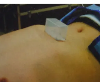
Figs. 1, 2 RPM 시스템의 block marker와 적외선 카메라
를 부착시켜 CT를 이용해 복부를 Scan하여 영상을 획득하
고 얻어진 영상을 RTP로 보내 치료계획을 수립한다. RTP
로 보내진 영상을 바탕으로 Gating System을 적용해 얻은
영상 중 호흡 주기가 가장 안정적인 50~55%의 Phases와 Full Phases의 scan 영상을 통하여 선량체적히스토그램의 선
량 값을 분석한다. Gating과 Non Gating에서 얻어진 종양조
직의 PTV와 GTV의 선량 값을 비교하여 분석하고 Gating
유무에 따른 정상조직의 중요 장기(spinal cord, kidney, stomach)의 선량 값을 비교하여 분석한다(Figs. 3, 4).
Fig. 1. The marker block of the RPM system.
결 과
1. 종양조직의 GTV와 PTV 선량 분석
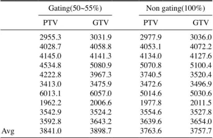
Table 1에서는 종양조직에 조사된 PTV 선량의 DVH 값 을 분석한 결과 Gating(50~55%) System을 적용했을 때 3841.0, Non Gating에서는 3763.6의 평균선량이 산출되어 졌다. GTV는 Gating System을 적용했을 때 3898.71, Non Gating일 때는 3757.74의 평균선량이 산출되었다. 따라서 Gating System 적용했을 때 종양조직에 더 많은 선량이 조 사되어 정확하고 정밀한 선량전달이 이루어진 치료계획으 로 평가된다. 2. 정상조직 중요장기의 DVH 값 분석 Table 2에서는 정상조직에 조사된 선량의 DVH 값을 분
석한 결과 Gating(50~55%) System 적용하여 Liver 주변의 결정장기는 Spinal Cord, Kidney(right, left), Stomach에서는 481.58, 244.34, 65.56, 888.63의 평균 선량 값이 산출되었다. Non Gating(100%)에서는 545.61, 276.19, 84.90, 1205.44의
평균 선량 값이 산출 되었다. 그리고 이를 백분율로 환산했
을 때, Gating 적용 시 정상조직이 받는 선량이 Non Gating일
때 보다 중요장기의 각각 12%, 12%, 23%, 27%가 선량이 줄
어 들어 정상조직에 도움이 된다는 것을 확인할 수 있었다.
3. Whole Liver의 PTV, GTV 최대값과 최소값 분석
4D simulation의 Gating System과 Non Gating 영상이 며 whole liver의 min, max, mean dose를 Figs. 5, 6에서 측 정하여 Table 3에 표시하였다. Table 3에서 보듯이 Gating System이 적용된 영상의 종양 조직선량의 평균 선량이 Non Gating에 비해 약 100cGy 정도 적은 것을 알 수 있다.
Table 3. Average values of min, max and mean dose in whole liver
Min dose Max dose Mean dose
Gating
(50~55%) 27.0 4114.4 1086.4
Non gating
(100%) 24.4 3993.8 1182.5
Note: The unite is the number of cGy.
Table 2. Abdomen treatment plan of CT simulation DVH average
value of normal tissue
Cord Right kidney Left kidney Stomach
Gating
system 5481.58 244.34 65.56 888.63
Non gating
system 545.61 276.19 84.90 1205.44
Note: The unite is the number of cGy.
Fig. 3. Range setting of 50~55% of gating system.
Fig. 4. Scan imaging of respiratory phases.
Table 1. DVH values of tumor tissue of abdomen treatment plan
of 4D-CT simulation
Gating(50~55%) Non gating(100%)
PTV GTV PTV GTV 2955.3 3031.9 2977.9 3036.0 4028.7 4058.8 4053.1 4072.2 4145.0 4141.3 4134.0 4127.6 4534.8 5080.9 5070.8 5100.4 4222.8 3967.3 3740.5 3520.4 3413.0 3475.9 3472.6 3496.9 6013.1 6057.0 5014.6 5030.6 1962.2 2006.6 1977.8 2011.5 3542.9 3524.2 3554.6 3527.8 3592.8 3643.2 3639.6 3654.0 Avg 3841.0 3898.7 3763.6 3757.7
고 찰
호흡을 고려한 4D RT는 종양조직의 방사선 흡수율을
높이고 정상조직의 선량을 가능한 최소화 시키고자 하
는 치료기술로 이러한 목적을 달성하기 위해서는 4차원
CT Simulation 치료계획이 필요하다(Brandner et al. 2006; Ramsy et al. 2006; Heinzerling et al. 2008). 특히 간, 폐 부위 와 같이 복부에 위치한 장기는 호흡으로 인한 움직임을 피 할 수 없다. 움직임이 나타나는 장기는 4차원 모의치료 영
상을 통하여 호흡주기를 추적하여 치료에 적용하는 RPM
System이나 환자고정용구가 반드시 필요하다. 호흡을 고
려한 치료는 환자의 자세와 Body fix가 호흡에 영향을 상
당히 미치고 있다는 보고가 있다(Balter et al. 1996, Balter et al. 1998; Rietzel et al. 2006). Body fix가 종양의 움직임
을 상하방향에서는 제한하는데 도움을 주고 환자의 움직임 을 좌우방향에서는 움직임을 제어하지 못한 것으로 연구되 었다(Gierga et al. 2005; Lim et al. 2007). 특히 4D RT 치료 를 하면 Liver의 경우 13.9%, Stomach 35.2%, Rt Kidney와 Spinal cord는 각각 61.8% 37.3%의 선량 흡수를 줄일 수 있 었다는 보고이다(Underberg et al. 2005; Keall et al. 2006). 4D RT 치료의 궁극적인 목표는 정확도를 높이기 위한 방법 으로 Target의 정확한 위치를 파악하고 계획된 선량을 타깃 에 정확히 전달하기 위해서는 영상유도 방사선 치료와 호흡 고려를 위한 치료가 결합되어야 한다는 것이다(Wagman et al. 2003). 본 논문에서는 방사선 치료 목표가 달성될 수 있 도록 4D RT 치료를 통하여 환자의 움직임과 영상을 유도하 는 치료를 통하여 종양선량보다는 정상조직을 보호하는 치 료계획을 실현하므로 유용한 치료계획임을 입증하고자 했 다. 향후 보다 정확성과 정밀도를 높이는 치료 연구가 계속 진행되어야 될 것이라고 사료된다.
결 론
본 연구는 4D Simulation에서 Gaing System을 이용하여 DVH값을 비교분석한 결과 다음과 같은 결론을 얻을 수 있 었다. 4D Simulation에서는 PTV와 GTV 선량이 환자의 상 태와 병소 부위에 대한 많은 영향을 받는데 Gating System 을 이용했을 때 PTV와 GTV값에 대한 종양조직의 치료보 다 정상조직 보호가 우선시 된다. 종양조직의 PTV와 GTV 값의 호흡동조시스템을 적용 유무에 따른 선량체적히스토 그램을 분석한 조사선량을 비교한 결과 종양조직의 조사선 량은 큰 변화가 없었다. 정상조직의 PTV와 GTV값의 호흡 동조시스템을 적용 유무에 따른 선량체적히스토그램을 분 석한 결과로 호흡동조시스템으로 분석한 결과 정상조직의 주요 장기에 대한 선량이 적게 조사되었음을 알 수 있다. 본 연구를 통하여 움직이는 장기나 환자 호흡에 영향을 받는 부위에 호흡동조시스템을 실시하여 종양조직의 치료에는 도움을 주고 정상조직에는 선량을 감소시켜 장해를 줄일 수 있는 치료가 필요하다고 사료된다.
참 고 문 헌
Balter JM, Lam KL, McGinn C, Lawrence T and Haken RT. 1998. Improvement of CT-based rreatment planning models of abdominal target using static exhale imaging. Int. J. Radi-at. Oncol. Biol. Phys. 141(4):939-943.
Balter JM, Ten RK, Lawrence T, Lam K and Robertson J. 1996. Uncertainties quantitation in CT-based radiation therapy
Fig. 5. Gating CT and DVH value.
treatment planning associated with patient breathing. Int. J. Radiat. Oncol. Biol. Phys. 36(1):167-174.
Brandner ED, Wu A, Chen H, Heron D, Kalnicki S, Koman-duri K, Gerszten K, Burton S, Ahmed I and Shou Z. 2006. Abdominal organ motion measured using 4D CT. Int. J. Radiat. Oncol. Biol. Phys. 65(2):554-560.
Geld YV, Senan S, Lagerwaard F, Underberg R, Slotman B and Tinteren HV. 2006. Evaluating mobility for radiotherapy plan-ning of lung tumors: a comparison of virtual fluoroscopy and 4D CT. Lung Cancer. 53(1):31-37.
Gierga DP, Brewer J, Sharp GC, Betke M, Willett CG and Chen GT. 2005. The correlation between internal and exter-nal markers for abdomiexter-nal tumors: implications for respi-ratory gating. Int. J. Radiat. Oncol. Biol. Phys. 61(5):1551-1558.
Heinzerling JH, Anderson JF, Papiez L, Boike T, Chien S, Zhang G, Abdulrahman R and Timmerman R. 2008. Four-Dimen-sional computed tomography scan analysis of tumor and organ motion at varying level of abdominal compression during stereotaxic treatment of lung and liver. Int. J. Radiat. Oncol. Biol. Phys. 70(5):1571-1578.
Keall PJ, Mageras GS, Balter JM, Emery RS, KM Foster, Ji-ang SB, Kapatoes JM, Low DA, Murphy MJ, Murray BR, Ramsey CR, Wong JW and Yorke E. 2006. The manage-ment of respiratory motion in radiation oncology report of AAPM TG 76. Med. Phys. 33(10):3874-3900.
Kubo HD and Hill BC. 1996. Respiration gated radiotherapy treatment: a technical study. Phys. Med. Biol. 41(1):83-91. Lim S, Park SH, Ahn SD, Suh Y, Shin SS, Lee SW, Kim JH,
Choi EK, Yi BY, Kwon SI, Kim S and Jeung TS. 2007.
Guiding curve based on the normal breathing as monitored by thermocouple for regular breathing. Med. Phys. 34(11): 4514-4518.
Lujan AE, Larsen EW, Balter JM and Ten Haken RK. 1999. A method for incorporating organ motion due to breathing into 3D dose calculations. MedPhys. 26(5):715-720. Nishioka T, Nishioka S, Kawahara M, Tanaka S, Shirato H,
Nishi K and Hiromura T. 2009. Synchronous monitoring of external/internal respiratory motion: validity of respi-ration-gated radiotherapy for liver tumors. Jpn J. Radiol. 27(7):285-289.
Rietzel E, Liu AK, Doppke KP, Wolfgang JA, Chen A, Chen G and Choi N. 2006. Design of 4D treatment planning target volumes. Int. J. Radiat. Oncol. Biol. Phys. 66(1):287-295. Underberg RWM, Largerward FJ, Slotman BJ and Senan S.
2005. Benefit of respiration gated stereotactic radiotherapy for stage lung cancer an analysis of 4D CT datasets. Int. J. Radiat. Oncol. Biol. Phys. 62(2):554-560.
Wagman R, Yorke E, Ford E, Giraud P, Mageras G, Minsky B and Rosenzweig K. 2003. Respiratory gating for liver tumors: use in dose escalation. Int. J. Radiat. Oncol. Biol. Phys. 55(3):659-668.
Wunderink W, Romero AM, Osorio E, de Boer H, Brandwijk R, Levendag P and Heijmen B. 2007. Target coverage in im-age-guided stereotactic body radiotherapy of liver tumors. Int. J. Radiat. Oncol. Biol. Phys. 68(1):282-290.
Received: 1 July 2018 Revised: 29 July 2018 Revision accepted: 5 August 2018