레진의 종류에 따른
Glass fiber post와 Core의 파절
연세대학교 대학원
치의학과
레진의 종류에 따른
Glass fiber post와 Core의 파절
지도 이 근 우 교수
이 논문을 석사 학위논문으로 제출함
2002년 6월 일
연세대학교 대학원
치의학과
심 동 욱
심동욱의 석사 학위논문을 인준함
심사위원 인
심사위원 인
심사위원 인
연세대학교 대학원
2002년 6월 일
감사의 글
먼저 제게 능력 주신 주님께 감사드립니다. 본 논문이 완성되기까지 늘 격려해 주시고 지도하여 주신 이근우 교수님께 깊은 감사를 드리며 논문 작성과 심사에 많은 조언과 배려를 해주신 김광만 교수님, 심준성 교수님, 그리고 항상 곁에서 도움을 주신 이호용 교수님, 정문규 교수님, 한동후 교 수님, 한종현 교수님, 최병갑 교수님, 문홍석 교수님께 감사드립니다. 또한 본 연구에 도움을 주신 치과 재료학 교실의 김경남 교수님, 이상배 선생님, 중앙 기공실의 우창우 선생님을 비롯한 보철과 의국원 여러분께 감사를 드 립니다. 언제나 사랑으로 돌보아주시는 부모님과 형 그리고 누나와 늘 곁에서 성 원하여 주시는 모든 분들과 이 작은 기쁨을 나누고 싶습니다. 2002년 6월 심동욱 드림목 차
그림 및 표 ··· ⅲ 국문 요약 ··· ⅳ I. 서 론 ··· 1 II. 연구 재료 및 방법 가. 연구 재료 ··· 4 나. 연구 방법 ··· 51. Glass fiber post의 표면 처리 ··· 5
2. 치아 시편의 제작 ··· 6 3. Post의 치관내 합착과 표준화 금속 주형의 제작 ··· 6 4. Core의 축성과 주조 post의 제작 ··· 7 5. 압축 파절 하중 실험용 레진 시편의 제작 ··· 8 6. Thermal cycling의 시행 ··· 8 7. 최대 파절 하중과 압축 파절 하중의 측정 ··· 8 8. 파절 양상의 관찰 ··· 9 9. 통계 분석 ··· 9 III. 연구 성적 ··· 11 가. 치아 시편의 최대 파절 하중의 측정 ··· 11 나. 레진 시편의 압축 파절 하중의 측정 ··· 12 다. 파절 양상의 육안 관찰 ··· 14
라. 파절 부위의 주사 전자 현미경 (SEM) 소견 ··· 14 IV. 총괄 및 고찰 ··· 16 V. 결 론 ··· 22 참고 문헌 ··· 24 사진 부도 ··· 32 영문 요약 ··· 35
그림 및 표 차례
Fig. 1. Schematic drawing of test specimen showing dimensions in mm ··· 7
Fig. 2. Schematic representation of main fracture lines
in catastrophic failure ··· 10 Fig. 3. The means and standard deviations of maximum fracture load
in the teeth specimens ··· 12
Fig. 4. The means and standard deviations of compressive fracture load in the resin specimens ··· 13
Table I. The resin cores used in this study ··· 4
Table II. The bonding systems used in this study ··· 5
Table III. The means of maximum fracture load of teeth specimens(N) ···· 11
Table IV. The means of compressive fracture load of resin specimens(N) ··· 13
국문 요약
레진의 종류에 따른 Glass fiber post와 Core의 파절
보다 자연스럽고 심미적인 치과 치료의 수요 증가로, 특히 심미적 재료의 기계 적 및 물리적 성질의 개선에 관심이 모아지고 있는 가운데, 최근 개발되어 근관
치료 후 심미성 회복을 위하여 쓰여지는 glass fiber post는 기존 post system에
비해 물리적 성질, 심미성, 치근 파절 및 수복물 파절의 위험성, 상방 core와의 결 합력, 방사선 불투과도 그리고 제거 및 재치료성 등 많은 부분에 있어 장점이 있 음이 보고되어 왔으며, 또한 생체 친화적이고 화학적 안정성이 높다. 본 연구의 목적은 glass fiber post에 가장 적합한 조성의 core용 레진을 알아보는 것이다.
Glass fiber post(ParaPost® Fiber White)와 각기 다른 조성의 core용 레진(ParaCore,
Z100TM, Rebilda®, Admira®)으로 자연치를 수복하고 만능 시험기로 하중을 부여한
후, 시편의 최대 파절 하중과 파절 양상을 조사하고 금속 주조 post와 core로 수 복한 군과 비교 연구를 통하여, 다음과 같은 결과를 얻을 수 있었다.
1. 치아 시편의 최대 파절 하중 측정 결과, ParaCore, Z100TM, Rebilda®, Admira®
순이었고, ParaCore와 Z100TM이 Rebilda®와 Admira®보다 유의차 있게 높았다.
(p<0.05).
2. 레진 시편의 압축 파절 하중의 평균값은 Z100TM, ParaCore, Rebilda®, Admira®
순이었고, Z100TM과 Rebilda®, Z100TM과 Admira® 및 ParaCore와 Admira® 사 이에 유의차가 있었다.(p<0.05).
3. 치아 시편의 파절 양상은 전체적으로는 post를 중심으로 세 부분으로 나타나는
가 type D이었으며, Rebilda®와 Admira®에서는 post의 파절없이 치아면과 평 행하게 나타난 core의 파절인 type B가 높게 나타났다(40%).
4. 금속 주조 post와 core로 수복한 치아는 높은 하중에도 post와 core의 파절을 보이지는 않았지만, 시편의 단면을 관찰한 소견에서는 치근 파절을 발견할 수 있었다.
이상의 결과를 토대로 glass fiber post를 위한 치아 색조의 레진 core용 재료로
는 ParaCore와 Z100TM이 추천되며, 앞으로도 glass fiber post와 core용 복합 레진
을 사용한 많은 연구가 필요할 것으로 사료된다.
핵심되는 말 : Glass fiber post, 레진 core, 금속 주조 post와 core, 최대 파절 하중, 압축 파절 하중, 파절 양상.
레진의 종류에 따른 Glass fiber post와 Core의 파절
연세대학교 대학원 치의학과
( 지도 이 근 우 교수 )
심 동 욱
I. 서 론
지난 20년동안 치과 재료는 수복 치료에 있어 비약적으로 발전되어 왔고, 보다 자연스럽고 심미적인 치과 치료의 수요 증가로 특히, 심미적 재료의 기계적 및 물 리적 성질의 개선에 관심이 모아지고 있다. 우식이나 외상으로 인해 심하게 손상된 치아는 일반적으로 근관 치료 후, 잔존 치근을 보존하고 상실된 치질을 회복하기 위해 post와 core를 사용하고 금관으로 수복한다. Post의 사용은 18세기 Fauchard가 나무로 만든 post를 사용한 이래로 다양한 형태와 재질의 post가 개발되어 왔는데, 초기의 wire형태의 post로부터 주 조 기술의 발달로 금속 주조 post가 사용되었고, 1970년대에는 금속 주조 post의 단점을 보완하기 위해 금 합금, 스테인레스 스틸 등을 재료로 한 금속 기성 post 가 개발되었으며, 80년대에는 치근 파절을 줄이기 위한 carbon fiber post와 90년대에는 심미성 증진을 위한 zirconium post가 개발되었다.1-6) 특히, 근관 치료된
전치부의 심미성 회복을 위한 완전 도재관 하방에 사용되는 기존의 금속 주조 post,
금속 기성 post, carbon fiber post 및 zirconium post 등은 치근 파절, post의 탈 락과 심미성의 한계가 상존하여 post의 기능성과 심미성을 개선시키려는 점에 연
구의 초점이 이루어져 왔는데, 금속 주조 post와 금속 기성 post는 강도는 뛰어나 나 색조와 광투과성이 떨어지며 치질과 물리적 성질의 차이로 치근 파절의 위험
이 있다.1-6) Zirconium post와 quartz fiber post는 치아 색조의 post로서, 세라믹
계열인 zirconium post는 색조와 광투과성은 우수하나 역시 치질과 상이한 물리적 성질로 인한 치근 파절의 위험성이 있고 실패시 제거가 힘들며, 다른 post들에 비 해 레진 core와의 접착력이 떨어진다. Quartz fiber post는 적절한 탄성으로 치근 파절의 위험성은 줄였으나, 탄소 입자로 인해 자체의 색조와 광투과성이 만족스럽
지 못하고 방사선 투과도가 강하다는 단점이 있다.2-4)
최근 개발되어 사용되고 있는 glass fiber post는 레진 기질 내에 생체 적합성을 가진 glass fiber와 filler를 삽입하여 만든 것으로, 기존 post system에 비해 물리 적 성질, 심미성, 치근 파절 및 수복물 파절의 위험성, 상방 core와의 결합력, 방사 선 불투과도 그리고 제거 및 재치료성 등 많은 부분에 있어 장점이 있음이 보고 되어 왔으며, 또한 생체 친화적이고 화학적 안정성이 높다. 특히 탄성계수, 항복 강도, 굴곡 강도 등 물리적 성질이 상아질과 유사하여 기존 post보다 치근 파절을 현저히 줄인다고 알려져 있다.2-6) 상실된 치관부를 대체하기 위해 post 상방에 축성하는 core용 재료는 아말감, glass ionomer cement reinforced with silver, hybrid glass ionomer, compomer 및 복합 레진 등이 있는데, 아말감은 물리적 성질이 우수하고 경제적이지만 느린 경화 시간, 치질과의 낮은 접합성, 부식 산물로 인한 주위 조직의 변색과 짙은 색 조 등의 단점이 있는 반면, glass ionomer cement reinforced with silver, hybrid glass ionomer 및 compomer 등은 경화 시간, 치질과의 접합성, 불소 유리에 의한 항우식성 및 색조 등에서 장점을 갖지만, 아말감과 복합 레진에 비해 물리적 성질 이 떨어진다. 그래서 완전 도재관의 하방에 사용되는 core용 재료로는 적절한 색 조와 상대적으로 나은 물리적 성질, 치질과의 접합성, 조작의 간편성과 소요 시간 등의 장점으로 복합 레진이 가장 추천되고 있다.2,7-12) Post와 core로 수복된 치아의 예후에 미치는 인자는 여러 가지이지만 잔존 치질 의 보존 정도 특히, 치관부 치질의 보존 정도에 가장 큰 영향을 받으며, 초기에는 post와 core가 근관 치료된 치아의 강화를 위해 시술되었으나 post와 core 자체는
치질의 강화에는 기능이 없고 치관부 수복에 있어 유지와 저항 형태를 부여하는 역할을 한다. 하지만 post와 core로 수복된 치아도 가끔 실패할 수 있고, 실패 양
상은 post 탈락, 치근과 post 및 core의 파절로 나타난다.13-18)
Fiber post 중에 carbon fiber post를 사용한 연구는 많이 보고되고 있으나, 근래 들어 개발된 glass fiber post를 이용한 연구는 아직 미흡한 실정이며, 자연치를 직 접 glass fiber post로 수복하여 실험한 연구는 지금까지 보고된 바 없다. 본 연구
는 근관 치료 후 심미성 회복을 위한 완전 도재관 하방에 사용되기에 가장 적절
한 post와 core 재료인 glass fiber post와 각기 다른 조성의 레진 core로 직접 자 연치를 수복하고 만능 시험기로 구강 내에서 받을 하중을 부여한 뒤, 레진의 종류 에 따른 각 시편의 파절시까지의 최대 하중 측정 및 파절 양상을 관찰하여, glass fiber post에 가장 적합한 조성의 레진 core 종류를 알아봄으로써 임상에서 glass fiber post 사용시에 적절한 레진 core 선택에 있어 도움이 되고자 한다.
II. 연구 재료 및 방법
가. 연구 재료
Glass fiber post로는 직경 1.25 mm의 red color coded ParaPost® Fiber White (Coltene Whaledent® Inc., Mahwah, U.S.A.)를 사용하였다.
Core용 레진으로 ParaCore(Coltene Whaledent® Inc., Mahwah, U.S.A.), Z100TM (3M Dental Products, St.Paul, U.S.A.), Rebilda®(VOCO, Cuxhaben, Germany) 및
Admira® (VOCO, Cuxhaben, Germany)를 사용하였다(Table I). 그리고 Core를 축
성할 치면은 레진 core 제품에 맞게 제조자가 추천하는 접착 system으로 처리하였 다(Table II).
Table I. The resin cores used in this study
Name Polymerization mode Main compositions Manufacturer
ParaCore Dual cure Bis-GMA, EGDMA, TMPTMA
Dibenzoyl peroxide
Coltene Whaledent®
Z100TM Light cure Bis-GMA, TEGDMA 3M
Rebilda® Self cure Bis-GMA, UDMA
Dibenzoyl peroxide VOCO
Admira® Light cure Ormocers®, Bis-GMA, HEMA VOCO
* Bis-GMA : Bisphenol A diglycidyl methacrylate EGDMA : Ethylenglycol dimethacrylate
TMPTMA : 1,1,1 Trimethylolpropan trimethacrylate TEGDMA : Triethylenglycol dimethacrylate
UDMA : Diurethane dimethacrylate
Table II. The bonding systems used in this study
나. 연구 방법
1. Glass fiber post의 표면 처리
먼저 알코올로 닦아내고, air water syringe로 1분간 세척했다. 치질과의 결합력
을 높이기 위하여 표면을 sandblasting과 etching 처리를 했는데,19) sandblasting은
유니트에 부착하여 사용하는 MiniblasterTM(Miniblaster, Israel)를 이용하여 50 µm
의 aluminium oxide를 post에서 3 cm 떨어진 거리에서 2초씩 네 방향에서 분사 하고 증기 세척을 하였다. Etching은 37% 인산으로 30초간 처리 후, air water syringe로 1분간 세척하였다.
Name bonding system Compositions
ParaCore ParaCore system
Etching gel (ETCHANT 15) ParaPost® Adhesive Conditioner A ParaPost® Adhesive Conditioner B
Z100TM ScotchbondTM
Etchant Primer / Adhesive Activator / Catalyst
Ceramic primer
Rebilda® Solobond Plus Etching gel (Vococid)
Primer / Adhesive
Admira® Admira® Bond Etching gel (Vococid)
2. 치아 시편의 제작 총 50개의 발거시 손상되지 않은 건전한 상악 소구치를 선택하여 sodium hypochlorite 용액에 담궈 깨끗이 하고, 모든 술식 과정 이외에는 식염수에 보관하 였다. 먼저 모든 치아들의 치관 부위를 water spray하에서 다이아몬드 디스크로 삭제 하고 45번 파일까지 근관을 성형했다. 치아를 sample holder에 위치하고 투명 아 크릴 레진(ORTHO-JET, Lang Dental Mfg. Co. Inc., Wheeling, U.S.A.)에 묻은 후, 절단면이 치아의 장축과 수직이 되도록 500-grit SiC paper로 표면을 다듬었다. 치
근 속에 위치할 post를 위한 공간은 직경 1.25 mm의 red color coded ParaPost®
standard drill을 사용하여 7 mm 길이로 형성하고 2.25% sodium hypochlorite와 식염수로 세척하였으며, 근관 충전은 시행하지 않았다.
그리고 표준화된 치수강 형성을 위하여 단경 1.5 mm, 장경 3.5 mm와 높이 2.5 mm의 타원기둥형 금속 template를 만들어, 각 치아의 치수강 부위에서 template가 정확히 위치되도록 water spray하에 다이아몬드 바로 치아 절단면 을 형성하였다.
3. Post의 치관내 합착과 표준화 금속 주형의 제작
표면 처리한 직경 1.25 mm의 glass fiber post를 총 길이가 11 mm가 되도록 잘 라 치관부의 post 길이가 4 mm가 되도록 하고, air와 paper point로 근관 내의 습기를 제거한 후, self etching ED primer(Kuraray Co. Ltd., Osaka, Japan)로 60 초간 처리하고 건조시켰다.
레진 시멘트(Panavia F®, Kuraray Co. Ltd., Osaka, Japan)를 lentulo spiral에 묻 혀 근관내에 적용하고 post 표면에도 살짝 도포한 후 근관 내로 합착하였다. 위치 한 post를 finger pressure로 누르면서 근관 이외 부위의 잉여분 시멘트를 브러쉬
로 제거하고 광중합기(XL3000, 3M Dental Products, St.Paul, U.S.A.)로 네 방향에 서 20초간 조사하여 완전히 경화시킨 후, 잔여 시멘트를 깨끗이 제거하였다.
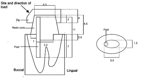
그리고 동일한 크기의 core를 축성하기 위하여 단경 3.5mm, 장경 5.5mm와 높이 4.5mm의 타원 기둥형의 금속 core를 주조하고 milling machine으로 마무리한 후에 금속 core 주조면 위에 Pattern resin(GC Corp., Tokyo, Japan)을 입힌 다음, 이 를 분리하고 주조하여 core를 복제할 수 있는 표준화 금속 주형을 제작했다(Fig. 1).
Fig. 1. Schematic drawing of test specimen showing dimensions in mm. a. Bucco-lingual view of the tooth specimen
b. Occlusal view of the tooth specimen
4. Core의 축성과 금속 주조 post의 제작 표준화 금속 주형을 이용하여 단경 3.5 mm, 장경 5.5 mm와 높이 4.5 mm의 동 일한 크기의 타원 기둥형의 레진 core를 2 mm 두께씩 적층 축성하였다. 중합 방 1 7 2 11 2.5 4 4.5
Site and direction of load Buccal Lingual Zig Resin core Post Post 3.5 1.5 4.5 3.5 1 7 2 11 2.5 4 4.5
Site and direction of load Buccal Lingual Zig Resin core Post Post 3.5 1.5 1 7 2 11 2.5 4 4.5
Site and direction of load Buccal Lingual Zig Resin core Post Post 3.5 1.5 4.5 3.5
식에 따른 광조사 시간이나 경화 시간은 제조자의 지시를 따랐다.
대조군인 금속 주조 post와 core를 위한 pattern 제작은 Spee-Dee Plastic pin (Pulpdent Corp. of America)과 Pattern resin(GC Corp., Tokyo, Japan)을 이용하 여 10개의 시편 모두에서 직접법으로 제작하여 Type IV 금 합금으로 주조하였다. 역시 동일한 크기의 치관부 core 형성을 위하여 표준화 금속 주형을 이용하였다.
금속 주조 post와 core의 표면을 sandblasting 처리를 하고 glass fiber post와 같
은 방법으로 레진 시멘트(Panavia F®, Kuraray Co. Ltd., Osaka, Japan)로 합착하
였다. 5. 압축 파절 하중 실험용 레진 시편의 제작 레진 core들의 압축 파절 하중을 측정하기 위하여 지름 3 mm, 높이 10 mm의 원기둥 모양의 레진 시편을 각 10개씩, 모두 40개를 제작하였다. 6. Thermal cycling의 시행 제작한 시편에 구강 내와 같은 온도 변화를 재현하기 위해 Thermocycling tester (Tokyo TL., Tokyo, Japan)로 thermal cycling을 시행하였다.7,20)
두 수조 안의 물의 온도는 각각 5℃와 55℃로 설정하고, 시편의 thermal cycling 은 1500 cycles을 시행하였으며, 각 수조에서의 dwell time과 수조 간의 transfer time은 모두 15초로 설정하였다.
치아 장축에 45도의 균일한 각도로 하중을 부여하기 위해, 치아 시편을 위치시 킬 special zig를 투명 아크릴 레진(ORTHO-JET, Lang Dental Mfg. Co. Inc., Wheeling, U.S.A.)으로 만들었다. 또 core의 치관부 2 mm를 덮는 core covering zig 를 제작하여(Fig. 1), 하중 부여 각도를 명확히 하고 하중 부여 부위의 국소적 하
중 집중을 막았다.21-23)
치아 시편 블록을 만능 시험기(Instron 6022, Instron Ltd, U.K.)에 위치하고 cross head speed를 1 mm/min으로 시편이 파절될 때까지 힘을 가한 후 post와 core의 최대 파절 하중을 측정하였다. 또 레진 시편들의 압축 파절 하중을 측정하 였다.
8. 파절 양상의 관찰
파절 하중 실험 후, 육안으로 파절 양상을 관찰하였고 시편의 레진 core 부위의 파절 양상에 따라 모든 시편을 type A에서부터 type E로 분류하였다(Fig. 2).
각 type에서 전형적인 치아 시편을 1개씩 골라 Low speed saw(ISOMET, BUEHLER, Lake Bluff, U.S.A.)로 협설 방향으로 post와 core의 중앙을 지나가도록 water spray 하에서 자른 후, 파절된 표면의 양상을 주사전자현미경(S-2700, HITACHI, Ltd., Japan)을 이용하여 200배로 확대하여 관찰하였다.
9. 통계 분석
각 치아 시편의 최대 파절 하중과 레진 시편의 압축 파절 하중에 대한 자료를 비모수 요인 분산 분석법인 Wilcoxon rank sum test로 실험군 간의 유의성을 확 인하고 Tukey's studentized range(HSD) test로 각 실험군 간에 어떠한 통계학적 유의차가 있는지를 알아 보았다. 모든 분석은 95%의 신뢰도로 검정하였다.
(a) type A (b) type B (c) type C
(d) type D (e) type E
Fig. 2. Schematic representation of main fracture lines in catastrophic failure.
(a) type A : The fracture of the core unrelated to the post.
(b) type B : The fracture of the core without the fracture of the post, the fracture line is parallel to the tooth cutting surface.
(c) type C : The two parted fracture of the core around the post. (d) type D : The three parted fracture of the core around the post. (e) type E : The fracture of the post and core.
III. 연구 성적
가. 치아 시편의 최대 파절 하중의 측정
Wilcoxon rank sum test 결과, 치아 시편의 최대 파절 하중 사이에 유의차가 있
었으며, ParaCore, Z100TM, Rebilda® 및 Admira®의 최대 파절 하중의 평균값은
ParaCore가 가장 높이 나타났고 다음이 Z100TM 순이었다. 금속 주조 post와 core
는 시편 모두가 하중이 1000N에 도달할 때까지 파절이 일어나지 않았다. Tukey
test 결과, ParaCore와 Rebilda®, ParaCore와 Admira®, Z100TM과 Rebilda® 및
Z100TM과 Admira® 사이에서 유의차가 있었다. ParaCore와 Z100TM 및 Rebilda®와
Admira® 사이에는 유의차가 존재하지 않았다(Fig 3, Table III).
Table III. The means of maximum fracture load of teeth specimens(N)
n Means ± SD (N) Tukey gruop
ParaCore 10 697.6 ± 87.7 A
Z100TM 10 586.7 ± 97.4 A
Rebilda® 10 365.5 ± 69.9 B
Admira® 10 354.4 ± 78.0 B
Cast post 10 1000≦
* n : number of specimen SD : standard deviation
A, B : statistical grouping for load value of materials(Kruskal-Wallis test & Tukey grouping)
Fig. 3. The means and standard deviations of maximum fracture load in the teeth specimens.
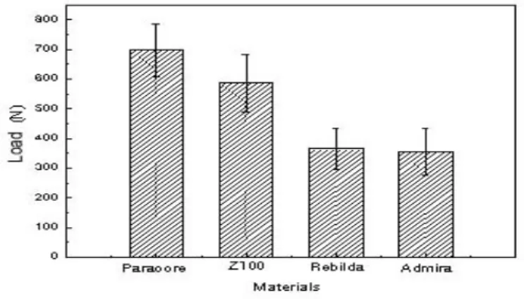
나. 레진 시편의 압축 파절 하중의 측정
Wilcoxon rank sum test 결과, 레진 시편의 압축 파절 하중 사이에 유의차가 존
재하였으며, ParaCore, Z100TM, Rebilda® 및 Admira®의 압축 강도의 평균값은
Z100TM이 가장 높았고 다음이 ParaCore 순이었다. Tukey test 결과, ParaCore와
Admira®, Z100TM과 Rebilda® 및 Z100TM과 Admira® 사이에서 유의차가 존재하였
다. ParaCore와 Rebilda®, ParaCore와 Z100TM 및 Rebilda®와 Admira® 사이에는 유의차가 존재하지 않았다(Fig. 4, Table IV).
Table IV. The means of compressive fracture load of resin specimens(N)
n Means ± SD (N) Tukey gruop
ParaCore 10 1121.1 ± 167.9 A B
Z100TM 10 1330.4 ± 172.7 A
Rebilda® 10 989.5 ± 152.5 C B
Admira® 10 864.5 ± 173.4 C
* n : number of specimen SD : standard deviation
A, B, C : statistical grouping for load value of materials(Kruskal-Wallis test & Tukey grouping)
significantly different between groups(p<0.05)
Fig. 4. The means and standard deviations of compressive fracture load in the resin specimens.
다. 파절 양상의 육안 관찰
치아 시편의 파절 양상에 따른 분포는 Table V와 같으며, 전체적으로는 post를 중심으로 세 부분으로 나타나는 core의 파절인 type D가 가장 높게 나타났고
(62.5%), ParaCore에서는 90%, Z100TM에서는 100%가 type D이었다. Rebilda®와
Admira®에서는 post를 지나가며 치아 절단면과 평행하게 나타난 core의 파절인
type B가 높게 나타났다(40%).
Table V. The distribution of fracture mode of teeth specimens
라. 파절 부위의 주사 전자 현미경 (SEM) 소견
Post와 관계 없는 core 자체의 파절인 type A(사진 부도 1)와 post를 중심으로 두 부분으로 나타나는 core의 파절인 type C에서 파절을 포함하지 않은 부위(사진 부도 5)를 보면, glass fiber post와 레진 core 사이의 결합이 견고한 것이 관찰된 다. 그리고 post를 지나가며 치아면과 평행하게 나타난 core의 파절인 type B의 post, 레진 core 및 상아질의 계면을 보면 레진 core와 상아질 사이는 분리되었지 만, glass fiber post와 레진 core 사이의 결합은 견고하며(사진 부도 2), 파절을 포 함한 부위들은 glass fiber post와 레진 core 사이의 결합이 분리된 것을 볼 수 있
type A type B type C type D type E
ParaCore 1 9
Z100TM 10
Rebilda® 4 2 3 1
다(사진 부도 3, 4). 또한 금속 주조 post와 core로 수복한 치아가 높은 하중에도 파절을 보이지는 않았지만, 시편의 단면을 관찰한 소견에서는 치근 파절을 발견할 수 있었다(사진 부도 6).
IV. 총괄 및 고찰
Glass fiber post는 최근에 개발된 재료로서 근관 치료 후, 보다 자연스럽고 심미
적인 완전 도재관 수복을 위해서 도재관 하방에 사용되어지는데, 기존 post system 에 비해 물리적 성질, 심미성, 치근 파절 및 수복물 파절의 위험성, 상방 core와의 결합력, 방사선 불투과도 그리고 제거 및 재치료성 등 많은 부분에 있어 장점이
있음이 보고되어 왔으며, 생체 친화적이고 화학적 안정성이 높다.2-6) 하지만, glass
fiber post도 단점은 존재하여, 사용시 구강내 타액과 접촉하면 post의 굴곡을 유 발하여 이차 우식의 위험과 레진 core의 탈락을 야기할 수 있는데, 이는 glass
fiber post가 수분 분해성에 민감하기 때문이다.24) 그러므로 임상에서 사용할 때에
도 주의 깊은 술식이 요구된다고 하겠다.
Post system을 선택할 때 고려 사항으로는 치근 파절과 post 자체 파절에 대한 저항력, 치아와의 유지력, 응력 분산, 색조와 광투과성 등의 심미성 및 여러 core 재료와의 적절한 결합력과 응집에 의한 post-core의 단일 단위로의 기능 등이 있 다.5,6)
Post와 core로 수복된 치아의 실패 양상은 금관과 post 탈락 같은 유지력 상실과
치근과 post 및 core의 파절로 나타난다.15,16) Hatzikyriakos 등은 post와 core로 수
복하고 금관을 장착한 후, 3년간 술후 임상 평가에서 실패 양상은 치근 파절 (2.6%), 치근과 post와 core의 인접 치아 조직의 우식적 파괴의 복합적 영향(1.9%),
금관과 post와 core 사이의 시멘트 상실(3.2%)로 나타났다고 보고했다.15) Torbjorner
등은 술후 4-5년간 임상 평가를 통해 post 실패의 가장 큰 요인을 유지력 상실로 보고했고, 하악보다 상악에서 더 높게 실패율이 나타났으며 특히, 상악 전치에서 가장 높았다고 발표했다.16) 상아질-치근 복합체의 수분 함유량은 근관 치료 상태와 과정에 따라 다르고 치 관부 상아질은 치근 상아질보다 상아 세관의 수가 2배 가량 많으며, 연령 증가에 따라 세관주위 상아질의 침착으로 수분 함유량이 줄어든다. 그리고 치료 과정에서
우식 제거, access cavity 및 근관 형성 중의 치근 상아질의 과다 제거 등 내외부 치질의 상실로 치아가 약해질 뿐만 아니라, 치주 인대 기능 저하로 인한 tactile
sensation의 감소로 과다 부하의 위험성이 있다.25-28) 이런 이유로 무수치의 파절
저항은 주로 잔존 치질량에 의해 영향 받는다.17,27-30) Tjan과 Whang 등은 post 형
성후, 2-3 mm의 잔존 치질량이 확보되면 치근 파절에 저항이 유의성있게 높아진
다고 보고하였고,29) Sorensen과 Engleman 등은 치아 파절에 저항하기 위해서는
metal collar ferrule보다 최소한 1 mm의 치관부 치질 확보가 중요하다고 하였으
며,30) Milot과 Stein 등은 대부분의 치질이 보존된 경우는 post의 선택이 치근 파
절의 저항에 거의 영향이 없다고 결론내렸다.17) 그리고 ferrule 효과가 post의 쐐기 효과나 치근 파절을 방지하는 기능에 대해 부정적인 견해가 발표됨으로써, 금속 주조 post와 core의 적응증은 거의 사라진 상태이다.30-32) 근관 치료를 한 치아를 수복하는 데 있어 잔존 치질의 보존도 하나의 목적이 다.33) 다른 실패보다 더 파괴적이고 거의 대부분이 발치까지 이르게 되는 치근 파 절보다는, 덜 심각하고 수복 가능한 상태로 남는 core 파절이 더 바람직한 실패 양상으로 생각할 수 있다.34) 금속 주조 post와 core로 수복된 치아가 상당히 큰 파
절 하중에 견디는 것은 사실이나,35) glass fiber post와 레진 core로 수복된 치아는
보다 낮은 하중을 견디지만 core의 파절로써 치질을 보존할 수 있다. 다시 말해,
core 파절이 치근 파절을 미리 예방한다는 것이다.34)
이번 연구 결과도 금속 주조 post와 core로 수복한 치아가 높은 하중에도 post 파절을 보이지는 않았지만, 시편의 단면을 관찰한 소견에서는 치근 파절을 발견할 수 있었으며(사진 부도 6), 복합 레진 파절이 치근 파절에 필요한 하중보다 낮은 하중에서 이루어졌다는 여러 실험 결과와 일치한다.34-36) 하지만, 치아의 비가역적 인 손상 없이 부러진 post를 재치료하는 것도 쉽지 않은 일이다. 그러나 glass fiber post는 재치료의 측면에서 이의 해결책을 제시해 주고 있다.35) 상실된 치관부를 대체하기 위해 post 상방에 축성하는 core용 재료는 아말감, glass ionomer cement reinforced with silver, hybrid glass ionomer, compomer 및 복합 레진 등이 있는데, 완전 도재관의 하방에 사용되는 core용 재료로는 적절 한 색조와 상대적으로 나은 물리적 성질, 치질과의 접합성, 조작의 간편성과 소요
시간 등의 장점으로 복합 레진이 가장 추천되고 있다.2,7-12) 하지만 이 재료도 낮은 탄성 계수로 인해 기계적 및 열적 자극에 의해 영구 변형이 올수 있고, 수분 흡수
에 의한 체적 안정성이 떨어지는 단점이 있어,2,12,23,37,38) core의 탈락과 수복물의 실
패를 가져올 수 있다. Core의 탈락은 미세누출, 재발성 우식과 근관 치료의 실패 를 초래한다. 그래서 titanium과 lanthanide 입자를 첨가하여 core용 복합 레진의 기계적 성질의 개선을 도모하기도 한다. 그렇지만 역시 잔존 치질량의 중요성이 간과되어선 안된다. 복합 레진 system은 자가 중합 방식이나 이원 중합 방식의 system이 추천된다. 왜냐하면, 광중합 방식의 복합 레진은 광조사기의 강도 저하, 불충분한 조사 시간 및 과도한 두께의 레진 적용 등으로 깊은 부위의 불완전한 중합의 위험이 있기 때문이다. 또 자가 중합 방식은 대개 paste type으로 보다 간편하고 흐름성이 좋 아 치질 내의 공간으로 쉽게 주입이 되고 모양 형성이 용이하다.8,39) Core용 재료를 선택할 때는 post의 응력 전달에 영향을 주기 때문에 응력의 양 과 형태도 고려해야 한다. 단단한 재료일수록 post로 가는 응력의 양은 줄고 응력 도 더 직접적으로 치근에 전달되며,23) 금속제 core는 치경부에 큰 응력을 야기하고 레진 core는 치근단부에 더 큰 응력을 야기한다고 알려져 있다.11,40) 그리고 post와 core용 재료 사이도 기계적 성질이 유사한 것이 좋은데, 이는 견고한 계면을 형성 하고 재료간의 응력 유발이 적어 미세누출과 실패 가능성을 줄일 수 있기 때문이 다.11) Glass fiber post는 무게비로 42%의 glass fiber, 29%의 filler 및 29%의 레진
으로 구성되므로,2) 복합 레진과의 화학적 결합도 기대할 수 있다. Trope 등은
post를 위한 공간 형성이 근관 치료를 받은 치아를 유의성 있게 약화시키며 post 가 이를 강화시키지는 못하지만, post 공간과 access cavity를 복합 레진으로 채우
면 강도를 개선할 수 있다고 발표했다.28) 레진 core와 상아질면 사이에 일어나는 미세누출은 복합 레진과 치질 사이의 부 착력 부족과 레진의 중합수축으로 인한 것으로, 이는 상아질 접착 system의 사용 으로 줄일 수 있다.13,40) 레진 core의 미세누출은 수분의 오염과 재료의 분해를 야 기하며,42) 미세누출로 인한 core의 결합력 저하는 core의 파절 가능성을 높인다.21) 그러므로 레진 core, 접착 system 및 상아질 간의 부가적 결합력을 개선함으로써
post와 core의 실패율을 줄일 수 있다.43) 레진과 치질의 결합력은 치면의 청결도 같은 외부 상태에도 영향을 받는데, 그 중에 임시 합착제의 잔류는 레진과 상아질 의 결합력을 저하시키는 요인으로 생각된다. 특히 유지놀을 함유한 임시 합착제는
레진 core를 연화시키고 약하게 하므로 주의해야 한다.44-47)
이번 연구에서 치아 시편 파절 부위의 주사 전자 현미경 사진을 보면 post와 관 계 없는 core 자체의 파절인 type A와 post를 중심으로 두 부분으로 나타나는 core의 파절인 type C에서 파절을 포함하지 않은 부위를 볼 때, glass fiber post와 레진 core 사이의 결합이 견고한 것이 관찰된다. 그리고 post의 파절없이 치아면 과 평행하게 나타난 core의 파절인 type B에서 post, 레진 core 및 상아질의 계면 을 볼 때, 레진 core와 상아질 사이는 분리되었지만, glass fiber post와 레진 core 사이의 결합은 견고하며, 파절을 포함한 부위들은 glass fiber post와 레진 core 사 이의 결합이 분리된 것을 볼 때(사진 부도 1), 레진 core의 파절시 가장 먼저 core 와 상아질이 분리되고 이어 glass fiber post와 레진 core 사이의 결합력이 약해지 면서, 마지막으로 core의 파괴가 나타난다고 생각된다. 이는 앞에 기술한 여러 실 험 결과와도 일치한다.13,21,41,43) 압축 강도와 인장 강도는 core 재료에서 중요한 인자로 생각된다. Core는 대개 큰 부피의 치질을 대신하므로 여러 방향에서 오는 저작압과 비기능적인 힘을 견 뎌야 한다.8) 재료의 성질 중에 인성(toughness)은 파절에 대한 저항을 나타내는 성 질로 파절을 유발하는 데에 필요한 에너지의 양으로 정의되는데, brittle한 재료가 crack의 진행에 의해 파절까지 가는 경향으로 나타난다. 이 성질은 crack의 기시에 대한 저항이 아니라, 이미 존재하는 crack이나 흠의 불안정한 진행에 대한 저항으 로 설명된다.48) 연구와 실험을 통해 복합 레진의 파괴 인성은 습기나 타액의 오염 에 의해 저하될 수 있다고 밝혀졌다.49-51) 그리고 Thermocycling은 복합 레진의 미 세누출, microcracking 및 표면의 분해에는 관여하나, 직접적인 재료의 파절 인성 의 저하를 유발하지는 않는 것으로 알려졌다.20) Cho 등은 실험을 통해 광중합형 복합 레진이 자가 중합형 복합 레진보다 압축 강도와 인장 강도 면에서 다소 나은 것으로 보고했다.8) 이는 압축 파절 하중 시험 에서 광중합형 복합 레진인 Z100TM이 자가 중합형 복합 레진인 Rebilda®보다 유
의차 있게 높은 결과와 일치한다. 이는 흐름성이 좋은 paste type인 Rebilda®의 filler 함량이 상대적으로 낮기 때문이 아닌가 생각된다. 또, Combe 등은 실험을 통해 ceramic filled composite가 거의 모든 기계적 성질에서 hybrid resin composite 보다 떨어졌으며, 특히 저작압의 영향과 관련된 압축 강도는 hybrid resin composite
에 비해 현저히 차이 나는 것으로 나타났다.9) 이는 최대 파절 하중과 압축 파절
하중 실험에서 ceramic filled composite인 Admira®가52-54) 다른 제품에 비해 유의
성 있게 낮은 수치가 나온 사실과도 일치하는데, 일반적으로 filler의 함유는 파괴 인성을 증가시키지만,49,50,55) 딱딱한 무정형의 입자가 비교적 부드러운 레진의 기질 에 섞이게 되면 오히려 파괴 인성이 감소하기도 하는데, 이는 입자 변형 에너지장 들이 겹치는 결과로 보여진다.56) 아마도 Admira®의 경우도 여기에 해당하지 않나 추측된다. 즉 Rebilda®와 Admira®의 비교적 낮은 압축 강도와 낮은 인성으로 인한 파절
강도의 저하 때문에 post 상방에 위치하는 core용 레진으로 사용할 때, post와 관 계없는 core 자체의 파절인 type A와 post를 지나가며 치아면과 평행하게 나타난 core의 파절인 type B가 많이 나타났고(Table V), 치아 시편의 최대 파절 하중도
낮았다고 볼 수 있으며, 상대적으로 높은 압축 강도를 가진 Z100TM과 ParaCore는 치아 시편도 높은 파절 하중을 보였다(Table IV). 본 연구는 치근을 바로 아크릴 레진 블록에 묻음으로써 견고한 아크릴 레진에 의한 치근 구조의 외부 강화를 막을 수는 없으므로, 파절 양상의 자연스러운 재현 에는 한계가 있을 수 밖에 없었다.32,34) 그러나 치주 인대를 재현하지 않은 이유는, 치아를 부드러운 탄성 고무 인상재를 얇게 바르는 방식의 재현 방법으로는 시편 에 하중 부여시 탄성층의 최대한 변형으로 치아 장축에 정확한 각도로 하중을 줄 수 없으며 탄성층 범위 내에서 회전력을 받을 수 있을 뿐만 아니라, 재료가 치주 인대 섬유의 기능과 배열 및 점탄성을 재현하기에는 한계가 있다.21) 또 여러 실험 을 통해 근관 충전이 치근의 강도에 거의 영향을 주지 않는다는 결과에서 근관 충전을 시행하지는 않았으나,57-59) 근관 충전 과정 중 유지놀이 함유된 sealer의 사 용이 레진 시멘트의 결합력을 저하시키는 것으로 알려져 있다.34,46) 이 실험은 치아에 장기적이고 주기적인 하중을 부여한 것이 아닌, 단기적이고
정적인 하중을 부여한 뒤에 실패 양상을 관찰한 것으로 부가적인 피로 하중을 고
려한 연구가 필요하리라 생각된다.21-23) 뿐만 아니라, 레진 시멘트와 금관 사이의
불충분한 결합력과 저작압이나 전단 압력 후에 레진 core의 변형이 glass fiber po st의 실패에 미치는 영향과 glass fiber post와 레진 core 사이의 결합력의 개선이 post의 실패를 줄일 수 있는 지의 여부를 보는 연구도 필요할 것으로 사료된다.
V. 결 론
최근 개발되어 근관 치료 후 심미성 회복을 위하여 쓰여지는 glass fiber post
(ParaPost® Fiber White)와 각기 다른 조성의 core용 레진(ParaCore, Z100TM, Rebilda®,
Admira®)으로 자연치를 수복한 후 만능 시험기로 하중을 부여하여 시편의 최대
파절 하중과 파절 양상을 알아보고 금속 주조 post와 core로 수복한 대조군과 비 교한 연구를 통하여 다음과 같은 결과를 얻었다.
1. 치아 시편의 최대 파절 하중 측정 결과, ParaCore, Z100TM, Rebilda®, Admira®
순이었고, ParaCore와 Z100TM이 Rebilda®와 Admira®보다 유의차 있게 높았다.
(p<0.05).
2. 레진 시편의 압축 파절 하중의 평균값은 Z100TM, ParaCore, Rebilda®, Admira®
순이었고, Z100TM과 Rebilda®, Z100TM과 Admira® 및 ParaCore와 Admira® 사 이에 유의차가 있었다.(p<0.05).
3. 치아 시편의 파절 양상은 전체적으로는 post를 중심으로 세 부분으로 나타나는
core의 파절인 type D가 가장 많았고(62.5%), ParaCore는 100%, Z100TM은 90%
가 type D이었으며, Rebilda®와 Admira®에서는 post의 파절없이 치아면과 평
행하게 나타난 core의 파절인 type B가 많이 나타났다(40%).
4. 금속 주조 post와 core로 수복한 치아는 높은 하중에도 post와 core의 파절을 보이지는 않았지만, 시편의 단면을 관찰한 소견에서는 치근 파절을 발견할 수 있었다.
이상의 결과를 토대로 glass fiber post를 위한 치아 색조의 레진 core용 재료로
는 ParaCore와 Z100TM이 추천되며, 앞으로도 glass fiber post와 core용 복합 레진
참고 문헌
1. Desort K.D. The prostodontic use of endodontically treated teeth : Theory and biomechanics of post preparation. J Prosthet Dent 49 : 203-6, 1983
2. Quintas A.F., Dinato J.C., Bottino M.A. Aesthetic posts and cores for metal free restoration of endodontically treated teeth. Pract Periodont Aesthet Dent 12 : 875-84, 2000
3. Roberto Martelli. Fourth-generation intraradicular posts for the aesthetic restoration of anterior teeth. Pract Periodont Aesthet Dent 12 : 579-84, 2000
4. Freedman G.A. Esthetic post-and-core treatment. Dental Clinics of North America. 45(1) : 103-16, 2001
5. Assif D., Bitenski A., Pilo R., Oren E. Effect of post design on resistance to fracture of endodontically treated teeth with complete crowns. J Prosthet Dent 69 : 36-40, 1993
6. Assif D., Oren E., Marshak B.L., Aviv I. Photoelastic analysis of stress transfer by endodontically treated teeth to supporting structure using different restorative techniques. J Prosthet Dent 61 : 535-43, 1989
7. Tirado J.I.M., Nagy W.W., Dhuru V.B., Ziebert A.J. The effect of thermocycling on the fracture toughness and hardness of core buildup materials. J Prosthet Dent 86 : 474-80, 2001
8. Cho G.C., Kaneko L.M., Donovan T.E., White S.N. Diametral and compressive strength of dental core materials. J Prosthet Dent 82 : 272-6, 1999
9. Combe E.C., Shaglouf A.M.S., Watts D.C., Wilson N.H.F. Mechanical properties of direct resin core buildup materials. Dent Mater 15 : 158-65, 1999
10. Levartovsky S., Goldstein G.R., Georgescu M. Shear bond strength of several new core materials. J Prosthet Dent 75 : 154-8, 1996
11. Yaman P., Thorsteinsson T.S. Effect of core materials on stress distribution of posts. J Prosthet Dent 68 : 416-20, 1992
12. Oliva R.A., Lowe J.A. Dimensional stability of composite used as a core material. J Prosthet Dent 56 : 554-61, 1986
13. Mannocci, F., Ferrari, M., Watson, T.F. Microleakage of endodontically treated teeth restored with fiber posts and composite cores after cycling loading : A confocal microscopic study. J Prosthet Dent 85 : 284-91, 2001
14. Margano S.M. Restoration of pulpless teeth : Application of traditional principles in present and future contexts. J Prosthet Dent 75 : 375-80, 1996
15. Hatzikyriakos A.H., Reisis G.I., Tsingos N. A 3-year postoperative clinical evaluation of posts and beneath existing crowns. J Prosthet Dent 67 : 454- 81, 1992
16. Torbjorner A., Karlsson S., Odman P.A. Survival rate and failure characteristics for two post designs. J Prosthet Dent 73 : 439-44, 1995
17. Milot P., Stein S. Root fracture in endodontically treated teeth related to post selection and crown design. J Prosthet Dent 68 : 428-35, 1992
18. Sorensen J.A., Martinoff J.T. Intracoronal reinforcement and coronal coverage : A study of endodontically treated teeth. J Prosthet Dent 51 : 780-4, 1984
19. 김 태형, 심 준성, 이 근우. Glass fiber post와 Composite resin core의 전단 결합강도연구. 연세대학교 치의학과 논문집 : in press
20. Gale M.S., Darvell B.W. Thermal cycling procedures for laboratory testing of dental restorations. J Dent 27 : 89-99, 1999
21. Huymans M.C., Peters M.C., Van der Varst P.G., Plasschaert A.J. Failure characteristics of endodontically treated premolars restored with a post and direct restorative materials. Int J Endodont 25 : 121-129, 1992
22. Huymans M.C., Peters M.C., Van der Varst P.G., Plasschaert A.J. Failure behavior of direct post and core restored premolars. J Dent Res 71(5) : 1145-50, 1992
23. Huymans M.C., Peters M.C., Van der Varst P.G., Plasschaert A.J. Failure behavior of fatigue-tested post and core. Int J Endodont 26 : 294-300, 1993
24. Vallitu P.K., Ruyter I.E., Ekstrand K. Effect of water storage on the flexural properties of E-glass and silica fiber acrylic resin composite. Int J Prosthodont 11 : 340-50, 1998
25. Gutmann J.L. The dentin-root complex : Anatomic and biologic considerations in restoring endodontically treated teeth. J Prosthet Dent 67 : 458-67, 1992
26. Helfer A.R., Melnick S., Schilder H. Determination of the moisture content of vital and pulpless teeth. Oral Surgery 34 : 62-5, 1972
27. Assif D., Gorfil C. Biomechanical considerations in restoring endodontically treated teeth. J Prosthet Dent 71 : 565-7, 1994
28. Trope M., Maltz D.O., Tronstad L. Resistance to fracture of restored endodontically treated teeth. Endodontics Dental Traumatology 1 : 108-11, 1985
29. Tjan A.H.L., Whang S.B. Resistance to root fracture of dowel channels with various thickness of buccal dentin walls. J Prosthet Dent 53 : 496-500, 1985
30. Sorensen J.A., Martinoff J.T. Clinically significant factors in dowel design. J Prosthet Dent 52 : 28-35, 1984
31. Loney R.W., Kotowicz W.E., McDowell G.C. Three-dimensional photoelastic stress analysis of the ferrule effect in cast post and cores. J Prosthet Dent 63 : 506-512, 1996
32. Sirimai S., Aiis D.N., Morgano S.M. An in vivo study of the fracture resistance and incidence of vertical root fracture of pulpless teeth restored with six post-and-core systems. J Prosthet Dent 81 : 262-9, 1999
33. Sorensen J.A., Engelman M.J. Ferrule design and fracture resistance of endodontically treated teeth. J Prosthet Dent 63 : 529-36, 1990
34. Fraga R.C., Chaves B.T., Mello G.S.B., Siqueira J.F. Jr. Fracture resistance of endodontically treated roots after restorations. J Oral Rehab 25 : 809-813, 1998
35. Sorensen J.A., Engelman M.J. Effect of post adaptation on fracture resistance of endodontically treated teeth. J Prosthet Dent 64 : 419-24, 1990
36. Bex R.T., Parker M.W., Judkins J.T., Pelleu G.B. Effect of dentinal bonded resin post-core preparations on resistance to vertical root fracture. J Prosthet Dent 64 : 412-5, 1990
37. Kovarik R.E., Breeding L.C., Caughman W.F. Fatigue life of three core materials under simulated chewing conditions. J Prosthet Dent 68 : 584-90, 1992
38. Hirasawa T. et al. Initial dimension change of composite in dry and wet conditions. J Dent Res 62 : 28-31, 1983
39. Plasmans P.J., Welle P.R., Vrijhoef M.M. In vitro resistance of composite resin dowel and cores. J Endodont 14 : 300-4, 1988
40. Ko C.C., Chu C.S., Chung K.H., Lee M.C. Effects of posts on dentin stress distribution in pulpless teeth. J Prosthet Dent 68 : 421-7, 1992
41. Tjan A.H.L., Grant B.E., Dunn J.R. Microleakage of composite resin cores treated with various dentin bonding systems. J Prosthet Dent 66 : 24-9, 1991
42. Freemann M.A., Nicholls J.I., Kydd W.L., Harrington G.W. Leakage associted with load fatigue-induced preliminary failure of full crowns placed over three different post and core systems. J Endodont 24 : 26-32, 1998
43. Rosentritt M. et al. Comparison of in vitro fracture strength of metallic and tooth-coloured posts and cores. J Oral Rehab 27 : 595-601, 2000
44. Watanabe E.K. et al. Temporary cements remnants as an adhesion inhibiting factor in the interface between resin cements and bovine dentin. Int J Prosthodont 10 : 440-52, 1997
45. Xie J., Powers J.M., McGuckin R.S. In vitro bond strength of two adhesives to enamel and dentin under normal and contaminated conditions. Dent Mater 9 : 295-9, 1993
46. Woody T.L., Davis R.D. The effect of eugenol-containing and eugenol-free temporary cements on microleakage in resin bonded restorations. Oper Dent 17 : 175-80, 1992
47. DeWald J.P., Moody C.R., Ferracane J.L. Softening of composite resin by moisture and cements. Quintessence Int. 19 : 619-21, 1988
48. Lloyd C.H. The fracture toughness of dental composites. II. The environment and tenperature dependence of the stress intensification factor (KIC). J Oral
49. Ferracane J.L., Antonio R.C., Matsumoto H. Variables affecting the fracture toughness of dental composites. J Dent Res 66 : 1140-5, 1987
50. Lloyd C.H., Lannetta R.V. The fracture toughness of dental composites. I. The development of strength and fracture toughness. J Oral Rehab 9 : 55-66, 1982
51. Lloyd C.H. The fracture toughness of dental composites. III. The effect of environment upon the stress intensification factor (KIC) after extended
storage. J Oral Rehab 11 : 3 93-8, 1984
52. Manhart J., Kunzelmann K.H., Chen H.Y., Hickel R. Polymerization contraction stress in light-cured packable composite resins. Dent Mater 17 : 253-59, 2001
53. Manhart J., Kunzelmann K.H., Chen H.Y., Hickel R. Mechanical properties and wear behavior of light-cured packable composite resins. Dent Mater 16 : 33-40, 2000
54. Manhart J., Kunzelmann K.H., Chen H.Y., Hickel R. Mechanical properties of new composite restorative materials. Int J Biomed Mater Res 53 : 353- 61, 2000
55. Pillar R.M., Vowles R., Williams D.F. The effect of enviormental aging on the fracture toughness of dental composites. J Dent Res 66 : 722-6, 1987
56. Goldman M. Fracture properties of composite and glass ionomer dental restorative materials. J Biomed Mater Res 19 : 771-83, 1985
57. Leary J.M., Aquilino S.A., Svare C.W. An evaluation of post length within the elastic limits of dentin. J Prosthet Dent 57 : 277-81, 1987
58. Trabert K.C., Caputo A.A., Abou-Rass M. Tooth fracture - A comparison of endodontic and restorative treatment. J Endodont 4 : 341-5, 1978
59. Weine F.S. Endodontic therapy. CV Mosby p144 : 1976
60. 이 영수, 강 익제. Quartz fiber post의 물리적 특성에 관한 연구. 대한치과 보철학회지 40(1) : 68-78, 2002
논문 사진 부도 설명
사진 부도 1. Type A의 파절에서 glass fiber post와 레진 core 사이의 계면 (×200)
사진 부도 2. Type B의 파절에서 glass fiber post, 레진 core 및 상아질 사이의 계면 (×200)
사진 부도 3. Type B의 파절에서 glass fiber post와 레진 core 사이의 계면 (×200)
사진 부도 4. Type D의 파절에서 glass fiber post와 레진 core 사이의 계면 (×200)
사진 부도 5. Type C 파절에서 파절을 포함하지 않은 부위의 glass fiber post와 레진 core 사이의 계면 (×200)
논문 사진 부도 ①
사진 부도 1 사진 부도 2 사진 부도 3 사진 부도 4논문 사진 부도 ②
사진 부도 5 사진 부도 6
* R : Resin core F : Fiber post D : Dentin C : Cast post
ABSTRACT
The fracture characteristics of Glass fiber post and Core
on using different types of core resin materials
Dong-Wook Shim. D.D.S.
Department of Dentistry, The Graduate School, Yonsei University
(Directed by Prof. Keun-Woo Lee, D.D.S., M.S.D., Ph.D.)
Increased demands for natural and esthetic dental treatment by patients have empahasized and enhanced importance in mechanical and physical properties of dental materials especially used in esthetic dentistry. Glass fiber post is one of recent developments to accommodate esthetic restoration for endodontically treated teeth. This has many advantages over conventional post system in physical properties, esthetic factor, risk of root and restoration fracture, adhesion to core, radio opacity, removal and retrievabilty, biocompatibility and chemical stability. This in vitro study was to evaluate the most effectual combination of resin for core material. Teeth specimens were made using glass fiber post(ParaPost® Fiber White) and different types of core resin materials (ParaCore, Z100TM, Rebilda® and Admira®) and were loaded to faiure in a universal test machine. The maximum fracture load and fracture mode were investigated in the specimens that were restored with resin and those of metal cast and core.
1. On measuring the maximum fracture load of teeth specimens, the order of maximum value was ParaCore, Z100TM, Rebilda®, Admira®. ParaCore and Z100TM showed a significant higher value than Rebilda® and Admira® (p<0.05).
2. The means compressive fracture load of resin specimens, the order was Z100TM, ParaCore, Rebilda®, Admira®, and there were significant differences between Z100TM and Rebilda®, Z100TM and Admira® and ParaCore and Admira®(p<0.05).
3. The fracture mode of teeth specimens showed the type D fracture, the three parted fracture of the core around the post was most common type of the fracture mode(62.5%). The type D fracture appeared at 100% of ParaCore and 90% of Z100TM. The type B fracture, the fracture of the core without the fracture of the post, the fracture line is parallel to the tooth cutting surface was most common at Rebilda® and Admira®(40%).
4. The teeth restored with metal cast post and core didn`t showed the fracture of post and core, in spite of high loads. However root fractures at the sectional view of specimens could be found.
In conclusion, referring to the values of maximum fracture load and mean compressive fracture load, ParaCore and Z100TM had high values and are recommended as tooth colored resin core material for glass fiber post. Further researchs using glass fiber post and composite resin for core material will be needed.
Key words : Glass fiber post, resin core, metal cast post and core, maximum fracture load, compressive fracture load, fracture mode.