상악 단일 치아 임프란트의
후향적 연구
연세대학교 대학원
치의학과
상악 단일 치아 임프란트의
후향적 연구
지도 문 익 상 교수
이 논문을 석사 학위논문으로 제출함
2003년 6월 일
연세대학교 대학원
치의학과
조 수 진
조수진의 석사 학위 논문을 인준함
심사위원________________인
심사위원________________인
심사위원________________인
연세대학교 대학원
2003년 6월 일
감사의 글
부족한 점이 많지만 늦게나마 논문을 완성하게 되었습니다. 한 편의 논문이 완성되기 까지 항상 격려해 주시고 이끌어 주신 문익상 교수님께 감사드리며, 깊은 관심과 자상하신 조언으로 일깨움을 주신 조규 성 교수님, 이근우 교수님께 감사드립니다. 그리고, 자료수집을 도와주었던 치주과 의국 여러분께도 감사드립니다. 항상 격려와 사랑을 베풀어주신 부모님께 이 논문을 드립니다. 2003년 6월 조 수 진목 차
국문요약 ··· iii I. 서 론 ··· 1 II. 연구대상 및 방법 ··· 3 1. 연구대상 ··· 3 2. 연구방법 ··· 5 1) 치아 상실 원인별 분류 ··· 5 2) 임프란트의 성공기준 ··· 6 3) 방사선학적 검사 ··· 6 4) 통계학적 분석 ··· 8 II. 연구결과 ··· 9 1. 치아 상실의 원인 ··· 9 2. 임프란트의 성공률 ··· 10 3. 변연골 소실에 관한 방사선학적 고찰 ··· 10 IV. 총괄 및 고찰 ··· 12 V. 결 론 ··· 17 참고문헌 ··· 19 Abstract ··· 26도표 및 사진 부도 목차
Table 1. Distribution of age and gender of patients ··· 3
Table 2. Distribution of implants according to position ··· 4
Table 3. Description of bone defect and specific surgery ··· 4
Table 4. Distribution of implants according to length and diameter ··· 5
Table 5-1. Reasons for upper anterior teeth replacement ··· 9
Table 5-2. Reasons for upper posterior teeth replacement ··· 9
Table 6. Description of complications ··· 11
Table 7. Marginal bone loss of upper anterior group and upper posterior group ··· 11
Table 8. Marginal bone loss of bone defect group and non-defect group ··· 11
Figure 1. Schematic drawing for the amount of marginal bone loss ··· 7
국문요약
상악 단일 치아 임프란트의 후향적 연구
상악에서 2단계 술식으로 machined Brånemark 임프란트를 사용한 단일 치아 임프란트 시술후 변연골 소실 변화량을 측정하여 전치부와 구치부에 서 변연골 소실량을 비교하고, 상악 전치부에서 열개, 천공의 골결손이 있 는 경우와 골결손이 없는 경우의 변연골 소실량을 비교하기 위해 1995년 1 월부터 상악 단일 치아 상실로 임프란트 매식 치료를 받은 환자 중 단일 치관수복물을 장착한 71명을 대상으로 연구하여 다음과 같은 결과를 얻었 다. 1. 상악 전치부 치아상실 원인별 분류시 외상의 빈도가 61%로 가장 높 게 나타났으며, 상악 구치부 치아상실 원인은 선천적 결손, 심한 치주질환, 외상 등으로 다양하게 나타났다. 2. 보철물 장착 1년 후 상악 전치부군에서는 총 41개 임프란트 중 1개에 서 누공을 동반한 임프란트주위염이 발생하였고, 상악 구치부군에서 총 32 개의 임프란트 중 2개가 실패하여, 1년 성공률은 상악 전치부군에서 97.56 %, 상악 구치부군에서 93.75 %였다. 3. 변연골 소실량은 보철물 장착 1년 후 상악 전치부군에서 0.44 ± 0.25 mm로, 상악 구치부군에서 0.57± 0.32 mm로 나타났으며, 통계학적으로 유의성 있는 차이가 있었다 (P<0.05). 4. 상악 전치부에서 보철물 장착후 1년간 열개와 천공의 골결손군은 0.40 ± 0.10 mm의 변연골소실이 나타났고 정상군은 0.48 ± 0.26 mm의 골소실 이 나타났으며, 통계학적으로 유의성 있는 차이는 없었다 (P>0.05). 이상의 결과에서 볼 때 상악 전치부가 구치부보다 변연골 소실이 더 적 게 나타났으며, 상악 단일 치아 상실시 해당 수여부의 상태를 고려한 적절 한 길이, 직경의 임프란트를 적절한 치유기간을 주고 기능적 부하를 시킨 경우, 단일 치아 임프란트 시술은 인접치아의 손상없이 치아를 수복할 수 있는 유용한 치료방법으로 사료된다. 핵심 되는 말 : 상악 전치부, 상악 구치부, 단일 치아 임프란트, 변연골 소 실, 골결손, 성공률, 후향적 연구
상악 단일 치아 임프란트의 후향적 연구 연세대학교 대학원 치의학과 (지도 문 익 상 교수) 조 수 진
I. 서 론
1960년대 초 골유착 임프란트가 소개된 이후, 완전 무치악 환자와 부분 무치악 환자에서 골유착 임프란트는 장기간 성공적으로 사용되어져 왔다. 1981년 Adell등은 완전 무치악 환자의 15년 동안의 임상연구에서 상악에서 81%, 하악에서 91%의 생존율을 보고하였고, 1999년 Lekholm등은 부분 무 치악 환자의 10년간 관찰기간에서 상악에서 90.2%, 하악에서 93.7%의 누 적성공률을 보고하였다. 완전무치악과 부분무치악에서 골유착 임프란트가 장기간의 높은 생존율을 보여준 이후, 단일치아 결손증례에서도 골유착성 임프란트를 이용하는 술식이 1986년 Jemt등에 의해 처음 시도되었다. 단일 치아 결손시 인접치가 건전하거나 아주 작은 수복물로 충전이 되있 는 경우, 단일 치아 임프란트 치료는 계속 가공 의치에 비해 인접치를 보 존할 수 있다는 장점이 있다. 단일 치아 임프란트의 누적 성공률에 관해 1995년 Andersson등은 98.5%의 3년간 누적 성공률을 보고 하였고, 1996년 Avivi-Arber와 Zarb등은 98%의 3년간 누적 성공률을 보고 하였으며, 1994 년 Cordioli등은 95.4%의 3년간 누적 성공률을 보고 하였다. 이와 같이 단 일 치아 임프란트에 관한 문헌들은 상하악 전체에 걸친 성공률을 보고하고있다. 그러나, 상악과 하악의 골질의 차이가 있으며, 상악에서도 전치부보 다 구치부의 골질이 더 떨어진다. 또한, 악궁 내에서 전치부에서 구치부로 갈수록 측두하악 관절과 가까워져서 저작력도 증가되며 상악 구치부의 경 우 해부학적으로 상악동의 위치 때문에 임프란트 매식체의 길이에 제한을 받는 경우도 있다. 1990년 van Steenberghe등은 임프란트의 실패율은 골질 과 상관관계가 있었다고 발표한 반면, 1998년 Wyatt과 Zarb등은 상악과 하악, 전치부와 구치부의 4부위로 구분하여 임프란트의 실패율을 비교한 결과 부위별 차이가 없었다고 하였다. 상악 전치부의 경우 치아 상실로 인한 협측골의 소실로 잔존치조골에 함 몰이 생기는 경우가 빈번한데, 보철적인 관점에서 이상적인 위치에 임프란 트 매식시 천공, 열개가 생기기 쉽다. 1995년 Dahlin등은 열개와 천공 같은 국소적인 골결손부에서 골유도 재생술식을 사용하여 성공적인 신생골 형성 을 보고 하였고 골량이 부족한 환자에서도 성공적인 임프란트 시술이 가능 하다고 하였다. 그러나. 1999년 Lekholm등은 임프란트 매식시 협측 나사산 이 1-5개 노출된 경우의 연구 결과, 기능 5년 후에도 연조직문제가 발생되 지 않았으며 골결손부의 점진적 골 흡수도 없었다고 하였다. 2002년 Naert 등은 부분 무치악에서 열개가 있는 경우 변연골 소실량이 더 크게 나타났 다고 하였으나 통계적 유의성은 없다고 보고 하였다. 본 연구의 목적은 상악에서 machined Brånemark 임프란트를 사용한 2단 계 술식으로 단일 치아 임프란트 시술후 1년간 전치부와 구치부의 변연골 소 실 변화량과 성공률을 비교하고자 하였고, 또한 상악 전치부에서 열개, 천공 의 골결손이 있는 경우와 골결손이 없는 경우의 변연골 소실 변화량을 비교 하였다.
II. 연구대상 및 방법
1. 연구대상
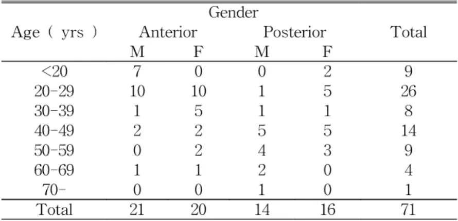
본 연구에서는 1995년 1월부터 단일 치아 상실로 연세대학교 치과 대학 부속병원과 영동세브란스병원 치주과에서 상악에 단일 치아 임프란트를 매 식하고 단일 치관수복물을 장착한 71명의 환자를 대상으로 하였다 (Table 1). 환자 중 2명은 연속되지 않은 부위에 2개씩 매식하여, 임프란트 개수는 총 73개 이었다 (Table 2). 상악전치부에 매식된 임프란트 41개를 상악 전 치부군으로, 상악구치부에 매식된 임프란트 32개를 상악 구치부군으로 하 였다. 또한 상악 전치부에서 열개와 천공이 발생한 13개를 골결손군으로 하고, 나머지를 정상군으로 하였다 (Table 3).Table 1. Distribution of age and gender of patients
Age ( yrs ) Gender Total Anterior Posterior M F M F <20 7 0 0 2 9 20-29 10 10 1 5 26 30-39 1 5 1 1 8 40-49 2 2 5 5 14 50-59 0 2 4 3 9 60-69 1 1 2 0 4 70- 0 0 1 0 1 Total 21 20 14 16 71 Anterior : upper anterior group
Table 2. Distribution of implants according to position
Position #17 #16 #15 #14 #13 #12 #11 #21 #22 #23 #24 #25 #26 #27 Number 0 4 5 7 5 3 15 12 6 0 7 6 3 0
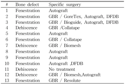
Table 3. Description of bone defect and specific surgery
# Bone defect Specific surgery 1 Fenestration Autograft
2 Fenestration GBR / GoreTex, Autograft, DFDB 3 Fenestration GBR / Bioguide, Autograft, DFDB 4 Dehiscence GBR /Collatape 5 Fenestration Autograft 6 Fenestration GBR / Collatape 7 Dehiscence GBR / Biomesh 8 Fenestration Autograft 9 Fenestration Autograft 10 Fenestration Autograft ,DFDB 11 Dehiscence No treatment 12 Dehiscence GBR / Biomesh,Autograft 13 Fenestration GBR / Resolute # Number of patient
GBR : Guided bone regeneration
DFDB : Pacific coast tissue bank, LA, USA
GoreTex® : W.L Gore and Associates, Flagstaff, AZ, USA Resolute® : W.L Gore and Associates, Flagstaff, AZ, USA Biomesh® : Samyang, Seoul, Korea
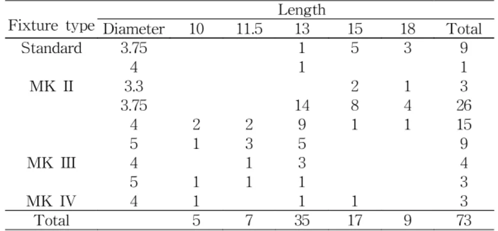
임프란트 매식은 각 환자의 해부학적 상황에 맞는 임프란트의 직경과 길 이를 결정하여 machined Brånemark 임프란트를 2단계로 단일 치아 임프 란트 매식법을 사용하여 시술하였다 (Table 4). 2차 수술은 1차 수술 후 약 6개월 경과 후 시행하였다. 2차 수술 후 2주정도의 연조직 치유기간을 거친 뒤 보철 지대주를 연결 하고 보철물을 장착하였다. 환자들은 적어도 1년에 1회 이상 주기적 내원 시 치태조절과 임상 및 방사선 사진 검사를 받았다.
Table 4. Distribution of implants according to length and diameter
Fixture type Length Diameter 10 11.5 13 15 18 Total Standard 3.75 1 5 3 9 4 1 1 MK II 3.3 2 1 3 3.75 14 8 4 26 4 2 2 9 1 1 15 5 1 3 5 9 MK III 4 1 3 4 5 1 1 1 3 MK IV 4 1 1 1 3 Total 5 7 35 17 9 73
2. 연구방법
1. 치아 상실 원인별 분류 치아 상실 원인은 외상, 치주질환, 선천적 상실, 기타 신경치료로 해결이 안되는 경우로 분류하였다.2. 임프란트의 성공기준
임프란트의 성공기준은 1986년 Albrektsson & Zarb가 발표한 다음과 같 은 기준에 준하여 조사하였다. 1) 각 임프란트가 임상 검사시 동요도가 없을 것. 2) 방사선학적으로 임프란트 주위 조직의 방사선 투과상이 없을 것. 3) 임프란트 매식 첫 1년 후 1mm이하, 매년 0.2mm이하의 수직골 소실. 4) 통증, 감염, 신경장애, 감각이상, 하악관 손상과 같은 지속적이고 비가 역적인 증상이나 징후가 없을 것. 3. 방사선학적 검사 모든 환자들은 표준화 장치인 XCP를 사용하여 초점-피사체간 거리가 16 inch인 장관 평행촬영법으로 구내 촬영하였다. 연구기시점이 되는 방사선 사진은 보철물 장착시 촬영한 사진을 사용하 였으며, 보철물 장착후 매년 촬영을 시행하고, 현상은 자동현상기를 이용하 였다.
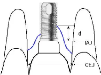
촬영된 사진은 슬라이드 입력용 scanner (Hewlet Packard Photosmart S20)를 사용하여 해상도 600dpi, 256 gray scale로 입력한 후, 개인용 컴퓨터 (Pentium 3, IBM호환)에서 각각의 방사선 사진을 디지털 이미지화 하였다. 측정 기준점은 임프란트 매식체와 지대주 원주의 연결부위로 결정하였으 며, 변연골의 높이는 임프란트-변연골 경계부의 흡수된 변연골 양상중 최 하방 기저부로 정하여 그 차이를 1024 X 768 pixel 해상도의 모니터 (15 inch LCD TFT)에서 Image tool (UTHSCSA V 3.0)을 이용하여 0.05mm
의 변연골 소실까지 계측하였다. 각 측정값은 근-원심부에서 측정하였으 며, 그 평균치를 선택하였다. 각 방사선상의 측정오차를 감안하여 임프란트 나사를 기준으로 방사선상과 실측비를 비교, 비례식으로 환산하여 실측값 을 계산하였고 (Figure 1), 상악 전치부군과 상악 구치부군의 보철물 장착 후 1년간의 변연골 흡수량을 비교하였다. 또한, 상악 전치부에서 골결손군 과 정상군의 보철물 장착후 1년간의 변연골 흡수량을 비교하였다.
Figure 1. Schematic drawing for the amount of marginal bone loss
IAJ : Implant Abutment Junction CEJ : Cemento Enamel Junction d : Amount of bone loss
4. 통계학적 분석
본 연구에서는 보철물 장착 1년 후 변연골소실량을 측정하여, 상악 전치 부와 구치부의 측정값의 유의차와, 골결손이 있는 경우와 정상군의 측정값 의 유의차를 통계프로그램 Eview 3에서 비모수 통계방법인 Wilcoxon Signed Ranks test로 검증하였다. 분석결과 얻어진 유의 확률값이 0.05 이 하일 경우 통계학적 유의성이 있는 것으로 판정하였다.
II. 연구결과
1. 치아 상실의 원인
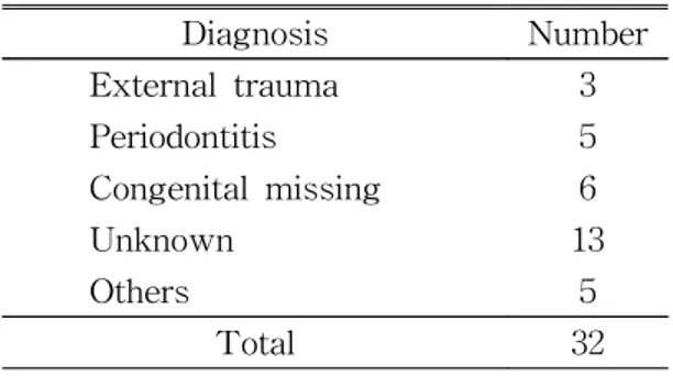
상악 전치부 치아 상실 원인별 분류시 외상이 61 %로 가장 높은 빈도로 나타났다 (Table 5-1). 상악 구치부의 치아 상실 원인은 선천적 결손, 심한 치주질환, 외상등 다양하게 나타났다 (Table 5-2).
Table 5-1. Reasons for upper anterior teeth replacement
Diagnosis Number External trauma 25 Periodontitis 2 Congenital missing 3 Unknown 8 Others 3 Total 41
Table 5-2. Reasons for upper posterior teeth replacement
Diagnosis Number External trauma 3 Periodontitis 5 Congenital missing 6 Unknown 13 Others 5 Total 32
2. 임프란트의 성공률
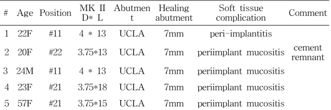
1년 성공률은 상악 전치부군에서 97.56 %, 상악 구치부군에서 93.75 % 였다. 보철물 장착 1년 후 상악 전치부군 에서는 1개의 임프란트에서 누공 을 동반한 임프란트 주위염이 발생하였고, 상악 구치부군에서 총 32개의 임프란트 중 2개가 보철물 장착 전 실패하였다. 2개 모두 2차 수술시 골유 착 실패로 제거하고 재수술하였다. 상악 전치부군에서 이후 정기 검진시 4 개의 임프란트에서 임프란트 주위 점막염이 발생하였고, 그 중 1개의 경우 는 잔존합착제로 인한 것이었다 (Table 6).3. 변연골 소실에 관한 방사선학적 고찰
변연골 소실량은 보철물 장착 1년 후 상악 전치부군에서 0.44 ± 0.25 mm, 상악 구치부군에서 0.57± 0.32 mm로 나타나, 통계학적으로 유의성 있 는 차이를 보였다 (P<0.05) (Table 7). 상악 전치부에서 보철물 장착후 1년간 열개와 천공의 골결손군은 0.40 ± 0.10 mm의 변연골소실을 보였고, 정상군은 0.48 ± 0.26 mm의 골소실을 보 였으며, 통계학적으로 유의성 있는 차이는 없었다 (P>0.05) (Table 8).Table 6. Description of complications # Age Position MK II D* L Abutmen t Healing abutment Soft tissue complication Comment 1 22F #11 4 * 13 UCLA 7mm peri-implantitis
2 20F #22 3.75*13 UCLA 7mm periimplant mucositis remnantcement 3 24M #11 4 * 13 UCLA 7mm periimplant mucositis
4 23F #21 3.75*18 UCLA 7mm periimplant mucositis 5 57F #21 3.75*15 UCLA 7mm periimplant mucositis
1. Chronic peri-implantitis with fistula was observed 1 year after delivery of crown.
2. 2 months after crown cementation with vitremer®, inflammation occurred due to cement remnant, which was removed by flap operation with vertical incision.
3. Peri-implant mucositis was observed 1 year after crown cementation. 4, 5. Peri-implant mucositis was observed after crown cementation.
Table 7. Marginal bone loss of upper anterior group and upper posterior group given as mean values ± SD
Marginal bone loss(mm)
Upper anterior Upper posterior 0.44± 0.25 0.57± 0.32
Table 8. Marginal bone loss of bone defect group and non-defect group given as mean values ± SD
Marginal bone loss(mm)
Bone defect Non-defect 0.40± 0.10 0.48± 0.26
IV. 총괄 및 고찰
임프란트 시술을 받은 완전무치악 환자의 평균연령은 Adell등(1990)의 연구에서 55.3세 인데 비해, 본 연구 대상인 상악 전치부의 단일 치아 임프 란트 시술을 받은 환자의 65%가 매식당시 나이는 30세 미만이었으며, 최 저 18세에서 최고령자는 66세이었고, 평균 29.8세였다. 전반적인 남녀 비율 은 비슷하게 나타났고, 30세 이하에서는 남자환자가 63% 이었다. 이는 Ekfeldt등(1994)의 연구결과와 유사하며 해당 연령층의 남자환자에서 외상 으로 인한 치아상실의 빈도가 높게 나타나는 것에 기인한다. 본 연구에서 도 상악 전치부 치아상실 원인별 분류시 외상이 61 %로 가장 높은 빈도로 나타났다. 상악 구치부의 단일 치아 임프란트 시술을 받은 환자의 평균나 이는 50.2세 이었다. 이는 상악 구치부의 치아 상실원인이 외상보다는 치주 질환이나 기타 다른 이유로 인한 치아상실이 더 많은 데 기인한다고 사료 된다. 본 연구에서 변연골 소실량은 보철물 장착 1년 후 상악 전치부군에서 0.44 ± 0.25 mm로, 상악 구치부군에서 0.57± 0.32 mm로 나타났다. 이것은 단일 치아 임프란트의 1년 후 변연골 소실에 관한 문헌 중 Jemt등(1993)의 0.8mm, Engquist등(1995)의 0.6mm, Malevez등(1996)의 0.8mm의 변연골 소실량 보다 유사하거나 적게 나타났으나, Scholander등(1999)의 0.15mm의 변연골 소실량보다 크게 나타났다. 상악 전치부와 상악 구치부의 변연골 소실량은 상악 구치부에서 더 크게 나타났으며, 통계적으로 유의한 차이가 있었다. 상악 구치부에서 변연골 흡수가 더 많이 일어난 것은 Schmitt등 (1993)에 의하면 상악 내에서도 전치부보다 구치부의 골질이 더 약하며 저 작력도 전치부에서 구치부로 갈수록 증가되기 때문이다. 이것은 1990년van Steenberghe등이 임프란트의 실패율은 골질과 상관관계가 있었다고 한 것과 유사하지만 반면, 1998년 Wyatt와 Zarb등은 상악과 하악, 전치부 와 구치부의 4부위로 구분하여 임프란트의 실패율을 비교한 결과 부위별 차이가 없었다고 하였다. 2002년 Naert등의 부분무치악 환자에서의 연구결 과, 지대주 연결 후 6개월 후 상악이 하악보다 골소실이 더 많이 일어났으 나, 이후 차이의 유의성이 없어졌다고 하였고, 전치부와 구치부 간의 골소 실차이는 유의성이 없었다고 보고하였다. 본 연구에서 보철물 장착후 1년간 열개와 천공의 골결손군은 0.40 ± 0.10 mm의 변연골소실을 나타냈으며 정상군은 0.48 ± 0.26 mm의 골소실 을 나타냈다. 골결손군과 정상군의 골소실량 차이는 통계적 유의성이 없었 다 (P>0.05). 2002년 Naert등은 부분 무치악에서 열개가 있는 경우 변연골 소실량이 더 크게 나타났다고 하였으나 통계적 유의성은 없다고 보고 하였 다. 열개와 천공의 골결손이 있는 경우 임프란트 매식시 차단막을 사용한 골유도 재생술식의 유용성은 여러 문헌상에서 보고 되었다 (Dahlin et al. 1988, Becker et al. 1990, Buser et al 1990). 1995년 Dahlin등은 열개와 천공 같은 국소적인 골결손부에서 골유도 재생술식을 사용하여 성공적인 신생골 형성을 보고 하였고 골량이 부족한 환자에서도 성공적인 임프란트 시술이 가능하다고 하였다. 그러나, 1999년 Lekholm등은 임프란트 매식시 협측 나사산이 1-5개 노출된 경우의 연구 결과, 기능 5년 후에도 연조직 문제가 발생되지 않았으며 골결손부의 점진적 골 흡수도 없었다고 하였다. 본 연구에서도 골결손군과 정상군의 골 소실량의 차이가 통계적 유의성이 없었다. 따라서 환자의 구강위생상태가 청결하게 유지될 수 있다면, 1차 수 술시 5개 이하의 협측 나사산이 노출된 경우 골유도 재생술시 발생할 수 있는 차단막 노출로 인한 감염 같은 합병증을 고려해 보았을 때 좀더 신중
한 기준 하에 차단막을 사용한 골유도 재생술 시술을 결정해야 된다고 사 료된다.
단일 치아 임프란트는 점진적으로 발전되고 있는 시술로 아직까지는 완전 무치악이나 부분 무치악 환자에서의 임프란트에 비해서 상대적으로 적은 수의 임프란트를 짧은 기간 동안 추시 관찰한 문헌들만 보고 되고 있다( Engquist et al. 1995 , Jemt et al. 1993, Haas et al. 2002 ). 또한, 단일 치 아 임프란트에만 적용되는 성공기준 자체가 없어서 무치악 환자에게 제시 되는 성공기준을 똑같이 적용하고 있다. 본 연구에서도 성공기준을 1986년 Albrektsson & Zarb가 발표한 임프란트의 성공기준에 준하여 조사하였다. 임프란트의 성공기준에서 연간 골소실이 0.2mm이하 이어야한다고 설정되 어 있는데, Avivi-Aber와 Zarb(1996)에 의하면 실제로 단일 치아 임프란트 의 경우 연간 골소실이 0.2mm에 훨씬 못 미치는 경우가 많으며, 이것은 양 옆에 인접자연치가 존재하기 때문에 완전 무치악 환자에서 나타나는 점진 적 골소실이 거의 발생하지 않기 때문이라고 하였다. 이전 문헌들에 의하면 단일 치아 임프란트의 성공률은 완전무치악이나 부분 무치악 환자보다 더 높게 보고 되고 있다 ( Andersson et al. 1998 , Avivi-Aber et al. 1996 , Cordioli et al. 1994 , Laney et al. 1994 , Wannfors et al 1999 ). 본 연구에 서 1년 성공률은 상악 전치부군에서 97.56 %, 상악 구치부군에서 93.75 % 였다. 상악 전치부의 높은 성공률은 Schmitt등(1993)에 의하면 다른 부위에 비해 골질이 양호하고 저작력이 적게 작용하기 때문이라고 하였다. 또한, 매 식된 임프란트는 대부분 양옆에 인접치아와 접해있어 Becker등(1995)에 의 하면 이들 인접치아가 임프란트에 과도한 저작력이 가해지는 것을 감소시 키며, 기능시 임프란트를 보호한다고 하였다. Albrektsson과 Zarb (1986)에 의하면 특정 임프란트 시스템이 성공적이
라고 평가되기 위해서는 최소한 5년 이상 최소한 2개 이상의 센터에서 최 소한 50명 이상의 환자를 대상으로 전향적인 연구가 시행되어야 한다고 하 였다. 따라서 상악 단일 치관 임프란트의 성공률을 도출해내기 위해서는 추가적인 기간의 연구가 더 필요하다. 본 연구에서 치료 후 합병증은 상악 전치부에서 합착제 용해로 인한 치 관 탈락이 22%로 가장 많이 발생하였다. 단일 치관 임프란트에 관한 Henry등(1996)의 연구에서는 초기 단일치관 임프란트 시술후 가장 빈번하 게 발생한 문제점이 지대주 원주 나사 풀림이었으나, CeraOne abutment시 술후 많이 개선되었다. 지대주 원주 나사풀림은 Estheticone® abutment를 사용했던 경우 1개에서 발생하여 지대주 원주를 교체하였다. 1986년 Jemt등은 단일 치아 임프란트 술식이 기존의 임프란트 개념을 치은연하로 변형시킨 것으로 수복물과 지대주 원주 사이의 변연을 치은연 상에 두는 기존 술식에 비해 치은건강에 불리하다고 하였다. 본 연구에서 도 상악 전치부의 경우 임프란트 주변 누공이나 임프란트 주위 점막염같은 연조직 문제가 기존의 고정성보철물로 수복한 임프란트에 비해 빈번하게 발생했다. 임프란트 주위 점막염이 발생한 경우 비외과적인 방법에 화학적 인 요법을 병행하여 치료하며, 이후에도 계속적으로 점막염이 지속된 경우 감염된 연조직을 외과적 방법으로 제거하는 것도 고려해야 한다. 그러나, 외과적으로 치은감염을 해결하는 경우 치은변연의 위치를 변경시키는 결과 를 초래할 수 있다. 본 연구에서는 지대주 원주 나사가 풀리지 않고 안정 적인 경우에서도 임프란트 주위 점막염이 발생한 경우를 관찰할 수 있었 다. 이것은 변연을 치은연하로 위치시키는 것 자체가 임프란트주변 조직을 자극하거나, 합착제가 완전히 제거되지 않은 경우와 연관되어 있었다. 1998 년 Abrahamsson등은 임프란트 주변 조직 부착에 지대주 원주의 종류가
미치는 영향에 관한 연구 결과, CP titanium이나 ceramic abutment에 비해 gold alloy abutment는 임프란트 주변 조직이 지대주 원주보다 더 하방의 매식체 주변에서 부착이 일어난다고 보고 하였다. 본 연구에서도 상악 전 치부에서 임프란트 주위 점막염이나 임프란트 주위염이 발생한 경우 UCLA abutment와 연관되 있었다. 본 연구는 machined Brånemark 임프란트를 사용한 상악 단일 치아 임 프란트를 대상으로 전치부와 구치부에서 보철물 장착 1년 후 변연골 소실 변화량의 차이와 성공률에 관해 알아보았고, 상악 전치부에서 골결손군과 정상군의 보철물 장착 1년 후 변연골 소실 변화량의 차이에 관해 비교해 보았다. 상악에서 단일 치아가 상실된 경우 해당 수여부의 상태를 고려한 적절한 길이, 직경의 임프란트를 적절한 치유기간을 주고 기능적 부하를 시킨 경우, 단일 치아 임프란트 시술은 인접치아의 손상없이 치아를 수복 할 수 있는 유용한 치료방법으로 사료되며, 상악 전치부와 구치부에서 단 일 치아 임프란트의 장기적인 성공률을 알아보기 위해 추후 계속적인 추시 검진이 필요하다고 사료된다.
V. 결 론
상악에서 2단계 술식으로 machined Brånemark 임프란트를 사용한 단 일 치아 임프란트 시술후 변연골 소실 변화량을 측정하여 전치부와 구치 부에서 변연골 소실량을 비교하고, 상악 전치부에서 열개, 천공의 골결손이 있는 경우와 골결손이 없는 경우의 변연골 소실량을 비교하기 위해, 1995 년 1월부터 상악 단일 치아 상실로 임프란트 매식 치료를 받은 환자 중 단 일 치관수복물을 장착한 71명을 대상으로 연구하여 다음과 같은 결과를 얻 었다. 1. 상악 전치부 치아상실 원인별 분류시 외상의 빈도가 61%로 가장 높 게 나타났으며, 상악 구치부 치아 상실 원인은 선천적 결손, 심한 치주질 환, 외상등 다양하게 나타났다. 2. 보철물 장착 1년 후 상악 전치부군에서는 총 41개 임프란트 중 1개에 서 누공을 동반한 임프란트주위염이 발생하였고, 상악 구치부군에서 총 32 개의 임프란트 중 2개가 실패하여, 1년 성공률은 상악 전치부군에서 97.56 %, 상악 구치부군에서 93.75 %였다. 3. 변연골 소실량은 보철물 장착 1년 후 상악 전치부군에서 0.44 ± 0.25 mm로, 상악 구치부군에서 0.57 ± 0.32 mm로 나타났으며, 통계학적으로 유 의성 있는 차이가 있었다 (P<0.05). 4. 상악 전치부에서 보철물 장착후 1년간 열개와 천공의 골결손군은 0.40± 0.10 mm의 변연골소실을 나타냈으며 정상군은 0.48 ± 0.26 mm의 골소 실을 나타냈으며, 통계학적으로 유의성 있는 차이는 없었다 (P>0.05 ). 이상의 결과에서 볼 때 상악 전치부가 구치부보다 변연골 소실이 더 적 게 나타났으며, 상악 단일 치아 상실시 해당 수여부의 상태를 고려한 적절 한 길이, 직경의 임프란트를 적절한 치유기간을 주고 기능적 부하를 시킨 경우, 단일 치아 임프란트 시술은 인접치아의 손상없이 치아를 수복할 수 있는 유용한 치료방법으로 사료된다.
참고문헌
Abrahamsson I, Berglundh T, Glantz PO, Lindhe J:The mucosal attachment at different abutments. An experimental study in dogs. J Clin Periodontol 25 (9): 721-727, 1998
Adell R, Lekholm U, Rockler B, Brånemark PI:A 15-year study of osseointegrated implants in the treatment of the edentulous jaw. Int J Oral Surg 10: 387-416, 1981
Adell R, Eriksson B, Lekholm U, Brånemark PI, Jemt T:A long term follow up study of osseointegrated implants in the treatment of totally edentulous jaws. Int J Oral Maillofac Implants 5 : 347-359, 1990
Albrektsson T, Zarb GA:Current interpretations of the osseointegrated response. Clinical Significance. Int J Prosthodont 6: 95-105, 1993 Andersson B, Ödman P, Lindvall AM:Single-tooth restorations on
osseointegrated implants:Results and experiences from a prospective study after 2-3years. Int J Oral Maxillofac Implants 10: 702-711, 1995
Andersson B, Odman P, Lindvall AM, Brånemark PI:Five year
prospective study of prosthodontic and surgical single-tooth implant treatment in general practice and at a specialist clinic. Int J Prosthodont 11: 351-355, 1998
Avivi-Aber L, Zarb GA:Clinical effectiveness of implant-supported single-tooth replacement. Int J Oral Maxillofac Implants 11:311-321,1996
Bahat O:Treatment Planning and placement of Implants in the posterior Maxilla:Report of 732 Consecutive Nobelpharma Implants. Int J Oral Maillofac Implants 8 : 151-161, 1993
Balshi TJ.:Candidates and Requirements for single tooth implant prosthesis. Int J Periodont Rest Dent 14: 317-331, 1994
Becker W, Becker BE, Handelsman M : Bone formation at dehisced dental implant sites treated with implant augmentation material : A pilot study in dogs. Int J Periodontics Restorative Dent 10: 92-101, 1990 Becker W, Becker BE : Replacement of maxillary and mandibular molars
with single endosseous implant restorations:A retrospective study. J Prosthetic Dent 74: 51-55, 1995
Book K, Karlsson S, Jemt T : Functional adaptation to full arch fixed prosthesis supported by osseointegrated implants in the edentulous mandible. Clin Oral Impl Res 3: 17-21, 1992
Brägger U : Use of radiographs in evaluating success, stability and failure in implant dentistry. Periodontology 2000 :17 : 77-88, 1998
Brånemark PI, Svensson B, van Steenberghe D : Ten-year survival rates of fixed prostheses on four or six implants ad modum Brånemark in full edentulism. Clin Oral Impl Res 6 : 227-231, 1995
Buser D, Brägger U, Lang NP, Nyman S : Regeneration and enlargement of jaw bone using guided tissue regeneration. Clin Oral Impl Res 1: 22-32, 1990
Cordioli G, Castagna S, Consolati E : Single-tooth Implant Rehabilitation. A Retrospective study of 67 implants . Int J Prosthodont 7:
525-531, 1994
Dahlin C, Lekholm U, Becker W, Becker B : Treatment of Fenestration and Dehiscence Bone defects around oral implants using the Guided Tissue Regeneration technique. A Prospective multicenter study. Int J Oral Maxillofac Implants 10: 312-318, 1995
Dahlin C, Linde A, Gottlow J, Nyman S : Healing of bone defects by
guided tissue regeneration. Plastic Reconstructive Surg 81(5):672-676, 1988
Danesh-Meyer MJ : Dental implants Part II : Guided Bone regeneration, Immediate implant placement, Peri-implantitis, Failing implants. J New Zealand Periodontol Nov 18-28, 1994
Ekfeldt, A., Carlsson, G.E. & Bӧrjesson, G.:Clinical evaluation of single- tooth restorations supported by osseointegrated implants:A retrospective study. Int J Oral Maxillofac Implants 9:179-183, 1994
Engquist B, Nilson H, Åstrand P.:Single tooth replacement by osseointegrated Brånemark implants. A retrospective study of 82 implants Clin Oral Impl Res 6: 238-245, 1995
Haas R, Polak C, Fürhauser R, Dörtbudak O, Watzek G : A long term follow up of 76 Brånemark single tooth implants . Clin Oral Impl Res 13:38-43, 2002
Hebel K, Gajjar RC : Cement-retained versus screw-retained implant restorations : Achieving optimal occlusion and esthetics in implant dentistry. J Prosthet Dent 77: 28-34, 1997
Henry PJ, Laney WR, Jemt T, Harris D, Krogh PHJ, Polizzi G, et al: Osseointegrated implants for single-tooth replacement:A prospective 5-year multicenter study. Int J Oral Maxillofac Implants 11: 450-455, 1996
Jemt T.:Modified single and short span restorations supported by osseointegrated fixtures in the partially edentulous jaw. J Prosthet Dent 55: 243-247, 1986
Jemt T, Lekholm U, Gröndahl K:A 3-year follow up study of early single implant restorations ad modum Brånemark. Int J Periodont Rest Dent 10:340-349, 1990
Jemt T, Pettersson P:A 3-year follow-up study on single implant treatment . J Dent 21 : 203-208, 1993
Jovanovic SA, Spiekermann H, Richter EJ : Bone regeneration around titanium dental implants in dehisced defect sites. A clinical study. Int J Maxillofac Implants 7: 233-245, 1992
Karlsson U, Gotfredsen K, Olsson C:Single tooth replacement by osseointegrated Astra Tech dental implants. A 2-year report. Int J Prosthodont 10: 318-324, 1997
Laney WR, Jemt T, Harris D, Henry PJ, Krogh PHJ, Polozzi G, Zarb GA, Herrmann I:Osseointegrated implants for single-tooth replacemen t:Progress report from a multicenter prospective study after 3 years. Int J Oral Maxillofac Implants 9: 49-54, 1994
Lekholm U, Gunne J, Henry P, Higuchi K, Steenberghe D: Survival of the Brånemark Implant in Partially Edentulous Jaws. A 10-year
Prospective Multicenter Study. Int J Oral Maxillofac Implants 14: 639-645, 1999
Lekholm U, Sennerby L, Roos J, Becker W : Soft tissue and Marginal bone conditions at osseointegrated implants that have exposed threads. A 5-year retrospective study. Int J Oral Maxillofac Implants 11: 599-604, 1996
Levine RA, Clem DS, Wilson TG, Saunders SL:A Multicenter Retrospective Analysis of the ITI Implant system used for single
tooth replacements. A preliminary results at 6 or more months of loading. Int J Oral Maxillofac Implants 12; 237-242, 1997
Lewis SG, Beumer J, Perri GR, Hornburg WP : Single tooth implant supported Restorations. Int J Oral Maxillofac Implants 3: 25-30, 1988
Mayer TM, Hawley CE, Gunsolley JC : The Single-tooth Implant : A viable alternative for single tooth replacement. J Periodontol 73: 687-693, 2002
Malevez CH, Hermans M, Daelemans PH:Marginal bone levels at Brånemark system implants used for single tooth restoration. The influence of implant design and anatomical region. Clin Oral Impl Res 7: 162-169, 1996
Naert I, Koutsikakis G, Quirynen M, Duyck I, van Steenberghe D, Jacobs R:Biologic outcome of implant-supported restorations in the treatment of partial edentulism. Part 2:A longitudinal radiographic evaluation. Clin Oral Impl Res 13: 390-395, 2002
Thilander B, Ödman J, Jemt T:Single implants in the upper incisor region and their relationship to the adjacent teeth. An 8 year follow up study. Clin Oral Impl Res 10: 346-355, 1999
Öhrnell LO, Hirsch J, Ericsson I, Brånemark PI:Single-tooth rehabilitation using osseointegration. A modified surgical and
prosthodontic appproach. Quint Int 19 : 871-876, 1988
Palmer RM, Floyd PD, Palmer PJ, Smith BJ, Johansson CB, Albrektsson T:Healing of implant dehiscence defects with and without expanded polytetrafluoroethylene membranes. A controlled clinical and histological study. Clin Oral Impl Res 5: 98-104, 1994
Priest G : Single tooth Implants and their role in preserving remaining teeth : A 10-year survival study. Int J Oral Maxillofac Implants 14:181-188
Roland M:How to treat ailing and failing implants. Implant Dent 1:25-33, 1992
Saadoun AP:Single tooth Peri-implant. Hard and soft tissue management for Aesthetic Results. Dental Implants, The art and science. Babbush CA 335-350
Scheller H, Urgell JP, Kultje C, Iven Klineberg. A 5-year multicenter study on Implant supported single crown restorations. Int J Oral Maxillofac Implants 13: 212-218, 1998
Scholander S:A retrospective evaluation of 259 single tooth replacement by the use of Brånemark Implants. Int J Prosthodont 12: 483-491, 1999
Schmitt A, Zarb G:The longitudinal clinical effectiveness of osseointegrated dental implants for single-tooth replacements. Int J Prosthodont 6: 197-202, 1993
Steven G, John Beumer III, George RP : Single tooth implant supported restorations. Int J Oral Maxillofac Implants 3: 25-30, 1988
Van Steenberghe D, Lekholm U, Bolender C, Fohmer T, Henry P,
Herrmann L:The applicability of osseointegrated oral implants in the rehabilitation of partial edentulism : A prospective multicenter study on 558 fixtures. Int J Oral Maxillofac Implants 5:272-281, 1990
Wannfors K, Smedberg JI : A prospective clinical evaluation of different single-tooth restoration designs on osseointegrated implants. A 3 year follow up of Brånemark implants. Clin Oral Implants Res 10: 453-458, 1999
Wyatt C, Zarb G:Treatment Outcome of Patients with implant-supported Fixed Partial Prostheses. Int J Oral Maxillofac Implants 13: 204- 211, 1998
Abstract
A Retrospective study on upper single tooth implants Soo-jin Jo, D.D.S.
Department of Dental Science, Graduate school, Yonsei University
(Directed by Associate Prof. Ik Sang Moon, D.D,S., M.S.D.,Ph.D.)
The aim of this retrospective study was to compare the amount of marginal bone loss between upper anterior area and upper posterior area with 71 upper single-tooth restorations on 2 stage machined Brånemark implants since Jan 1995. The second aim was to compare the bone defect group which had dehiscence and fenetration and the others in the upper anterior region. The results were as follows.
1. The most frequent reason of missing tooth in the upper anterior region was trauma by 61%. While upper posterior region showed various reasons such as congenital missing, advanced periodontitis, trauma. 2. Peri-implantitis with fistula occurred 1 of 41 implants in the upper anterior group in 1 year after loading and 2 of 32 implants in the upper posterior group failed before loading. The 1 year success rate of upper anterior group was 97.56 %, and 93.75 % for upper posterior group.
3. The mean marginal bone loss in the upper anterior group was 0.44 ± 0.25 mm, while 0.57± 0.32 mm in the upper posterior group. There was stastically significant difference in the amount of mean marginal bone loss (P<0.05).
4. The mean marginal bone loss of bone defect group was 0.40± 0.10 mm at one year, and 0.48± 0.26 mm for the control group. No statistically significant difference of mean marginal bone loss was showen between bone defect group and the others at implantation.
According to the results, the upper anterior region showed less marginal bone loss than the upper posterior region. In case of missing single upper tooth, careful consideration on recipient residual ridge to determine proper implant diameter and length, sufficient healing time, proper loading would lead to implant success. Single tooth implants in the maxilla seemed to be an alternative to fixed partial dentures without damage to adjacent teeth.
Key words:Upper anterior, Upper posterior, Single tooth implant, Marginal bone loss, Bone defect, Success rate, Retrospective study