Long-Term Comparison of Fixed-Bearing and
Mobile-Bearing Total Knee Replacements in Patients
Younger Than Fifty-one Years of Age with Osteoarthritis
Young-Hoo Kim, MD, Jun-Shik Kim, MD, Jin-Woo Choe, MD, and Hyoung-Jin Kim, MD
Investigation performed at the Joint Replacement Center of Korea, Ewha Womans University School of Medicine, Seoul, Republic of Korea
Background: There is limited information comparing the results of fixed-bearing total knee replacement and mobile-bearing total knee replacement in patients with osteoarthritis who are younger than fifty-one years and who have a fixed-bearing implant in one knee and a mobile-bearing implant in the other. The purpose of this study was to compare our long-term clinical and radiographic results of fixed-bearing total knee replacement and mobile-bearing total knee replacement in a group of patients from this population.
Methods: We prospectively compared the results of 108 patients with osteoarthritis who were younger than fifty-one years (mean age, forty-five years) who had received a fixed-bearing prosthesis in one knee and a rotating platform mobile-bearing prosthesis in the other. The mean follow-up was 16.8 years (range, fifteen to eighteen years). The patients were assessed clinically and radiographically. Knee motion and function were assessed as a primary outcome. Patients were assessed with questionnaires, and each knee was assessed separately.
Results: Although there was significant improvement in both groups of knees, there was no significant difference between the groups (i.e., fixed-bearing and mobile-bearing knees) with regard to the mean postoperative knee motion (126° and 128°, respectively; p = 0.79), the mean Knee Society knee clinical score (95 and 94 points, respectively; p = 0.79), or the Knee Society knee functional score (84 and 85 points, respectively; p = 0.19) at the latest follow-up. In the fixed-bearing group, one knee was revised because of infection, two for aseptic loosening of the tibial component, and two because of wear of the tibial polyethylene insert. In the rotating platform mobile-bearing group, two knees were revised because of instability and one because of infection. The Kaplan-Meier survivorship for revision at 16.8 years of follow-up was 95% (95% confidence interval, 91 to 100) for the fixed-bearing prosthesis and 97% (95% confidence interval, 93 to 100) for the rotating platform mobile-fixed-bearing prosthesis. Conclusions: Long-term results of both fixed and mobile-bearing total knee arthroplasties were encouraging in patients who were younger than fifty-one years of age with osteoarthritis. However, we found no superiority of the mobile-bearing total knee prosthesis over the fixed-bearing total knee prosthesis.
Level of Evidence: Therapeutic Level II. See Instructions for Authors for a complete description of levels of evidence.
A
lthough total knee arthroplasty is performed with in-creasing frequency in younger (i.e., fifty-five years of age or less) and more active patients, very few long-termstudies have been performed in this younger patient population1-4 . Most midterm and long-term studies on total knee arthroplasty in younger patients have included a relatively large percentage of Disclosure: None of the authors received payments or services, either
directly or indirectly (i.e., via his or her institution), from a third party in support of any aspect of this work. None of the authors, or their insti-tution(s), have had any financial relationship, in the thirty-six months prior to submission of this work, with any entity in the biomedical arena that could be perceived to influence or have the potential to influence what is written in this work. Also, no author has had any other rela-tionships, or has engaged in any other activities, that could be perceived to influence or have the potential to influence what is written in this work. The complete Disclosures of Potential Conflicts of Interest submitted by authors are always provided with the online version of the article.
A commentary by James A. Shaw, MD, is linked to the online version of this article at jbjs.org.
patients with rheumatoid arthritis who were somewhat inac-tive1-4
. There is relatively little information on cohorts of younger patients with osteoarthritis5-10
. Moreover, the majority of these studies on osteoarthritis were small, retrospective case series of fixed-bearing total knee arthroplasty performed before 1996— years in which the polyethylene was sterilized in air as opposed to in an inert environment. These data have limited applicability to contemporary fixed-bearing and mobile-bearing total knee im-plants with higher-quality polyethylene.
A mobile-bearing total knee prosthesis was introduced to minimize interface stresses between the implant and bone. One commonly stated reason for using a mobile-bearing total knee arthroplasty was that it allows younger patients to be more active and it reduces articular and backside wear of the tibial polyethylene bearing11
. Despite the theoretical advantages (i.e., less polyethylene wear and a lower rate of osteolysis) that have been purported to be associated with use of a mobile-bearing total knee prosthesis rather than a well-designed fixed-bearing total knee implant, these advantages have not been proven in young patients12-14
.
The purpose of this prospective randomized study was to evaluate the long-term clinical and radiographic results of
fixed-bearing total knee arthroplasty and mobile-bearing total knee arthroplasty in patients with osteoarthritis who were younger than fifty-one years of age and had a fixed-bearing implant in one knee and a mobile-bearing implant in the other.
Materials and Methods
Demographics
B
etween April 1993 and March 1996, we prospectively enrolled 114 patients (228 knees) who had bilateral primary tricompartmental osteoarthritis of the knee severe enough (Ahlb¨ack grade III to V)15to warrant simultaneous bilateral sequential total knee arthroplasties. All patients were younger than fifty-one years of age and agreed to participate in the study (Fig. 1). None of the 114 patients died during the interim, but six of the study patients (twelve knees) were lost to follow-up. Therefore, 108 patients (216 knees) were included in the study. The six patients did not have any associated problems when lost to follow-up. The study was registered in the ClinicalTrials.gov Protocol Regis-tration System (trial number, NCT01361152) and was approved by our institutional review board, and all patients provided informed consent. Randomization between the use of an anatomic modular (AMK; DePuy, Warsaw, Indiana) fixed-bearing prosthesis or low contact stress rotating plat-form (LCS RP; DePuy) mobile-bearing prosthesis was determined from a se-quential pool based on a table of randomized numbers. Each of the 108 patients received an AMK fixed-bearing total knee component on one side and an LCS mobile-bearing total knee component on the contralateral side. Eighty-threeFig. 1
patients (166 knees) were women, and twenty-five (fifty knees) were men. The mean age of the patients at the time of the operation was forty-five years (range, twenty-nine to fifty years). The mean height was 158 cm (range, 148 to 188 cm), and the mean weight was 64 kg (range, 50 to 88 kg). Thirty-six patients (33%) in the AMK group and twenty-eight (26%) in the LCS RP group had a prior arthroscopic debridement.
Prosthesis
The AMK prosthesis used in this study was a posterior cruciate-retaining de-sign, and the LCS RP prosthesis was a posterior cruciate-sacrificing design. Femoral and tibial components in both the AMK and LCS groups had a pol-ished cobalt-chromium articular surface (average roughness, <0.1 mm). The mean thickness of the tibial polyethylene insert was 12 ± 2 mm in both groups. The resin type of tibial polyethylene insert was 4150 resin. All tibial polyeth-ylene inserts were sterilized with gamma radiation in a vacuum in both groups. The mean shelf age of the tibial polyethylene insert was 0.7 years in both groups. Surgical Technique
All bilateral simultaneous sequential total knee arthroplasties were performed by the senior author (Y.-H.K.) on the same day. A pneumatic tourniquet at a pressure of 250 mm Hg was inflated after exsanguination of the limb with an Esmarch bandage. In all knees, an anterior midline skin incision (10 to 12 cm in length) was used, followed by a medial parapatellar capsular incision. In the knees with an AMK prosthesis, we prepared the femur first and then the tibia. In the knees with an LCS RP prosthesis, we prepared the tibia first and then the femur. The anterior cruciate ligament was excised and the posterior cruciate ligament was retained in the AMK group. Both anterior and posterior cruciate ligaments were excised in the LCS RP group. Ligament balancing was done, and an attempt was made in both groups to resect 10 mm of tibial bone to achieve a surface that was perpendicular to the shaft of the tibia in the coronal plane with 7° posterior slope in the sagittal plane. The distal part of the femur was resected with an attempt to achieve femorotibial alignment of 6° valgus in the coronal plane in both groups. All patellae were resurfaced with use of a polyethylene patellar prosthesis in both groups. All implants were cemented after pulsed lavage, drying, and pressurization of cement.
Rehabilitation
Starting with the second postoperative day, patients used a continuous passive-motion machine for passive range-of-passive-motion exercises twice daily for thirty minutes. Also on the second postoperative day, they started active range-of-motion exercises and began standing at the bedside or walking with the aid of crutches or a walker twice daily for thirty minutes each time under the su-pervision of a physical therapist. Patients used crutches or a walker with full weight-bearing for six weeks and used a cane when needed thereafter. Clinical Evaluation
Clinical evaluations were done at three months after the operation, at one year after the operation, and then yearly thereafter. The mean follow-up period was 16.8 years (range, fifteen to eighteen years). At the time of each follow-up, all clinical data were recorded and compiled by a clinical fellow (J.-W.C.) who was not part of the operative team and was blinded to allocation. We obtained the Knee Society knee score16, Hospital for Special Surgery knee score17, and Western Ontario and McMaster Universities Osteoarthritis Index (WOMAC) score18separately for each knee. We found that it was relatively easy for patients to identify the degree of pain in each knee. We inquired with regard to the degree of stiffness with use of the WOMAC instrument separately for each knee. We also asked about function separately for each knee, although it was difficult for patients to distinguish the function in each knee. However, they were able to distinguish the degree of impairment of function in each knee. The active range of motion of each knee, both with the patient in the supine position and while bearing weight, was determined with use of a standard (60 cm) clinical goni-ometer before the operation and at the time of the review. The patients were told to bend the knees as much as possible while lying in a supine position and
in a squatting position. The flexion contracture, flexion angles, and knee motion were measured for all patients on two occasions by two of the authors (Y.-H.K. and J.-S.K.), each of whom was blinded to the type of implanted prosthesis. When the measured knee motion was different between the two observers, the values were averaged and that number was reported19. The level of activity was assessed with use of the Tegner and Lysholm activity score20. Radiographic Evaluation
Anteroposterior radiographs with the patient standing (including the hip and the ankle) and with the patient lying supine, lateral radiographs, and skyline patellar radiographs were made preoperatively and at each follow-up visit and were assessed for alignment of the limb (tibiofemoral angle), position of the components, level of the joint line, and presence and location of radiolucent lines at the bone-cement interface according to the recommendations of the Knee Society16. Anteroposterior standing radiographs were used to determine any sequential change in the alignment of the limb that might be attributable to polyethylene wear and/or loosening of the implant. Supine anteroposterior radiographs were used to determine the presence of a radiolucent line more precisely. Skyline patellar radiographs were examined for the presence of pa-tellar tilt, subluxation, or dislocation. All radiographs were obtained under fluoroscopic guidance to control rotation of the knee. Radiographic data at the time of each follow-up were analyzed and recorded by another clinical fellow (J.-W.C.) who was not part of the operative team. However, this assessment was not blinded to allocation because the radiographic appearances of the two implants differ.
Computed Tomographic Scan Evaluation
At the latest follow-up, all patients underwent a computed tomography (CT) scan (with a multislice scanner) to determine the rotational alignment of the components and the presence of osteolysis. The scan sequence was between 10 cm above the superior pole of the patella and 10 cm below the tibial tuberosity, with use of contiguous 2.5-mm slices. Rotational alignment of the femoral com-ponent was determined by measuring the angle between the line joining the medial and lateral epicondyles of the femur and that joining the posterior margins of the femoral component. Rotational alignment of the tibial com-ponent was assessed by measuring the angle between the line connecting the tibial tuberosity anteriorly and the site of insertion of the posterior cruciate ligament posteriorly and the anteroposterior line passing although the center of the anterior and posterior margins of the tibial component. Osteolysis was defined as any nonlinear region of periprosthetic cancellous bone loss with delineable margins. One author (J.-W.C.) examined all CT scans. The intra-observer kappa statistic for the radiographic and CT examinations was 0.94. Statistical Analysis
An a priori power calculation was performed with use of a clinically relevant difference in range of motion of 5° and a standard deviation of 10°. For an effect size of 20% in functional outcome, as measured with use of a validated in-strument such as the linear analog scale assessment (in which a = 0.05 and b = 0.80), calculations revealed that ninety patients would be needed in each group. In addition to the required number of ninety subjects, twenty-four more pa-tients were recruited to allow for possible attrition. The changes in the Knee Society and Hospital for Special Surgery knee scores and WOMAC scores were evaluated with use of a paired t test. Range of motion of the knee was compared between the two groups with use of a paired t test, as were complication rates and radiographic data. All statistical analyses were performed with use of a two-tailed t test. The level of significance was set at p < 0.05. A survivorship analysis was performed to determine the cumulative rate of survival of the implant during the period of the study21and was reported with 95% confidence in-tervals (CIs). The end point for the analysis was aseptic loosening and revision surgery for any reason.
Source of Funding
Results
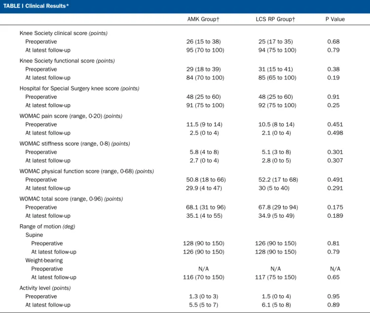
Clinical Results Knee Scores (Table I)
T
he Knee Society clinical score and Hospital for Special Surgery knee scores did not differ significantly between the two groups either preoperatively (p = 0.68 and 0.91, re-spectively; paired t test) or postoperatively (p = 0.79 and 0.25, respectively; paired t test). In the AMK group (fixed-bearing), the mean postoperative Knee Society clinical score was 95 points (range, 70 to 100 points) and the Hospital for Special Surgery score was 91 points (range, 75 to 100 points). In the LCS RP group (mobile-bearing), the mean postoperative Knee Society clinical score was 94 points (range, 75 to 100 points) and theHospital for Special Surgery score was 92 points (range, 75 to 100 points).
Range of Motion (Table I)
Preoperatively, the mean knee flexion contracture was 15° (range, 0° to 28 °) in the AMK group and 12° (range, 0° to 33°) in the LCS RP group. The mean preoperative range of motion in the supine position was 128° (range, 90° to 150°) in the AMK group and 126° (range, 90° to 150°) in the LCS RP group. This difference was not significant (p = 0.81, paired t test). At three months, no knee had a measurable flexion contracture. The mean range of motion in the supine position at the time of final follow-up was 126° (range, 90° to 150°) in the AMK group
TABLE I Clinical Results*
AMK Group† LCS RP Group† P Value
Knee Society clinical score (points)
Preoperative 26 (15 to 38) 25 (17 to 35) 0.68
At latest follow-up 95 (70 to 100) 94 (75 to 100) 0.79
Knee Society functional score (points)
Preoperative 29 (18 to 39) 31 (15 to 41) 0.38
At latest follow-up 84 (70 to 100) 85 (65 to 100) 0.19
Hospital for Special Surgery knee score (points)
Preoperative 48 (25 to 60) 48 (25 to 60) 0.91
At latest follow-up 91 (75 to 100) 92 (75 to 100) 0.25
WOMAC pain score (range, 0-20) (points)
Preoperative 11.5 (9 to 14) 10.5 (8 to 14) 0.451
At latest follow-up 2.5 (0 to 4) 2.1 (0 to 4) 0.498
WOMAC stiffness score (range, 0-8) (points)
Preoperative 5.8 (4 to 8) 5.1 (3 to 8) 0.301
At latest follow-up 2.7 (0 to 4) 2.8 (0 to 5) 0.307
WOMAC physical function score (range, 0-68) (points)
Preoperative 50.8 (18 to 66) 52.2 (17 to 68) 0.491
At latest follow-up 29.9 (4 to 47) 30 (5 to 40) 0.291
WOMAC total score (range, 0-96) (points)
Preoperative 68.1 (31 to 96) 67.8 (29 to 94) 0.175
At latest follow-up 35.1 (4 to 55) 34.9 (5 to 49) 0.189
Range of motion (deg) Supine
Preoperative 128 (90 to 150) 126 (90 to 150) 0.81
At latest follow-up 126 (90 to 150) 128 (90 to 150) 0.79
Weight-bearing
Preoperative N/A N/A N/A
At latest follow-up 116 (70 to 150) 117 (75 to 150) 0.65
Activity level (points)
Preoperative 1.3 (0 to 3) 1.5 (0 to 4) 0.95
At latest follow-up 5.5 (5 to 7) 6.1 (5 to 8) 0.89
*AMK= anatomic modular fixed-bearing, LCS RP = low contact stress rotating platform, WOMAC = Western Ontario and McMaster Universities Osteoarthritis questionnaire, and N/A= not available (preoperative weight-bearing range of knee motion was not measured due to pain). †The values are given as the mean and standard error of the mean.
and 128° (range, 90° to 150°) in the LCS RP group. This dif-ference was not significant (p = 0.79, paired t test).
WOMAC Score (Table I)
Preoperative WOMAC scores (mean score, 68.1 points [range, 31 to 96 points] in the AMK group and 67.8 points [range, 29 to 94 points] in the LCS RP group; p = 0.175) were improved signifi-cantly (mean score, 35.1 points [range, 4 to 55 points] in the AMK group and 34.9 points [range, 5 to 49 points] in the LCS RP group; p = 0.189) in both groups at the time of the latest follow-up. Activity Score (Table I)
The Tegner and Lysholm preoperative activity scores (mean score, 1.3 points [range, 0 to 3 points] in the AMK group and 1.5 points [range, 0 to 4 points] in the LCS RP group) were improved significantly (mean score, 5.5 points [range, 5 to 7 points] in the AMK group and 6.1 points [range, 5 to 8 points] in the LCS RP group) in both groups at the time of the latest follow-up. Radiographic Results
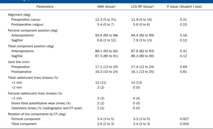
There were no significant differences between the two groups with regard to the alignment of the knee, the position of the femoral and tibial components in the coronal and sagittal planes, or the preoperative and postoperative joint line (p > 0.05 for all; paired t
test). The alignment of the knee was a mean of 5.4° of valgus in the AMK group and 5.6° of valgus in the LCS RP group. Twelve knees (11%) in the AMK group and fourteen knees (13%) in the LCS RP group had tibial radiolucent lines (<1 mm in width) in zone 1. Two knees (2%) in the AMK group had radiolucent lines (>2 mm in width) around the tibial component, and the component was loose. Three knees (3%) in the AMK group and four knees (4%) in the LCS RP group had femoral radiolucent lines (<1 mm in width) in zone 1. Two knees (2%) in the AMK group, but no knee (0%) in the LCS RP group, had gross tibial polyethylene insert wear. The CT scans showed no significant difference in the ex-ternal rotation of the femoral or tibial components of either de-sign. Radiographic and CT scans showed tibial osteolysis in two knees (2%) in the AMK group, but no tibial osteolysis in the LCS RP group. No knee in either group had femoral or patellar oste-olysis. No knee in either group had patellar dislocation, loosening, or patellar clunk syndrome (Table II).
Revision Operations
In the AMK group, five revisions (5%) were performed. One knee (1%) was revised because of infection, two (2%) for aseptic loosening of the tibial component, and two (2%) for wear of the polyethylene tibial insert. In the LCS RP group, three revisions (3%) were performed. Two knees (2%) were revised because of
TABLE II Radiographic Results*
Parameters AMK Group† LCS RP Group† P Value (Student t test)
Alignment (deg)
Preoperative (varus) 12.3 (5 to 21) 11.9 (0 to 15) 0.21
Postoperative (valgus) 5.4 (0 to 7) 5.6 (0 to 6) 0.15
Femoral component position (deg)
Anteroposterior 93.6 (85 to 98) 94.4 (90 to 99) 0.16
Sagittal 6.8 (2 to 12) 7.9 (3 to 13) 0.12
Tibial component position (deg)
Anteroposterior 88.1 (83 to 92) 87.6 (82 to 93) 0.31
Sagittal 87.5 (80 to 91) 86.3 (80 to 90) 0.12
Joint line (mm)
Preoperative 17.1 (13 to 25) 17.4 (12 to 24) 0.69
Postoperative 16.3 (10 to 24) 16.1 (13 to 25) 0.81
Tibial radiolucent lines (knees) (%)
<1 mm 12 (11) 14 (13)
>2 mm 2 (2) 0 (0)
Femoral radiolucent lines (knees) (%)
<1 mm 3 (3) 4 (4)
Gross tibial polyethylene wear (knee) (%) 2 (2) 0 (0) Osteolysis (knee) (%) (radiographic and CT scan) 2 (2) 0 (0) Rotation of the components by CT (deg)
Femoral component 3.4 (3 to 5) 3.3 (3 to 5) 0.927
Tibial component 2.6 (2 to 3) 2.4 (2 to 3) 0.916
*AMK= anatomic modular fixed bearing, LCS RP = low contact stress rotating platform, and CT = computed tomography. †The values are given as the mean and standard error of the mean.
instability of the knee, and one (1%) because of infection five years after surgery.
Survivorship Analysis
The Kaplan-Meier survivorship21
analysis of implants showed that the rate of survival was 95% for the AMK prosthesis (95% CI, 91 to 100) and 97% for the LCS RP prosthesis (95% CI, 93 to 100) at 16.8 years postoperatively when revision was defined as the end point. When aseptic loosening was defined as the end point, the survival rate was 98% in the AMK group and 100% in the LCS RP group (95% CI, 94 to 100 for both) at 16.8 years postoperatively (Fig. 2).
Discussion
T
his randomized prospective comparison of the results of fixed-bearing total knee arthroplasty and mobile-bearing rotating platform total knee arthroplasty in young patients was initiated to answer important questions about function, wear, osteolysis, and survivorship after total knee arthroplasty. We are not aware of any prior randomized prospective studies in which overall function and outcome following use of a mobile-bearing total knee arthroplasty were directly compared with the results associated with a fixed-bearing total knee arthroplasty inpatients with osteoarthritis younger than fifty-one years of age. In a longer-term randomized study comparing fixed-bearing and mobile-bearing total knee arthroplasties in elderly patients (mean age, 69.8 years), Kim et al. found no significant differ-ence between the two groups in terms of clinical and radio-graphic results, prevalence of wear of polyethylene tibial insert, osteolysis, revision rates, and survivorship22
. A multicenter ran-domized trial comparing fixed-bearing and mobile-bearing total knee arthroplasties in 250 knees followed for a maximum of two years23
and studies comparing a fixed-bearing design with a rotating-platform design in the same patients who had un-dergone a bilateral total knee arthroplasty24,25
have shown no significant difference in various outcome measures at the time of short-term follow-up (less than six years). Moreover, a single-center randomized trial comparing fixed (all-polyethylene tibial component) and mobile-bearing total knee arthroplasty designs in 312 knees followed for a minimum of two years26
showed no significant difference in various measures at the time of the short-term follow-up (less than four years).
Our study documented gratifying results of total knee arthroplasties in which either a fixed-bearing or a mobile-bearing prosthesis was used. No significant clinical advantage could be demonstrated between a fixed-bearing and a
mobile-Fig. 2-A Fig. 2-B
Fig. 2-C
Radiographs of both knees of a forty-five-year-old woman with primary three-compartmental osteoarthritis. Fig. 2-A Preoperative standing anteroposte-rior radiograph of both knees, demonstrating bone-on-bone contact between the medial femoral condyles and medial tibial plateaus. Fig. 2-B Antero-posterior radiograph of both knees at fifteen years after surgery, revealing that the anatomic modular fixed-bearing (left) knee prosthesis and the low contact stress rotating platform mobile-bearing (right) knee prosthesis are embedded rigidly. No radiolucent lines or osteolysis are demonstrated about the tibial components in either knee, and no gross wear of the polyethylene tibial insert is visualized in either knee. Fig. 2-C Lateral ra-diographs of the same knees show the absence of radiolucent lines or osteolysis around the femoral, tibial, and patellar components in either knee. (The radiograph of the left knee in Fig. 2-C has been flipped for the sake of better comparison.)
bearing prosthesis. We focused on good cementing technique with use of pulsed lavage and cement pressurization, correct flexion and extension gaps, and well-balanced ligaments to achieve a high success rate at 16.8 years after the operation.
Failure because of wear of the polyethylene tibial insert or osteolysis has been reported at very low rates in clinical series of contemporary fixed-bearing and mobile-bearing total knee ar-throplasties27-30
. Our findings of a low prevalence of osteolysis in both groups may be related to a preponderance of female patients, the use of a polished cobalt-chromium tibial baseplate to reduce backside wear of the insert, a polyethylene tibial insert sterilized by gamma radiation in a vacuum, and the short shelf life of the tibial insert. It is possible that the follow-up was not sufficiently long to reveal osteolysis. The concept that a mobile-bearing total knee prosthesis is associated with less wear and a lower prevalence of osteolysis than a well-designed fixed-bearing total knee pros-thesis remains to be proven in the longer-term follow-up.
A fixed-bearing total knee prosthesis cannot be fully con-forming without being exceedingly constrained to axial rotation, transferring large rotational stresses to the cement-bone interface. A mobile-bearing total knee prosthesis can overcome this conflict between conformity and axial constraint by allowing uncon-strained axial rotation with fully conforming articulations, thus reducing the axial stress to the cement-bone interface12,31
. Previ-ous studies of fixed-bearing total knee prostheses demonstrated that as conformity increased to minimize wear, theoretically more axial torque was applied to the cement-bone interface, which was liable to loosen the prosthesis32
. In our study, the prevalence of radiolucent lines around the tibial component was 13% (fourteen knees) in both groups. Two knees in the AMK fixed-bearing group but none in the LCS RP mobile-bearing group had a complete radiolucent line wider than 2 mm around the tibial component. Therefore, axial stress to the cement-bone interface of the tibial component appeared to be equivalent in both groups. In our series, we were able to use a mobile-bearing total knee prosthesis in every primary total knee arthroplasty selected by the process of randomization, irrespective of the range of knee deformity and the range of instability.
Although total knee arthroplasty is well accepted for pa-tients who are older than sixty-five years, its use is controversial for younger patients. Duffy et al.2
reported a 95% survival rate at fifteen years in patients who were younger than fifty-five years,
and, in a similar age group, Diduch et al.5
reported an 87% survival rate at eighteen years, with use of revision as an end point. Dixon et al.33
reported a 92.6% survival rate at fifteen years in patients with an average age of sixty-seven years, using revi-sion as an end point. Dalury et al.34found a slight age-related difference. They reported that the ten-year survival rate was 97.5% for patients who were younger than fifty-five years and 98.1% for older patients. They concluded that the survival rate was slightly lower in the younger group. Kim et al.30
reported that the survivorship of fixed-bearing total knee prostheses was 97.1% at 12.6 years in patients with an average age of 58.6 years and that the survivorship of mobile-bearing total knee pros-theses was 97.1% at 14.1 years in patients with an average age of 55.7 years. Our survival rates are substantially better than the reported survival rates for total knee arthroplasty in younger patients.
The main strength of our study is the large number of patients from a single institution and the relatively long follow-up. The main limitation was the dissimilarity of the prostheses, in that the geometry of the femoral, tibial, and patellar com-ponent differed, one implant was a posterior cruciate-retaining knee and the other was a posterior cruciate-substituting knee, and one prosthesis was a fixed-bearing total knee implant and the other was a mobile-bearing total knee implant.
In conclusion, our study demonstrated that the clinical and radiographic results of both fixed-bearing and mobile-bearing total knee implants were encouraging over a long-term follow-up period in patients younger than fifty-one years of age with osteoarthritis. However, we found no evidence to prove the superiority of the mobile-bearing total knee implant over the fixed-bearing total knee implant.n
Young-Hoo Kim, MD Jun-Shik Kim, MD Jin-Woo Choe, MD Hyoung-Jin Kim, MD
The Joint Replacement Center of Korea at Ewha
Womans University MokDong Hospital, 911-1, MokDong, YangChun-Ku, Seoul, Republic of Korea (158-710). E-mail address for Y.-H. Kim: [email protected]
References
1. Crowder AR, Duffy GP, Trousdale RT. Long-term results of total knee arthroplasty in young patients with rheumatoid arthritis. J Arthroplasty. 2005;20(7 Suppl3):12-6. 2. Duffy GP, Trousdale RT, Stuart MJ. Total knee arthroplasty in patients 55 years old or younger. 10- to 17-year results. Clin Orthop Relat Res. 1998;356:22-7. 3. Gill GS, Chan KC, Mills DM. 5- to 18-year follow-up study of cemented total knee arthroplasty for patients 55 years old or younger. J Arthroplasty. 1997;12:49-54. 4. Hofmann AA, Heithoff SM, Camargo M. Cementless total knee arthroplasty in patients 50 years or younger. Clin Orthop Relat Res. 2002;404:102-7. 5. Diduch DR, Insall JN, Scott WN, Scuderi GR, Font-Rodriguez D. Total knee re-placement in young, active patients. Long-term follow-up and functional outcome. J Bone Joint Surg Am. 1997;79:575-82.
6. Lonner JH, Hershman S, Mont M, Lotke PA. Total knee arthroplasty in patients 40 years of age and younger with osteoarthritis. Clin Orthop Relat Res. 2000;380:85-90. 7. Mont MA, Lee CW, Sheldon M, Lennon WC, Hungerford DS. Total knee arthro-plasty in patients </=50 years old. J Arthroarthro-plasty. 2002;17:538-43.
8. Stern SH, Bowen MK, Insall JN, Scuderi GR. Cemented total knee arthroplasty for gonarthrosis in patients 55 years old or younger. Clin Orthop Relat Res. 1990;260:124-9. 9. Tai CC, Cross MJ. Five- to 12-year follow-up of a hydroxyapatite-coated, cement-less total knee replacement in young, active patients. J Bone Joint Surg Br. 2006;88: 1158-63.
10. Duffy GP, Crowder AR, Trousdale RR, Berry DJ. Cemented total knee arthroplasty using a modern prosthesis in young patients with osteoarthritis. J Arthroplasty. 2007;22(6 Suppl 2):67-70.
11. Insall JN. Adventures in mobile-bearing knee design: a mid-life crisis. Orthope-dics. 1998;21:1021-3.
12. Buechel FF, Pappas MJ. Long-term survivorship analysis of cruciate-sparing versus cruciate-sacrificing knee prostheses using meniscal bearings. Clin Orthop Relat Res. 1990;260:162-9.
13. Jordan LR, Olivo JL, Voorhorst PE. Survivorship analysis of cementless meniscal bearing total knee arthroplasty. Clin Orthop Relat Res. 1997;388:119-23.
14. Ritter MA, Campbell E, Faris PM, Keating EM. Long-term survival analysis of the posterior cruciate condylar total knee arthroplasty. A 10-year evaluation. J Arthro-plasty. 1989;4:293-6.
15. Ahlb¨ack S. Osteoarthrosis of the knee. A radiographic investigation. Acta Radiol
Diagn (Stockh). 1968;Suppl 277:7-72.
16. Insall JN, Dorr LD, Scott RD, Scott WN. Rationale of the Knee Society clinical rating system. Clin Orthop Relat Res. 1989;248:13-4.
17. Insall JN, Ranawat CS, Aglietti P, Shine J. A comparison of four models of total knee-replacement prostheses. J Bone Joint Surg Am. 1976;58:754-65.
18. Bellamy N, Buchanan WW, Goldsmith CH, Campbell J, Stitt LW. Validation study of WOMAC: a health status instrument for measuring clinically important patient relevant outcomes to antirheumatic drug therapy in patients with osteoarthritis of the hip or knee. J Rheumatol. 1988;15:1833-40.
19. Kim YH, Choi Y, Kwon OR, Kim JS. Functional outcome and range of motion of high-flexion posterior cruciate-retaining and high-flexion posterior cruciate-substituting total knee prostheses. A prospective, randomized study. J Bone Joint Surg Am. 2009; 91:753-60.
20. Tegner Y, Lysholm J. Rating systems in the evaluation of knee ligament injuries. Clin Orthop Relat Res. 1985;198:43-9.
21. Kaplan EL, Meier P. Nonparametric estimation from incomplete observations. J Am Statist Assoc. 1958;53:457-81.
22. Kim YH, Yoon SH, Kim JS. The long-term results of simultaneous fixed-bearing and mobile-bearing total knee replacements performed in the same patient. J Bone Joint Surg Br. 2007;89:1317-23.
23. Wylde V, Learmonth I, Potter A, Bettinson K, Lingard E. Patient-reported out-comes after fixed- versus mobile-bearing total knee replacement: a multi-centre randomised controlled trial using the Kinemax total knee replacement. J Bone Joint Surg Br. 2008;90:1172-9.
24. Ranawat AS, Rossi R, Loreti I, Rasquinha VJ, Rodriguez JA, Ranawat CS. Comparison of the PFC Sigma fixed-bearing and rotating-platform total knee arthro-plasty in the same patient: short-term results. J Arthroarthro-plasty. 2004;19:35-9. 25. Bhan S, Malhotra R, Kiran EK, Shukla S, Bijjawara M. A comparison of fixed-bearing and mobile-fixed-bearing total knee arthroplasty at a minimum follow-up of 4.5 years. J Bone Joint Surg Am. 2005;87:2290-6.
26. Gioe TJ, Glynn J, Sembrano J, Suthers K, Santos ER, Singh J. Mobile and fixed-bearing (all-polyethylene tibial component) total knee arthroplasty designs. A pro-spective randomized trial. J Bone Joint Surg Am. 2009;91:2104-12.
27. Kim YH, Kook HK, Kim JS. Comparison of fixed-bearing and mobile-bearing total knee arthroplasties. Clin Orthop Relat Res. 2001;392:101-15.
28. Price AJ, Rees JL, Beard D, Juszczak E, Carter S, White S, de Steiger R, Dodd CA, Gibbons M, McLardy-Smith P, Goodfellow JW, Murray DW. A mobile-bearing total knee prosthesis compared with a fixed-bearing prosthesis. A multicentre single-blind randomised controlled trial. J Bone Joint Surg Br. 2003;85:62-7.
29. Woolson ST, Northrop GD. Mobile- vs. fixed-bearing total knee arthroplasty: a clinical and radiologic study. J Arthroplasty. 2004;19:135-40.
30. Kim YH, Choi Y, Kim JS. Osteolysis in well-functioning fixed- and mobile-bearing TKAs in younger patients. Clin Orthop Relat Res. 2010;468:3084-93.
31. Buechel FF Sr, Buechel FF Jr, Pappas MJ, Dalessio J. Twenty-year evaluation of the New Jersey LCS Rotating Platform Knee Replacement. J Knee Surg. 2002;15:84-9. 32. Werner F, Foster D, Murray DG. The influence of design on the transmission of torque across knee prostheses. J Bone Joint Surg Am. 1978;60:342-8.
33. Dixon MC, Brown RR, Parsch D, Scott RD. Modular fixed-bearing total knee arthroplasty with retention of the posterior cruciate ligament. A study of patients followed for a minimum of fifteen years. J Bone Joint Surg Am. 2005;87:598-603. 34. Dalury DF, Barrett WP, Mason JB, Goldstein WM, Murphy JA, Roche MW. Mid-term survival of a contemporary modular total knee replacement: a multicentre study of 1970 knees. J Bone Joint Surg Br. 2008;90:1594-6.