서
론
PICA(posterior inferior cerebellar artery) 동맥류는 두개강 내 동맥류 중 약 0.49에서 3%정도로 비교적 드물 다.8)13) 이 중에서도 대부분은 VA-PICA(vertebral artery-posterior inferior cerebellar artery)의 인접부위에서 발
원위부 후하소뇌동맥 뇌동맥류의 임상분석
연세대학교 의과대학 신경외과, 영상의학과
백인현∙박근영∙이재환∙허승곤∙김동준
*∙김동익
*∙이규창
Clinical Analysis of Distal Posterior Inferior Cerebellar Artery Aneurysm
In Hyun Baek, MD, Keun Young Park, MD, Jae Whan Lee, MD,Seung Kon Huh, MD, Dong Joon Kim, MD*, Dong Ik Kim, MD*, Kyu Chang Lee, MD Department of Neurosurgery and Diagnostic Radiology*, Yonsei University College of Medicine
ABSTRACT
Objective : This study was designed to determine the clinical characteristics of patients with aneurysms that are located at the distal posterior inferior cerebellar artery (dPICA). Patients & Methods : From September 1976 to June 2007, 54 consecutive patients with PICA aneurysms were treated at our institute. Among them, 19 patients had PICA aneurysms distal to the junction of the vertebral artery-PICA. We retrospectively reviewed the database and imaging studies as sources of information for analysis. Results : Five patients were male and 14 patients were female. The mean age was 44.6 years old (range: 23-70). Sixteen patients had ruptured lesions: 1 patient was Hunt and Hess Grade I, 4 were Grade II, 5 were Grade III, 4 were Grade IV and 2 were Grade V. Intraventricular hemorrhage or intracerebral hemorrhage was identified in 5 patients on the initial computed tomography (CT). Three patients had unruptured lesions. The locations of aneurysm were the lateral medullary segment in 10 patients, the tonsillomedullary segment in 1 patient, the telovelotonsillar segment in 5 patients and the cortical segment in 3 patients. Most aneurysms (17) were the saccular shape. Seventeen aneurysms were small and 2 were large or giant. The mean diameter of aneurysm was 6.5 mm (range: 2.0-28.0) and the mean diameter of the ruptured aneurysm was 4.8 mm (range: 2.0-12.0). Two patients had mirror aneurysms. Post-hemorrhagic hydrocephalus was identified in 10 patients on the initial CT and shunt surgery was performed on 3 patients. The obliteration methods of the aneurysms were microsurgery in 15 patients (midline suboccipital approach: 9, lateral suboccipital approach: 6) and endovascular surgery in 4 patients (therapeutic distal PICA occlusion: 3, intra-aneurysmal coiling: 1). Early surgery was performed on 2 patients, intermediate surgery (days between rupture: 4-10) was performed on 4 patients and delayed surgery was performed on 10 patients. The mean post-treatment follow up period was 49.5 months (range: 7-156). The clinical outcome was assessed using the modified Glasgow Outcome Scale. All the patients showed favorable outcomes. Five patients suffered from treatment-related complications (a CSF collection requiring wound revision for dura repair: 2, shunt surgery: 1 and transient hemiparesis due to impairment of the blood flow distal to the aneurysm: 2).Conclusions : In our series, distal PICA aneurysms had the characteristics of a female predominance, they more often presented with intraventricular hemorrhage and the rupture was of a relatively small size. Both microsurgery and endovascular surgery can be troublesome due to the small size, wide neck and tortuosity of the proximal parent artery and the location of aneurysms at a branching site. The surgeons should be careful for preserving vessel patency and insuring watertight dura repair. (Kor J Cerebrovascular Surgery 10(3):465-472, 2008)
KEY WORDS : Intracranial aneurysm∙Posterior Inferior Cerebellar artery∙Aneurysm surgery∙Endovascular treatment
논문접수일 : 2008년07월 31일 심사완료일 : 2008년08월 22일 교신저자 : 이재환, 120-752 서울시 서대문구 신촌동 134 연세대학교 의과대학 신경외과학교실 전화 : (02) 2228-2150�전송 : (02) 393-9979 E-mail : leejw@yuhs.ac * 본 논문의 요지는 2008년 춘계신경외과학회에서 구연되었음.
Table 1.
Clinical features of 19 patients with distal PICA
*
aneurysm
Patients
Aneurysm
Associated pathological condition
Case Age Sex FG* Clinical Location � Site§ Rupture Pathological Size Aneurysm Associated Hydrocephalus ICH* Treatment Complication Outcome$ No* (yrs) Grade� type (mm) No* aneurysm 1 1 2 3 m 4 3 1 R ruptured saccular 4 1 Moderate -Lat. Suboccipital good 1 2 3 0 m 3 4 1 R ruptured saccular 5 1 Mild -Lat. Suboccipital good 1 3 4 7 f 3 3 1 R ruptured saccular 2 1 Mild -Mid. Suboccipital good 1 4 5 1 f 4 5 1 R ruptured saccular 4 3 Rt.SCA Marked Yes Endo occlusion good Lt.PICA 1 5 4 5 f 2 3 1 L ruptured saccular 5 1 Marked -Coiling, GDC good 1 6 2 7 m 2 2 1 R ruptured saccular 4 1 -Endo occlusion good 1 7 7 0 f 3 2 1 R ruptured saccular 4 1 Mild -Lat. Suboccipital good 1 8 6 4 f 3 3 1 L ruptured saccular 3 1 -Lat. Suboccipital good 1 9 5 9 f 3 4 1 R ruptured saccular 5 1 Mild -Lat. Suboccipital good 10 50 m 3 2 1 L ruptured saccular 3 1 -Lat. Suboccipital C SF* collection good 11 70 f 4 4 2 L ruptured saccular 3 2 Rt.PICA Mild -Mid. Suboccipital CSF collection good 12 43 m 1 1 3 L ruptured fusiform 6 1 -Mid. Suboccipital good 13 40 f 1 2 3 R ruptured saccular 5 1 -Mid. Suboccipital good 14 47 f 4 3 3 L ruptured saccular 3 1 Mild -Mid. Suboccipital good 15 52 f -0 3 L unruptured saccular 3 1 -Mid. Suboccipital good 16 28 f -0 3 L unruptured saccular 4 1 -Mid. Suboccipital CSF collection fair 17 57 f 4 4 4 R ruptured saccular 3 1 Mild -Mid. Suboccipital good 18 51 f 3 5 4 R ruptured dissecting -2 -Endo occlusion good 19 37 f -0 4 L unruptured saccular 25 1 -Mid. Suboccipital good
* PICA = posterior inferior cerebellar artery; No = number; FG
= Fisher grade on brain computed tomography; ICH = intracerebra
l hemorrhage; CSF = cerebrospinal fluid.
� Hunt and Hess grade: 0 = unruptured; 1 = asymptomatic or mild headache and slightly nuchal rigidity; 2 = cranial nerve palsy, moderate-to-severe headache, nuchal rigidity; 3
= mild focal deficit, lethargy, confusion; 4 = stupor, moderate
d-to-severe hemiparesis, early decerabrate rigidity; 5 = deep c
oma, decerebrate rigidity, moribund appearance.
�
Aneurysm location: 1 = lateral medullary segement; 2 = tonsill
omedullary segement; 3 = telovelotonsillar segement; 4 = cortic
al segment
§
Site: R = right, L = left.
Treatment: lat. suboccipital = l ateral suboccipital craniotomy and clipping; m id. suboccipital = m idline suboccipital cranioto my and clipping; endo = Endovascular; GDC =
Guglielimi detachable coil. $ Outcome
at last visit; excellent = no deficit/back to premorb id condition; good = cranial nerve deficit or mild neurological deficit but able to work; fair = cranial nerve deficit or
mild neurological deficit, independent but unable to work; poor
생하는 근위부 PICA(pPICA) 동맥류이다. VA와 인접하지 않 은 말단부에서 일어나는 경우를 원위부 PICA(dPICA) 동맥류 라고 하며, 이는 pPICA동맥류 보다는 적은 분포를 보이며 0.28~1.4%의 발생률로 보고되고 있어7)8)12)15) 임상적인 특징 및 표준화된 치료 방법의 기준이 명확하지가 않다. 본 논문에서는 지금까지 본원에서 치료된 dPICA 동맥류 및 환자의 임상적인 특징을 확인하고, pPICA 동맥류와의 차 이를 분석하고자 한다.
대상 및 방법
본원에서 1976년 9월부터 2007년 6월까지 치료받은 3380 명의 환자 중에 PICA동맥류로 치료를 받은 54명의 환자를 대상으로 하였다. pPICA군은 VA-PICA이행부위와 anterior medullary 분절에서 기시하는 동맥류를 가진 환자 를 대상으로 하였고 35명이 포함되었다. dPICA군은 lateral medullary 분절 이후에 동맥류를 가진 환자를 대상으로 19 명이 포함되었다. 연구에 대상으로 선택된 환자의 의무기록, 방사선학적 영상, 수술 중의 소견 및 데이터베이스를 후향적 으로 검토하였으며, 수술 후로부터 평균 추적기간은 49.5개 월(7~156개월)이었다. PICA 동맥류를 가진 모든 환자는 치료 를 받기 전에 뇌지주막하 출혈의 진단을 위하여 뇌 전산화단층 촬영(CT, computed tomography)이 시행되었고, 뇌동맥류의 위치, 크기 및 모양을 확인하기 위하여 뇌혈관조영술이 이루어 졌다. PICA 동맥류의 위치는 Hudgins가 보고한 분류를 통하여 anterior medullary, lateral medullary, tonsillomedullary, telovelotonsillar 및 cortical 분절로 나누었다.8)진단 당시 시행된 뇌 CT를 통하여 뇌지주막하 출혈의 Fisher 등급을 분류 하였으며, 동반된 뇌내출혈, 뇌실출혈, 수두증의 여부를 확인 하였다. 임상적 결과는 치료 후 6개월이 되는 시점에서 modified Glasgow Outcome Scale(mGOS)로9) 평가하였
다. dPICA 군과 pPICA 군을 비교 분석하기 위하여 나이와 뇌동맥류 크기의 비교분석에 스튜던트 T 검정(Student T test)을 사용하였고, 동맥류의 위치와 진단 당시 CT에서 수두 증은 카이제곱 검정(Chisquare test)을 사용하였다. 그리고, 성별, 동맥류 파열, 동반 동맥류, 치료의 방법 및 시술 후 합 병증은 피셔 검정(Fisher’s exact test)를 사용하였다. 또한 진단 당시 CT에서 Fisher 등급, Hunt-Hess 등급 및 시술에 대한 결과는 Mann-Whitney U 검정을 시행하였다. 각 분석 에서 유의도(probability; p)는 0.05미만을 통계적으로 의미 가 있는 것으로 해석하였다.
결
과
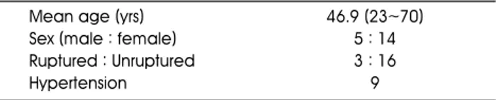
1976년 9월부터 2007년 6월까지 3380례의 동맥류를 가진 환자에서 PICA 동맥류는 54례(1.63%)로 이 중에 19례가 dPICA 동맥류이었으며 전체 동맥류의 0.56%를 보였다 (Table 1). 여성이 15례로 4례인 남성 보다 많았으며 평균 연Table 2. Characteristics of the patients with distal PICA�
aneurysm
Mean age (yrs) 46.9 (23~70)
Sex (male : female) 5 : 14
Ruptured : Unruptured 3 : 16
Hypertension 9
* PICA = posterior inferior cerebellar artery
Table 4. Characteristics of the distal PICA�aneurysm Location
Location lateral medullary 10 tonsillomedullary 1 televelotonsillar 5 cortical 3 Shape saccular 17 fusiform 1 dissecting 1 Site left 9 right 10 Size small 17 large 1 giant 1
* PICA = posterior inferior cerebellar artery
Table 5. Obliteration methods of aneurysm
Microsurgery 15
clipping 12
wrapping and clipping 3
Endovascular surgery 4
therapeutic distal PICA occlusion 3 intra-aneurysmal coiling 1 Table 3. The relationship between HHG and FG
HHG/FG UR 1 2 3 4 Total UR 3 3 1 1 1 2 1 1 2 4 3 1 2 2 5 4 2 2 4 5 1 1 2 Total 3 2 2 7 5 19
* HHG = Hunt and Hess grade; FG = Fisher grade; UR = unruptured aneurysm
령은 44.6세(23~70세)였으며, dPICA 동맥류 중에서 16례는 파열 되었고, 3례는 비파열 동맥류였다(Table 2). 진단 당시 Fisher 등급 3이 파열된 dPICA 환자 중 7례(36.8%)로 제일 많았으며 Hunt-Hess 등급은 등급 3이 5례로 가장 많았다 (Table 3).
PICA 분절에서 dPICA 동맥류의 분포하는 부위는 Table 4에서 보듯이 lateral medullay 분절에서에서 가장 많았다. 동맥류의 모양은 낭성(saccular) 형태가 17례로 가장 많았고, 박리(dissection) 형태도 1례에서 있었다. 동맥류의 크기는 작은 동맥류가 17례였고, 거대동맥류도 1례에서 볼 수 있었다 (Table 4). 진단 당시 CT 촬영에서 수두증이 동반된 경우는 9례가 있었고, 뇌출혈을 동반한 경우도 1례에서 볼 수 있었다 (Table 1). 동맥류가 진단이 되고 수술까지 들어가는 데 걸린 날을 보면 2일 이전에 수술을 한 경우는 2례에서 볼 수 있지 만, 10일 이후에 수술을 한 경우는 10례에서 볼 수 있었다. 수 술은 정중앙후두하접근법을 통한 경우와 측방후두부하접근을 한 경우가 각각 9례 및 6례였고, 중재적시술을 한 경우는 4례 였다(Table 5). dPICA 동맥류에 대한 수술의 합병증은 수술 후 뇌척수액고 임증이 3례에서 있었고, 일시적인 편마비가 2례에서 있었다. 수술 후에 지속적인 수두증으로 뇌실복강내 단락술은 3례에서 시행하였으며 치료에 대한 결과는 18례에서 호전되었다.
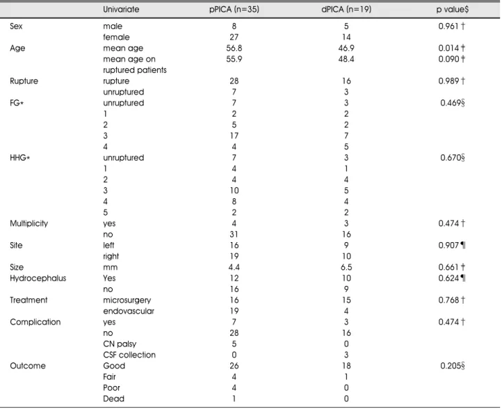
pPICA 군은 전체 35례로 dPICA 군보다 많았다(Table 6). dPICA 군은 pPICA 군보다 어린 나이에서 발생이 되었고
Table 6. Univariate analysis between pPICA and dPICA*
Univariate pPICA (n=35) dPICA (n=19) p value$
Sex male 8 5 0.961�
female 27 14
Age mean age 56.8 46.9 0.014�
mean age on 55.9 48.4 0.090� ruptured patients Rupture rupture 28 16 0.989� unruptured 7 3 FG* unruptured 7 3 0.469§ 1 2 2 2 5 2 3 17 7 4 4 5 HHG* unruptured 7 3 0.670§ 1 4 1 2 4 4 3 10 5 4 8 4 5 2 2 Multiplicity yes 4 3 0.474� no 31 16 Site left 16 9 0.907¶ right 19 10 Size mm 4.4 6.5 0.661� Hydrocephalus Yes 12 10 0.624¶ no 16 9 Treatment microsurgery 16 15 0.768� endovascular 19 4 Complication yes 7 3 0.474� no 28 16 CN palsy 5 0 CSF collection 0 3 Outcome Good 26 18 0.205§ Fair 4 1 Poor 4 0 Dead 1 0
* pPICA = proximal posterior inferior cerebellar artery aneurysm group; dPICA = distal posterior inferior cerebellar artery aneurysm group; FG = Fisher grade; HHG = Hunt and Hess grade
� Fisher`s exaxt test, � Student T test, §Mann-Whitney U test ¶ Chi-square test, $ probability < 0.05
(p=0.014), Fisher 등급 및 Hunt-Hess 등급은 dPICA 군에 서 통계적인 차이를 보이지는 않았다. 그리고 성별, 파열의 유 무, 동반된 뇌동맥류, 크기, 수두증의 동반여부, 치료방법, 합 병증 발생률 및 치료결과에서는 두 군간의 통계적 차이를 보 이지는 않았다. 하지만, pPICA 군은 수술 후에 2례에서 뇌신 경마비가 발생을 하였다. 반면에 dPICA군은 3례에서 수술 후 뇌척수액고임증이 발생하였다.
증
례
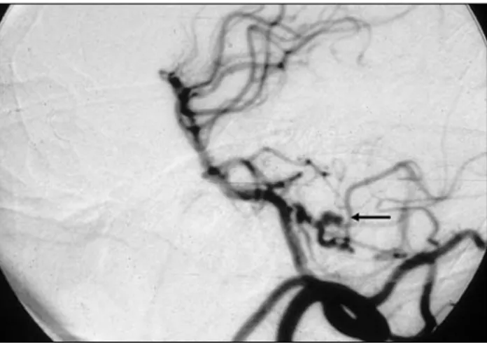
증례 1 64세 여자환자는 갑자기 발생된 두통 및 의식저하로 내원 하여 촬영한 CT에서 Fisher 등급 3의 뇌지주막하 출혈이 있 었고, 뇌혈관조영술을 통하여 왼쪽 lateral medullary분절에 3mm 크기의 낭성 형태의 동맥류를 진단 받았다(Fig. 1). 입 원 후 3일 째, 측방후두하접근법을 통한 결찰술을 시행 받았 고, 특별한 합병증 없이 퇴원하였다. 증례 2 70세 여자환자는 갑자기 발생된 의식저하로 응급실 내원하 여 촬영한 CT에서 Fisher등급 4의 뇌지주막하 출혈, 뇌실출 혈을 동반한 경미한 수두증이 관찰되었다. 뇌혈관조영술을 통 하여 오른쪽 tonsillomedullary분절에 낭성 형태의 4mm 크 기를 보이는 동맥류를 진단 받았다(Fig. 2). 입원 후 4일 째, 정중앙후두하접근법을 통하여 결찰술을 시행 받았고, 수술 후 뇌척수액고임증이 발생하였으나 입원 기간 중에 호전되었고 임상증상도 호전되었다. 증례 3 28세 여자환자는 지속적인 두통으로 검사 중에 우연히 발견 된 뇌 동맥류로 뇌혈관조형술을 시행하였고, telovelotonsillar 분절에 3.7mm의 낭성 형태의 동맥류를 진단받았다(Fig. 3). 정중앙후두하접근법을 통한 동맥류 결찰술을 시행받았고, 수 술 후에 뇌척수액고임증이 있어 재수술을 하였으나 경막재건 술에 실패하여 뇌실복강내단락술을 시행받았다. 증례 4 57세 여자환자는 갑작스런 의식저하로 응급실에 내원하여 시행한 CT에서 Fisher등급 4의 뇌지주막하출혈이 있었고, 뇌실출혈 및 경미한 뇌수두중이 관찰되었다(Fig. 4A). 입원 후 시행한 뇌혈관조영술에서 cortical 분절에 3.0mm 크기를 가지는 낭성 형태의 동맥류를 진단받았으며(Fig. 4B), 입원 후 11일 째 정중앙후두하접근법을 통한 동맥류 결찰술을 시행 받았다. 수술 후에 임상증상은 호전되었고 퇴원하였다.Fig. 1. The vertebral angiogram of a 64-years-old woman with subarachonoid hemorrhage of Fisher grade 3 on admission shows PICA aneurysm in lateral medullary segment. Black arrow indicates the aneurysm.
Fig. 2. The vertebral angiogram of a 70-years-old woman with subarachnoid hemorrhage of Fisher grade 4 on admission shows PICA aneurysm in tonsillomedullary segment, Black arrows indicate the aneurysm.
Fig. 3. The vertebral angiogram of a 28-years-old woman with incidentally found aneurysm shows PICA aneurysm in telovelotonsillar segment, Black arrows indicate the aneurysm.
고
찰
PICA에서 기시하는 동맥류는 흔하지 않으며, 대부분의 경 우에 VA-PICA이행부에서 발생한다.6) dPICA는 pPICA와
비교하여 드물며 전체 동맥류 중에서 0.28~1.4%의 발생률을 보인다.1)7)8)15)이처럼 발생빈도가 적고 지금까지 보고된 것으로 보면, 200례 정도이기 때문에 dPICA동맥류 및 환자의 특징 들에 대한 이해가 부족하며, 표준화된 치료 방법이 제시되지 못하고 있다.1)2)5)7)11)12)14)16)17) 본 연구에서 dPICA에서 기시하는 동맥류의 발생률은 전체 동맥류 중에서 0.56%를 차지하고 있으며 PICA동맥류 (1.06%) 보다 적은 발생률을 보였다. PICA분절 중에 lateral medullary 분절에서 10례(52.6%)로 가장 많았으며, telovelotonsillar 분절은 5례(26.3%)로 그 다음이었다. Lewis 등이 이전에 보고한 148례의 분절별 분포의 의하면, lateral medullary 분절에서 18%, tonsillomedullary에서 는 10%, telovelotonsillar의 경우에는 57%로 가장 많았 다.12)본 연구에서 동맥류의 모양은 낭성 형태가 가장 많았으 며 지금까지 보고된 연구도 낭성 형태가 가장 많았다.7)12)17)본 연구에서 방추 형태가 1례, 박리 형태가 1례에서 있었는데, 방 추 형태는 박리와 관련이 있는 것으로 보인다.12)본 연구에서 동맥류의 크기는 5mm이하의 작은 동맥류가 11례가 있었고, 평균 6.5mm였다. 다른 연구에 의하면 4.1, 7.6, 및 9.8mm 로 본 연구와 비교가 된다.7)12)17) 본 연구의 결과에 의하면, dPICA군에서 여성이 73.7%로 남성과 비교하여 많았는데, 50%부터 75%로 보고된 결과 보 다는 많거나 비슷하였다. 따라서 dPICA동맥류는 남성보다는 여성에게서 발생률이 높다는 것을 알 수가 있다. 그리고 평균 나이는 46.9세로 51.0, 57.7, 및 61.5세로 보고된 다른 연구 와 비교하면 본 연구의 환자군이 어린 것을 볼 수 있다.7)12)17) dPICA군에서 동맥류가 파열된 경우에 31.5%에서 뇌내출 혈 및 뇌실출혈이 동반되어 있었고 수두증도 60%에서 동반되 었는데, 다른 연구에서는 파열된 PICA동맥류중의 95%에서 수두증이 있다고 보고하고 있으며, dPICA동맥류가 파열된 경우에 뇌실출혈, 수두증 및 뇌실외배액술(extraventricular drainage)이 흔한 것을 볼 수 있다.10)12) 치료는 수술과 중재적시술이 각각 15례 및 4례에서 시행이 되었는데, 수술의 경우에 결찰술을 한 경우는 12례 및 wrapping을 통한 결찰술을 한 경우가 3례였으며 정중앙후두 하접근법을 통한 수술이 9례로 측방후두부하접근법으로 수술 을 한 6례 보다 많았다. 수술적인 치료가 중재적시술보다 많 은 것은 다른 연구의 결과7)12)17)와 동일하며 중재적시술이 적은 것은 뇌신경손상가능성은 적다고 보고됨에도 불구하고16)19) 개 두술에 의한 혈종의 제거가 수두증을 완화시키고, 후두와 감 압이 될 수 있고, 중재적시술을 통한 접근이 어렵기 때문으로 보인다.4)17) 하지만 본 연구에서 중재적 시술을 통한 4례에서 결과는 특별한 합병증이 보고되지 않았을 뿐더러 예후가 좋았 다. 물론 수술적인 치료의 결과도 좋았으나, 치료 후에 합병증 으로 뇌척수액고임증이 3례에서 발생하였다. 뇌척수액고임증 은 후두와 종양 수술의 경우에 14.3%에서 발생할 수 있다고 보고 되었으며, 측방후두하접근이 정중앙후두하접근에 의한 개두술 보다 더 많다고 보고하고 있다.18)20)그러나, 본 연구에 서는 정중앙후두하접근법을 통하여 2례에서 뇌척수액고임증 이 발생하였고, 측방후두하접근법에서는 1례가 발생하였다. 뇌척수액고임증은 수술 후에 이환율 및 치료비용 증가와 관련 이 있으므로 방수가 되도록 경막을 조심스럽게 닫는 것이 중 요할 뿐만 아니라 근육, 근막 또는 지방 등의 자가조직 이식을 이용하거나 젤라틴(gelatin) 또는 콜라젠 스폰지(collagen sponge)등을 이용하여 경막을 강화하는 등의 뇌척수액 누출 방지를 위하여 노력이 필요하겠다.3) pPICA와 dPICA동맥류에서 환자의 특징, 동맥류의 특징, 임상양상을 비교 하였다. 두 군간에 나이는 dPICA동맥류가 pPICA동맥류보다는 어린 것을 볼 수 있다(ρ=0.014). 하지만, 파열된 동맥류 사이에서 나이는 두 군간에 통계적 의미가 없 었다 (ρ=0.090). 다른 연구에서 dPICA 동맥류 집단의 평균 연령이 51.0, 57.7 및 61.5세인 점을 고려할 때,7)12)17)dPICA 와 pPICA의 연령적인 차이는 없는 것 같다. 성별, 동맥류의
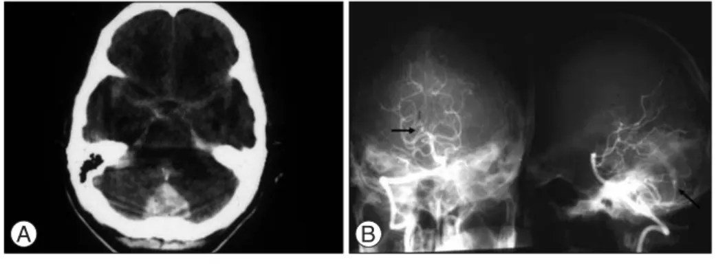
Fig. 4. A : Brain CT image of a 57-years-old woman shows subarachnoid hemorrhage with intraventricular hemorrhage within fourth ventricle. B : The vertebral angiogram shows PICA aneurysm in cortical segment. Black arrows indicate the aneurysm.
위치, 크기, 모양도 두 군간에 통계적 유의성을 보이는 차이는 없었으며, Fisher등급 및 Hunt-Hess등급에서도 통계적 차 이를 보이지는 않았다. 진단 당시 시행한 뇌 CT상에서도 의 미 있는 차이를 보이지는 않았다. 이러한 점에서 pPICA와 dPICA의 환자의 특징, 동맥류의 특징 및 임상증상의 발현에 는 큰 차이가 없다는 것을 확인할 수 있다. 하지만, 측방후두 부하접근법을 통한 수술의 시행이 용이한 pPICA동맥류의 경 우에 수술 후 뇌신경 마비의 발생이 5례에서 있었고, 다른 저 자들도 측방후두부하접근법을 통한 수술을 하는 dPICA의 lateral medullary 분절의 경우에 수술 후 뇌신경마비가 발 생할 수 있다는 점을 고려할 때,7)pPICA의 수술에서 뇌신경 마비 발생빈도가 높다고 추정할 수 있다. 반면에, dPICA동맥 류에서 수술 후 뇌척수고임증이 있었고, 이로 인하여 재수술 을 한 경우도 1례에서 있었지만, pPICA동맥류의 경우에는 뇌 척수액고임증이 발생한 예는 없었다. 따라서 정중앙후두하접 근법을 통하여 수술을 시행하는 경우가 많은 dPICA동맥류의 경우에 뇌척수액고임증이 많은 것이라고 추정해 볼 수 있다. 양 군간에 수술의 빈도와 중재적시술의 빈도는 통계적인 차이 를 보이지는 않았다 (ρ=0.768). 하지만, dPICA동맥류의 경우 에 수술적인 치료가 79%로 중재적시술 보다는 많았다. 일부 저자들은 중재적시술의 접근이 용이한 점, 뇌신경손상을 적게 유발하는 점 및 뇌간으로 들어가는 작은동맥의 손상이 적은 점을 내세우면서 수술적인 치료보다는 중재적 시술을 먼저 고 려해야 한다고 주장을 하고 있다.16)19) 하지만, 원위부에 위치 한 PICA동맥류의 경우에는 사행성 및 원위부의 얇은 혈관으 로 중재적 접근이 까다롭다고 주장하기도 한다.4)대부분의 저 자들은 중재적시술이 위와 같은 장점이 있을지라도 뇌지주막 하출혈과 동반된 뇌실출혈 및 수두증을 해결할 수 없으므로 수술적인 치료와 수술을 통한 혈종의 일부 제거가 수두증의 해결에 도움을 주며, 뇌실외배액술 및 뇌실복강내 단락술의 빈도를 줄일 수 있으므로 수술적인 치료를 적극적으로 해야 한다고 주장하고 있다.17) 하지만, 본 연구에서는 dPICA동맥 류의 치료에서 중재적 시술의 경우에 합병증 없었을 뿐만 아 니라 예후도 좋았고, 수술적인 치료 후에 3례에서 뇌척수액고 임증이 발생을 하였으므로, 중재적시술을 통한 접근이 가능한 dPICA동맥류의 치료에 중재적시술을 고려하는 것도 의미가 있다고 생각한다. Lewis 등은 해부학적으로 PICA를 근위부, 이행부 및 원위 부로 구별을 하여 근위부에 anterior 및 lateral medullary 분 절이 포함되며, 뇌간으로 들어가는 관통동맥들이 있기 때문에 수 술 시 에 PICA를 보 존 해 야 하 고 , 이 행 부 에 는 tonsillomedullary이며, 가급적 보전하는 것이 유리하며, 원위 부에는 telovelotonsillar와 cortical 분절이 포함되는데, 뇌 간으로 가는 관통동맥이 없고, 혈관 손상에 의한 소뇌경색 가 능성이 떨어지므로 수술 중에 희생할 수 있다고 보고하였다.12)
본 연구에서 dPICA동맥류에서 lateral medullary분절에 서 기시하는 빈도가 62.5%로 다른 보고에 비하여 빈도가 높 았다. 대부분의 저자들은 lateral medullary 분절의 동맥류 를 치료하기 위한 수술접근법으로 측방후두하접근법을 추천 하고 있으며,7)12) 본 연구에서도 10례의 환자중에 6례에서 측 방후두하접근법을 통한 수술을 시행하였고, 1례에서만 정중 앙후두하접근법을 통하여 수술을 시행하였으며, 나머지 3례 는 중재적시술을 통한 치료를 시행하였다. 우리의 의견으로는 Lewis가 분류한 근위부에 있는 dPICA동맥류에 해당하는 lateral medullary 분절의 동맥류인 경우에 수술로 인한 뇌 신경마비 및 뇌간으로 들어가는 관통동맥의 손상가능성이 있 고, 중재적시술을 통한 접근이 다른 dPICA분지와 비교하여 용이하기 때문에 중재적 시술을 통한 코일 색전술을 고려할 수 있겠다. 또한 본 연구에서 pPICA 동맥류의 수술에서 뇌신 경마비가 있었듯이 pPICA에 근접한 lateral medullary 분 절의 경우에 수술 후 발생할 수 있는 잠재적 뇌신경마비 및 뇌 간기능손상 가능성이 있고, 뇌간으로 들어가는 관통동맥의 손 상가능성은 있으나 중재적시술을 통한 동맥류의 선택적 코일 색전술이 어려울 경우에는 수술적인 치료를 적극 고려해야 할 것으로 보인다. 하지만, 중재적시술을 통한 동맥류의 선택적 색전술과 수술적 치료 모두 힘들 것으로 판단이 될 때에는 모 동맥의 혈류 보존이 중요하고, 관통동맥의 손상가능성은 있지 만 본 연구의 2례에서 볼 수 있듯이 중재적 시술을 통한 dPICA 를 폐색시키는 방법을 마지막으로 고려해 볼 수 있겠다. Lewis 가 분류한 이행부와 원위부에 해당하는 tonsillomedullary, televelotonsillar, cortical 분절의 경우에는 중재적시술의 접근이 어려우나 수술적 치료 후 발생할 수 있는 뇌척수액고 임증이 없으므로 dPICA의 폐색이 가능할 경우에 중재적시술 을 고려해 볼 수 있는 치료 방법이지만, 수술적인 접근이 비교 적 용이하고, 모동맥의 혈류 보존이 가능하며, 수술에 의한 혈 종 제거로 수두증 완화를 기대할 수 있으므로 수술적인 치료 를 적극적으로 고려해야 하겠다. 하지만, 수술적인 치료의 경 우에 뇌척수액고임증의 발생가능성이 있으므로 경막을 닫을 때, 방수를 위한 노력이 필요할 것으로 보인다. pPICA 동맥류 수술의 경우에 2례에서 뇌신경마비가 발생 하였고, dPICA 치료 후 2례에서 일시적인 편마비가 있었으 나 수술 후 시행한 뇌 CT상에서는 경색소견을 보인 예는 없 었다. 또한, dPICA에서는 호전이 94.7%였지만, pPICA의 경 우에 72.2%이고 예후가 불량한 경우가 11.1%가 있었기 때문 에 통계적인 차이는 보이지 않더라고 pPICA의 치료예후가 dPICA 보다는 불량하다고 생각해 볼 수 있다. 다른 연구에서 도 dPICA 동맥류의 치료가 비교적 예후가 좋다고 보고하고 있다. 본 연구에서는 dPICA 동맥류 치료가 어렵고, 치료방법
의 결정을 위하여 진단 후 10일 이후에 치료가 이루어진 경우 가 10례에서 있었지만, 지금까지 논의를 토대로 dPICA 동맥 류의 진단이 이루어지면, 적극적인 치료가 필요할 것으로 보 인다.
결
론
전체 동맥류 중에서 흔하지 않게 발생되는 dPICA동맥류는 임상적으로 환자와 동맥류의 특징에서 pPICA와 큰 차이를 보이고 있지 않다. 하지만, 치료 후 임상결과는 pPICA와 비 교하여 보다 좋으며 수술에 의한 합병증이 뇌신경마비 및 뇌 간경색과 같은 중한 경우보다는 뇌척수액고임증과 같은 경한 경우가 많다. 따라서 dPICA동맥류에서 lateral medullary 분절을 제외하고는 수술적인 치료를 적극적으로 고려하는 것 이 좋으며, 수술 시에 뇌척수액누출의 방지 및 모동맥의 혈류 보존을 위하여 보다 노력을 해야 하겠다. 중심 단어 : 뇌동맥류∙후하소뇌동맥∙뇌동맥류 수술 뇌혈관 내 수술.REFERENCES
11) Ali MJ, Bendok BR, Tawk RG, Getch CC, Batjer HH. Trapping
and revascularization for a dissecting aneurysm of the proximal posteroinferior cerebellar artery: technical case report and review of the literature. Neurosurgery 51: 258-62; discussion: 262-63, 2002
12) Anegawa SHT, Torigoe R, Nakagawa S, Furukawa Y, Tomokiyo M. Aneurysms of the distal posterior inferior cerebellar
artery-analysis of 14 aneurysms in 13 cases. No Shinkei Geka 29(2): 121-9, 2002
13) Booqaarts JD, Grotenhuis JA, Bartels RH, Beems T. Use of a
novel absorbable hydrogel for augmentation of dural repair: results of a preliminary clinical study. Neurosurgery 57(suppl 1):146-51, 2005
14) Bradac GB, Bergui M. Endovascular treatment of the posterior
inferior cerebellar artery aneurysms. Neuroradiology 46(12): 1006-11, 2004
15) Fujimura M, Nishijima M, Midorikawa H, Umezawa K, Hayashi T, Kaimori M. Fatal rupture following intra-aneurysmal
embolization for the distal posterior inferior cerebellar artery aneurysm with parent artery preservation. Clin Neurol Neurosurg 105: 117-20, 2003
16) Gács G, Vi´nuela F, Fox AJ, Drake CG. Peripheral aneurysms of
the cerebellar arteries. Review of 16 cases. J Neurosurg 58:63-8, 1983
17) Horiuchi T, Tanaka Y, Hongo K, Nitta J, Kusano Y, Kobayashi S.
Characteristics of distal posteroinferior cerebellar artery aneurysms. Neurosurgery 53: 589?95; discussion 595-6, 2003
18) Hudgins RJ, Day AL, Quisling RG, Rhoton AL Jr, Sypert GW, Garcia-Bengochea F. Aneurysms of the posterior inferior
cerebellar artery. A clinical and anatomical analysis. J Neurosurg 58: 381-7, 1983
19) Jennett B, Bond M. Assessment of outcome after severe brain
damage. Lancet 1: 480-4, 1975
10) Kallmes DF, Lanzino G, Dix JE, Dion JE, Do H, Woodcock RJ et al. Patterns of hemorrhage with ruptured posterior inferior
cerebellar artery aneurysms: CT findings in 44 cases. AJR Am J Roentgenol 169: 1169-71, 1997
11) Kim K, Kobayashi S, Mizunari T, Teramoto A. Aneurysm of the
distal posteroinferior cerebellar artery of extracranial origin: case report. Neurosurgery 49: 996-8; discussion: 998-9, 2001
12) Lewis SB, Chang DJ, Peace DA, Lafrentz PJ, Day AL. Distal
posterior inferior cerebellar artery aneurysms: clinical features and management. J Neurosurg 97: 756-66, 2002
13) Locksley HB. Report on the Cooperative Study of Intracranial
Aneurysms and Subarachnoid Hemorrhage: Section V, Part 1?Natural history of subarachnoid hemorrhage, intracranial aneurysms and arteriovenous malformations. Based on 6368 cases in the Cooperative Study. J Neurosurg 25:219-39, 1966
14) Mansmann U, Lasjaunias P, Meisel HJ. Treatment of patients with
cerebral arteriovenous malformations. Radiology 223: 879-80; author reply 880-1, 2002
15) Meisel HJ, Mansmann U, Alvarez H, Rodesch G, Brock M, Lasjaunias P. Cerebral arteriovenous malformations and
associated aneurysms: analysis of 305 cases from a series of 662 patients. Neurosurgery 46: 793-800, 2000
16) Mukonoweshuro W, Laitt RD, Hughes DG. Endovascular
treatment of PICA aneurysms. Neuroradiology 45: 188-92, 2003
17) Orakcioglu B, Schuknecht B, Otan N, Khan N, Imhof HG, Yonekawa Y. Distal posterior inferior cerebellar artery
aneurysms: Clinical characteristics and surgical management. Acta Neurochir (Wien) 147:1131-9, 2005
18) Santamarta D, Blazquez JA, Maillo A, Munoz A, Morales CF.
Analysis of cerebrospinal fluid related complications (hydrocephalus, fistula, pseudomeningocele and infection) following surgery for posterior fossa tumor, Neurocirugia (Astur) 14(2):117-26, 2003
19) Tikkakoski T, Leinonen S, Siniluoto T, Koivukangas J. Isolated
dissecting aneurysm of the left posterior inferior cerebellar artery: endovascular treatment with a Guglielmi detachable coil. AJNR Am J Neuroradiol 18: 936-8, 1997
20) Yamakami I, Serizawa T, Yamaura A, Nakamura T. Cerebrospinal
fluid leak after cranial base surgery. No Shinkei Geka 24(1):29-33, 1996