Vol. 47, No. 5, pp. 688 - 697, 2006
[6]-Gingerol, a major phenolic compound derived from ginger, has anti-bacterial, anti-inflammatory and anti-tumor activities. While several molecular mechanisms have been de-scribed to underlie its effects on cells in vitro and in vivo, the underlying mechanisms by which [6]-gingerol exerts anti-tu-morigenic effects are largely unknown. The purpose of this study was to investigate the action of [6]-gingerol on two human pancreatic cancer cell lines, HPAC expressing wild-type (wt) p53 and BxPC-3 expressing mutated p53. We found that [6]-gingerol inhibited the cell growth through cell cycle arrest at G1 phase in both cell lines. Western blot analyses indicated that [6]-gingerol decreased both Cyclin A and Cyclin-dependent kinase (Cdk) expression. These events led to reduction in Rb phosphorylation followed by blocking of S phase entry. p53 expression was decreased by [6]-gingerol treatment in both cell lines suggesting that the induction of Cyclin-dependent kinase inhibitor, p21cip1, was p53-indepen-dent. [6]-Gingerol induced mostly apoptotic death in the mutant p53-expressing cells, while no signs of early apoptosis were detected in wild type p53-expressing cells and this was related to the increased phosphorylation of AKT. These results suggest that [6]-gingerol can circumvent the resistance of mutant p53-expressing cells towards chemotherapy by inducing apoptotic cell death while it exerts cytostatic effect on wild type p53-expressing cells by inducing temporal growth arrest.
Key Words: [6]-gingerol, pancreatic cancer, G1 phase,
apoptosis, AKT
INTRODUCTION
Various plant-derived compounds or
phytoche-micals are known to have ability to interfere with carcinogenesis and tumorigenesis. There are many evidences that many phytochemicals, such as epigallocatechin-3-gallate (EGCG), genestine, tan-geretin, silymarin, silibinin, and quercetin, can inhibit the proliferation or survival of various cancer cells and also induce cell cycle arrest.1-5
Plant of ginger (Zingeiber officinale Roscoe,
Zingiberaceae) family is one of the most highly consumed dietary substances in the world. In China and Malaysia, the rhizome of ginger has been used in traditional oriental herbal medicine for the management of common cold, digestive dis-orders, rheumatism, neuralgia, colic and motion-sickness.6 The oleoresin from rhizome of ginger
contains pungent ingredients including gingerol, shoagol, and zingerone.7 Recently, these phenolic
substances have been found to possess many in-teresting pharmacological and physiological acti-vities. Of these, [6]-gingerol (1-[4'-hydroxy-3'-methoxyphenyl]-5-hydroxy-3-decanone), the major pungent principle of ginger, has anti-oxidant, anti-inflammation and anti-tumor promoting acti-vities.7
Anti-cancer and/or chemopreventive activities of [6]-gingerol have been reported. For instance, [6]-gingerol inhibited pulmonary metastasis in mice bearing B16F10 melanoma cells through the activation of CD8+ T cells.8It also inhibited tumor
promotion of ICR mice induced skin tumor by tumor promoter (TPA),9 and blocked the
azoxy-methane-induced intestinal carcinogenesis in rodents.10 [6]-Gingerol interfered with
EGF-in-duced transformation of mouse epidermal JB6 cell line, and reduced the activation of Activator Pro-tein-1 (AP-1), which plays a critical role in tumor
[6]-Gingerol Induces Cell Cycle Arrest and Cell Death of
Mutant p53-expressing Pancreatic Cancer Cells
Yon Jung Park,1 Jing Wen,1 Seungmin Bang,2 Seung Woo Park,2 and Si Young Song2
1Brain Korea 21 Project for Medical Science, 2Internal Medicine and Institute of Gastroenterology, Yonsei University College
of Medicine, Seoul, Korea.
Received February 1, 2006 Accepted May 4, 2006
This work was supported in part by the Brain Korea 21 Project for Medical Science.
Reprint address: requests to Dr. Si Young Song, Department of Internal Medicine, Yonsei University College of Medicine, 134 Shinchon-dong, Seodaemoon-gu, Seoul 120-752, Korea. Tel: 82-2-2228-1957, Fax: 82-2-2227-7900, E-mail: [email protected]
promotion.11 Moreover, [6]-gingerol exerted
in-hibitory effects on the cell viability and DNA synthesis, also induced apoptosis of human promyelocytic leukemia HL-60 cells.12 Recently, it
has been reported that ginger root extracts and gingerol inhibit the growth of Helicobacter pylori CagA+ strains, which has a specific gene linked to the development of gastric premalignant and malignant lesions.13 It suggests that ginger and
gingerol have effects of chemoprevention to the gastric-intestinal cancers.
Pancreatic ductal adenocarcinoma (pancreatic cancer) is the fifth leading cause of cancer death in Korea. Pancreatic cancer is a fatal disease, with a 5-year survival rate of less than 5%.14 In the
majority of cases (> 80%), at first diagnosis, pancreatic cancer has already become metastatic so that conventional treatment regimens provide minimal, if any, clinical benefit in prolonging life or ameliorating the negative prognosis of this disease.15 Resistance of recurrent disease to
cytotoxic drugs is the principal factor limiting long-term treatment success against pancreatic cancer. The oncogenesis of pancreatic cancer in particular appears to favor the development and subsequent expansion of cell clones that are resistant to apoptotic triggers. The basis for failed apoptosis and more specifically the cause of chemotherapy resistance in pancreatic cancer is multifactorial. Molecular mechanisms implicated to date include expression of P-glycoproteins and other multidrug resistance proeins, mutations of K-ras and p53, and high-level expression of Bcl-2 and other inhibitors of apoptosis. 16-18Therefore, an
important research objective is the identification of lead compounds that circumvent the resistance mechanisms that limit the success of conventional drugs.
Although anticancer activities of ginger extract and constituents have been examined, the under-lying mechanism has not been clarified yet. The purpose of this work was to develop an under-standing of [6]-gingerol's effects on pancreatic cancer cells to begin to determine its therapeutic value in preventing or treating this disease. Therefore we examined the anticancer activities of [6]-gingerol, and investigated its mechanism in two different pancreatic cancer cell lines, HPAC with wt p53 or BxPC-3 with mutant p53.
[6]-Gingerol's antiproliferative effects are associated with changes in the expression or phosphoryla-tion of Cyclin A, Cdks, p21 and Rb, and most commonly, causes cell cycle arrest at the G1 phase. We found that [6]-gingerol is capable of inducing cell death in the mutant p53-expressing cells, while arresting mutant p53-expressing cells at G1 phase. Thus, [6]-gingerol can circumvent drug resistance induced by p53 mutations.
MATERIALS AND METHODS Chemicals and cell culture
A purified preparation of [6]-gingerol (> 98.0% pure) was purchased from Wako Pure Chemicals (Osaka, Japan). It was dissolved in sterile DMSO at a stock concentration of 50 mM and stored at -20 . Human pancreatic cancer cells, BxPC-3 and HPAC, were obtained from the American Type Culture Collection (Manassas, VA, USA) and main-tained in RPMI1640 and DMEM/F12 medium respectively, containing 10% fetal bovine serum. Both cell lines were incubated at 37 in a humidified atmosphere with 5% CO2.
Assessment of cell viability
The viability of the cells was measured by the MTT [3,(4,5-dimethylthiazol-2-yl)-2,5-diphenyl tetra-zolium bromide] (Sigma, St. Louis, MO, USA) assay. The cells (4 × 10³cells/well) were seeded
into 96-well plates, after 24 hr they were treated with various concentration of [6]-gingerol or vehicle alone (0.1% DMSO) in serum containing media. After incubation for indicated times, MTT solution was added to the plate at a final con-centration of 0.5 mg/mL. The cells were incubated for 4 hr in dark at 37 . The resulting MTT-pro-ducts were dissolved by DMSO and determined by measuring the absorbance at 570 nm with ELISA reader (Molecular Devices, Sunnyvale, CA, USA). Each point represents the mean of tripli-cated experiments.
Cell cycle analysis
described.19Briefly, cells were seeded into 100 mm
dishes, after 24 hr treatment with the indicated concentrations of [6]-gingerol for indicated hours. Following treatment, cells were trypsinized, and washed twice with cold PBS (pH 7.4). The pellet was fixed with cold ethanol (70%) for 12 hr at 4 and washed with cold PBS. Then, they were in-cubated with RNase (200 g/mL final concentraμ -tion) and stained with Propidium Iodide (100 g/μ mL final concentration) for 1 hr and analyzed by flow cytometry. Flow cytometry was performed on a FACSCalibur system equipped with argon-ion laser (Becton Dickison Immunocytometry system, San Jose, CA, USA). Percentages of cells in each phase were calculated using Cell Modfit software programs (Becton Dickinson).
Assessment of apoptosis
To identify cells undergoing apoptosis, Annexin V-FITC Apoptosis Detection Kit (BD Pharmingen, Franklin Lakes, NJ, USA) was used. Cells were harvested at different intervals after [6]-gingerol treatment, containing floating and adherent cells. After washing with cold PBS (pH 7.4), the cells were stained and analyze by the flow cytometry. For each tube, 20,000 cells were immediately mea-sured on a FACSCalibur flow cytometer. The quantitative analysis was performed with Win MDI program version 2.8 (provided by the Flow Cytometry Core Facility, The Scripps Research Institute, La Jolla, CA, USA).
Protein extraction and Western blot analysis
Whole cell lysates, used to determine the levels of various proteins, were prepared by following method. In brief, the pancreatic cancer cells were seeded onto culture-dishes, after 24 hr incubation with [6]-gingerol for the indicated times. The cells were washed twice with cold PBS (pH 7.4) and added cell lysis buffer (70 mM beta-glycerol-phosphate (pH 7.2), 0.6 mM sodium vanadate, 2 M MgCl2, 1 mM Ethylene glycol-bis
(2-aminoe-thylether-)N,N,N',N'-tetraacetic acid (EGTA), 1 mM dithiothreitol (DTT), 0.5% Triton X-100, 0.2 mM phenylmethylsulfonyl fluoride (PMSF), 1X Protease Inhibitor; Leupeptin, Pepstatin, Aprotinin, and Antipain each 5 g/mL). Cell lysates wereμ
then centrifuged at 13,200 rpm for 30 min at 4 . The protein concentrations were measured by the Bradford dye-binding protein assay, using bovine serum albumin (Sigma) as a standard. For Western blot analysis, protein samples (30 g) were soluμ -bilized by boiling in sample buffer and subjected to SDS-PAGE followed by electrotransfer onto nitrocellulose membrane (Amersham Life Science, Buckinghamshire, UK). Blots were blocked for 2 hr at rom temperature with 5% nonfat dry milk and incubated at 4 overnight with the fol-lowing antibodies: rabbit polyclonal anti-Cyclin A, rabbit polyclonal anti-Cyclin D1, rabbit polyclonal anti-Cyclin E, rabbit polyclonal anti-Cdk 2 and rabbit polyclonal anti-Cdk 4 (Delta Biolabs, Camp-bell, CA, USA), goat or rabbit polyclonal anti-pRb (Ser 780) (BioSource, Camarillo, CA, USA), mouse monoclonal anti-Rb, rabbit polyclonal anti-Cdk 6, rabbit polyclonal anti-p21, mouse monoclonal anti-p53, rabbit polyclonal anti-phosphatidylino-sitol-3 kinase (PI3K) p85 subunit, goat polycloα -nal anti-ERK1, mouse monoclo-nal anti-pERK, mouse monoclonal anti-phospho-c-jun-N-terminal kinase (pJNK), goat or chicken polyclonal anti-AKT1, rabbit polyclonal anti-pAKT1/2/3 (Ser 473), and mouse monoclonal anti-K-Ras (Santa Cruz Biotechnology Inc., Santa Cruz, CA, USA). The membranes were washed three times and incubated with horseradish peroxidase-conju-gated species-appropriate secondary antibodies (Santa Cruz). After additional washing, they were developed with enhanced chemiluminescence reagents (Amersham Life Science), and exposed to films in a dark room.
Statistical analysis
When appropriate, statistical significance was tested using a two tailed Student's t-test; p 0.05 was considered significant. All values shown are means with the corresponding standard error.
RESULTS
[6]-Gingerol inhibits cell growth on both BxPC-3 and HPAC cells
[6]-gingerol on two pancreatic cancer cells expressing either wt (HPAC) or mutant p53 protein (BxPC-3), we treated cells with [6]-gingerol ranging from 50 to 800 M. As shown in Fig. 1, cells treated withμ different concentrations of [6]-gingerol for 0, 24, 72, and 120 h resulted in the growth inhibition in a dose- and time-dependent manner. [6]-Gingerol at the highest concentration (800 M) had similarμ mean cytotoxicity over the 72 h of 89.4% and 91.2% on BxPC-3 and HPAC cells, respectively. There was no significant difference in IC50values
between BxPC-3 cells and HPAC cells (387.4 Mμ and 405.3 M at day 3, respectively,μ p< 0.006). IC25 values were 186.1 and 146.3 M for BxPC-3μ
and HPAC, respectively.
[6]-Gingerol induces cell cycle arrest
Although it was apparent that [6]-gingerol was growth inhibitory to the pancreatic cancer cell lines, the sustained effects on culture growth raised suspicion that it might also affect the proliferation of surviving cells. To test this pos-sibility, we examined whether [6]-gingerol affects cell cycle progression. BxPC-3 and HPAC cells were treated with 400 M of [6]-gingerol (approxiμ -mate IC50 at day3). As shown in Fig. 2,
[6]-gingerol caused BxPC-3 cells to accumulate in G1-phase (59.2% in [6]-gingerol-treated vs. 42.3% in control) within 24 hr at the expense of cells mainly in S phase. Furthermore, this was
associ-ated with the induction of apoptosis as repre-sented by the large sub-G1 peak. On the other hand, cell cycle arrest of HPAC by [6]-Gingerol
Fig. 1. Effect of [6]-gingerol on survival of BxPC-3 and HPAC cells. [6]-Gingerol inhibited growth of both BxPC-3 and HPAC cells in a dose-dependent manner. Cells were treated with different concentrations of [6]-gingerol. Control cells (empty circle) were treated with vehicle alone (0.1% DMSO). Cells were harvested at day 1, 3 and day 5 and cell viability was determined by the 3-(4,5-dimethylthiazol-2-yl)-2,5-diphenyltetrazolium bromide (MTT) assay as described in Materials and methods. Percent viability was calculated by comparison with controls. Values shown as the mean ± SEM obtained from four independent experiments.
Fig. 2. [6]-Gingerol induces cell cycle arrest in both BxPC-3 and HPAC cell lines. Exponentially growing cells were exposed to either 0.1% DMSO (control) or [6]-gingerol (400 M) for 24, 48 or 72μ hrs. Cells were then harvested, washed in PBS, and fixed in 70% ethanol. DNA content was evaluated with propidium iodide staining and fluorescence measured and analyzed as described in Materials and Methods. Data are repre-sentative of three independent experiments.
was less dramatic in HPAC cells where cellular accumulation in G0/G1 phase was slightly higher in [6]-gingerol treated group (64.6%) compared to the control (54.6%). In contrast to BxPC-3 cells, we did not detect an increase in HPAC cells with hypodiploid DNA content, suggesting that apoptotic cell death was not induced. At 72 hr, HPAC cells partially recovered from [6]-gingerol-induced growth arrest suggesting the operation of compensatory mechanism(s).
[6]-Gingerol caused changes in various cell cycle related protein expressions
We examined the effect of [6]-gingerol on the levels of G1 phase-regulatory proteins by Western blot analyses (Fig. 3). Both BxPC-3 and HPAC cells were treated with 400 M of [6]-gingerol andμ harvested in every 12 hr. In BxPC-3 and HPAC cells, expression levels of various proteins were changed by [6]-gingerol. In BxPC-3, [6]-gingerol induced decrease in Cyclin A, Cdk 2, Cdk 4 and Cdk 6 expression and increase in Cyclin D1 expres-sion. [6]-Gingerol decreased the Cyclin A and Cdk 6 expression, but not Cdk 2 and Cdk 4, in HPAC cells. Phosphorylated Rb protein was decreased in both BxPC-3 and HPAC cells by [6]-gingerol.
Next, we studied the difference in basal and [6]-gingerol-induced levels of p53 and p21cip1,
between wt and mutant p53-expressing cells. In line with previous report,20the basal levels of p53
were elevated in mutant p53-expressing cells. [6]-Gingerol reduced the p53 levels of wt, but less significantly of mutant, p53-expressing cells (Fig. 4). Expression of p21cip1 was increased by
[6]-gingerol in both BxPC-3 and HPAC cells. It is well known that induction of p21cip1 is associated with
the expression of p53.21 The treatment of
[6]-gingerol reduced the p53 level in the both cells implying that the overexpression of p21cip1 by
[6]-gingerol might be caused by p53-independent events in both cell lines.
[6]-Gingerol induces apoptosis in BxPC-3 cell
Next, we assessed potential differences between the [6]-gingerol-induced cell death in wt and mutant p53-expressing cells. Since mutations in p53 often compromise the ability of cells to un-dergo apoptosis, we analyzed the mode of cell death involved. As can be seen in Fig. 5, early apoptosis (Annexin-V+/propidium iodide-) could be detected in the mutant p53-expressing BxPC-3 cells 24 hr after treatment with [6]-gingerol. The percentage of early-apoptotic cells and late apoptotic/necrotic cells (Annexin-V+/propidium iodide+) continued to rise, reaching a peak at 72 hr. On the other hand, [6]-gingerol did not induce death in wild type p53 expressing HPAC cells. HPAC cells displayed few early apoptosis and late apoptotic/necrotic fractions until 72 hr suggesting
Fig. 3. Western blot analyses of the cell cycle regulatory proteins. Both BxPC-3 and HPAC were treated with 400 M of [6]-gingerol, and harvested every 12 hr, and levels μ
of Cyclins, Cdks, Rb and pRb were determined by Western blotting. The upper band of Rb indicates phosphorylated form. pRb antibody detects specifically on Ser-780 phophorylated Rb. Data are representative of three inde-pendent experiments. Actin was used as a standard for each sample.
Fig. 4. Effects of [6]-gingerol on the expression of p21cip and p53. Both BxPC-3 and HPAC were treated with 400 M of [6]-ingerol, and harvested every 12 hr. The levels μ
of p21cip1, a Cyclin dependent kinase inhibitor, and p53 changed by [6]-Gingerol in BxPC-3 and HPAC cells. Three replicate experiments were done with similar results.
that HPAC cells are highly resistance to [6]-gingerol induced apoptosis. This observation cor-responds to the data of cell cycle analysis where HPAC cells had minimal sub-G1 population upon [6]-gingerol treatment even after 72 hr.
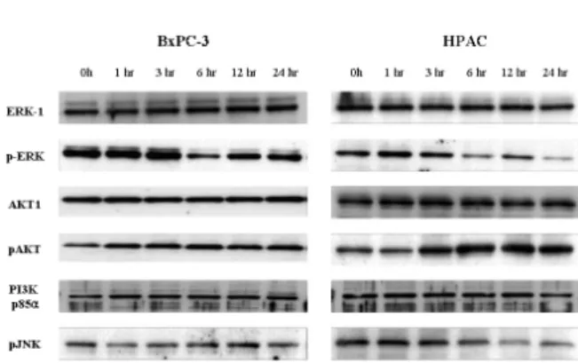
[6]-Gingerol affects AKT activation
Based on these findings, we sought to examine whether [6]-gingerol-induced apoptotic cell death was accompanied by the activation of common pro-apoptotic signaling pathways. The difference in [6]-gingerol response of these two cell lines may be caused by the altered PI3K/AKT path-ways which play important roles in cell cycle progression and cell survival.22 [6]-Gingerol was found to inhibit nuclear factor kappa B (NF-kB),
AP-1, cyclooxygenase-2 (COX-2) and p38 mitogen activated protein kinase (MAPK).23 Thus, we ex-amined the levels of pERK, pJNK and PI3K/AKT pathway proteins by Western blot analysis (Fig. 6). The down regulation of pJNK and pERK MAPK was time-dependently induced by [6]-gingerol in both cell types. In both cell lines, there was no change of PI3K regulatory subunit, p85 , levels by [6]-gingerol. While [6]-gingerolα did not change phosphorylation of AKT protein in BxPC-3 cells, active phosphorylated AKT (pAKT) started to appear 3 hr after incubation and increased further up to 24 hr in [6]-gingerol-treated HPAC cells. These results imply that [6]-gingerol-induced apoptosis in HPAC cells (p53 wild type) is suppressed via the PI3K/AKT pathway.
Fig. 5. Differential mode of death induced by [6]-Gingerol in pancreatic cancer cells expressing wt versus mutant p53. Cells were treated with 400 M of [6]-gingerol for 24, 28 and 72 hrs. Annexin-V and PI staining revealed increasingμ percentage of Annexin-positive and PI-positive cells with increasing time of [6]-gingerol in BxPC-3 cells. On the other hand, [6]-gingerol treatment on HPAC cells showed no changes in cell viability under the identical culture condition. The X and Y axis represents annexin V-FITC and Propidium Iodide (PI) fluorescence respectively. Population in lower-left part (Annexin V-FITC and PI negative) is viable, and lower-right part (Annexin V-FITC positive and PI negative) is undergoing apoptosis. Cells observed in Annexin V-FITC and PI positive (upper-right and upper-left parts) indicates either late stage of apoptosis or dead cells. The percentage of each part is calculated in the bottom table. The figure is representative of three independent experiments.
DISCUSSION
Phenolic compounds comprise one of the largest and most ubiquitous groups of plant metabolites. They are formed to protect the plant from photo-synthetic stress, reactive oxygen species (ROS), wounds, and herbivores.24 The most commonly
contained ones in foods are flavonoids and phenolic substances. Hence, phenolic compounds take important parts of the human diet. In addition, current interest is raised up by many observations that dietary phenolic compounds have various activities such as antioxidant, anti-inflammation and anti-carcinogenesis.
In this present study, we first investigated the effects of [6]-gingerol, a phenolic substance de-rived from ginger roots, on two pancreatic cancer cell lines. We found that [6]-gingerol inhibited the cell growth, disrupted the cell cycle progression in both HPac cells (wild type p53) and BXPC-3 cells (mutant p53 protein), and also induced apoptosis in BxPC-3 cells. Interestingly, it is noticeable that normal cell showed highly resistance to the cytotoxic effects of [6]-gingerol. RIE (rat intestinal epithelial cell) showed 50 percents growth inhibi-tion at over 900 M (data not shown) of [6]-μ gingerol for 3 days treatment. This selectivity may
be the great advantage of [6]-gingerol for the therapeutic or preventative use. While there were some reports that various phenolic substances induce cell cycle arrest in some phases,1-5 this is
the first report that reveals on [6]-gingerol effect upon cell cycle in cancer cell lines. Western blot analyses indicated that [6]-gingerol decreased the expression of Cyclin A and Cdks including Cdk2, Cdk4, and Cdk6 in BxPC-3. Also, Cyclin A and Cdk 6 expression levels were decreased in HPAC. Thus, the reduction of Cyclin or Cdk expressions results the blocking of Cyclin-Cdk complexes formation and that lowers the level of phospho-Rb. Since Rb proteins remain in unphosphorylated form, E2F cannot be activated and the cells fail to enter the S phase. Cyclin D1 might be the cause of apoptotic cell death when overexpressed.25,26
Alternatively, increase of Cyclin D1 in BxPC-3 cells may be a feedback response to drug-induced cell cycle arrest.
p53 is a tumor suppressor gene encoding a transcription factor. Its tumor-suppressive activity involves inhibition of cell proliferation through cell cycle arrest and/or apoptosis. Mutation in p53 occurs in more than half of human cancers.27 Cells
harboring mutated p53 lose the ability to elicit the enzymatic DNA repair cascade, to inhibit cell proliferation and to induce programmed cell death, resulting in induction of uncontrolled pro-liferation and malignancy.28,29 Tumors consisting of mutant p53-expressing cells exhibit high resis-tance to radiation and chemotherapeutic drugs. Circumventing this abnormal resistance is a major challenge in cancer therapy.20,30 In the present
study, we showed that [6]-gingerol exerted its cytotoxic activy toward cancer cells harboring mutant p53. Based on in vitro and in vivo studies, the Cip/Kip family including p21Cip1 were
ini-tially thought to interfere with the activation of G1/S phase related Cyclin/Cdk complexes. The Cyclin-dependent kinase inhibitor p21cip1 is a
major transcriptional target of the tumor-suppres-sor p53.21 BxPC-3 cells have point mutated p53
proteins and the HPAC cells have the wild type p53 proteins. While the basal levels of p53 were elevated in mutant p53-expressing cells as previously reported,20the level of p53 is decreased
by [6]-gingerol in both cell lines. Thus, p21cip1 in-duction by [6]-gingerol was not necessarily
depen-Fig. 6. Western bolt analysis for MPAK and PI3K/AKT
pathways in [6]-gingerol-treated pancreatic cancer cells. The BxPC-3 (A) and HPAC (B) were incubated with 400 M of gingerol for indicated times. Cells were lysed and μ
the cell lysates (30 g) were resolved by SDS-PAGE. AKT,μ ERK and JNK activation was analyzed by Western blotting with anti-phospho-AKT, ERK, anti-phosph-JNK or anti-p85-specific antibodies as indicated. Equal protein loading was confirmed by probing the membranes with antibodies detecting the respective unphosphorylated proteins (ERK and AKT). Three replicate experiments were done with similar results.
dent on p53 wild-type status since we detected p21 induction in mutated p53 cells.
It has been reported Ras signaling through the Raf/MAPK pathway also elevates levels of p21cip1
in some cell types.31-33 However it is still unclear
whether the over-expression of p21cip1 by [6]-gingerol is related with Ras signaling activations. A number of phytochemicals, including EGCG,34
tangeretin,3 genestein and silymarin4 have been
shown to induce cell cycle arrest accompanied by increased p21cip1 expression, an important Cdk
inhibitor in G1 and S phase. [6]-Gingerol also increased the level of p21cip1 in both cell lines. The
overexpression of p21cip1 facilitated cell cycle
arrest effects of [6]-gingerol. Unlike other Cyclins, Cyclin D1 level was increased in BxPC-3. It is pro-bably as a feedback response to G1 arrest. More recent study, however, has altered this view and revealed that even p21cip1 proteins are specific
inhibitors of Cyclin E- and A-dependent Cdk2, they act as positive regulators of Cyclin D-depen-dent kinases.31 Thus, the increase of Cyclin D1
level in BxPC-3 cells may be the consequence of the overexpression of p21cip1. However, it is un-clear why there was no change of Cyclin D1 level in spite of G1 phase arrest and overexpression of p21cip1 in HPAC cells. The fact that [6]-gingerol
has different effects on the cell cycle in different cell lines is intriguing and suggests that the ability of [6]-gingerol to affect the cell cycle may be dependent on other genetic alterations that the tumor harbors. For example, the levels of Cyclins, Cyclin-dependent kinases, their inhibitors, or the status of tumor suppressor genes such as p53 and Rb, that are all involved in cell cycle regulation, may determine whether a chemical inhibitor (drug) results in cell cycle arrest or not.
Exposure of mammalian cells to growth factors or genotoxic stress elicits a variety of cellular responses, including the activation of protein kinase cascades involving ERKs, stress-activated protein kinases (SAPK/JNK) and p38 MAPK.35 Therefore, we determined the effect of [6]-gingerol on the activation of extracellular signal-regulatted protein kinase-1/2 (ERK1/2), p38 MAPK, and JNK, which are representative MAPKs involved in a wide array of cellular signaling cascade. PI3K/ AKT pathway has an important role in preventing cells from undergoing apoptosis and contributing
to the pathogenesis of malignancy.36 More
re-cently evidences have suggested that this pathway is also associated with the regulation of cell cycle progression.22 The anti-cancer activities of [6]
gingerol could be associated with a control of signal transduction of PI3K/AKT pathway and this led us to investigate the change of the survival pathway-associated proteins. In the both cells [6]-gingerol could not affect the expression of the regulatory subunit of PI3K, p85 . However,α [6]-gingerol increased phosphorylation of AKT, which is regulated by PI3K, in only the HPAC cells. Activated AKT is known to promote the cell survival by anti-apoptoic mechanism.37
Additio-nally, activated AKT also phosphorylates and inactivates the proapoptotic protein, BAD.22,36,38 In
HPAC cells, the increase of phospholated AKT might protect apoptosis, despite of cell cycle arrest by [6]-gingerol. On the other hand, there was no change in phosphorylation of AKT by [6]-gingerol-treated BxPC-3 cells and thus failed to counteract the gingerol-induced apoptotic cell death.
In conclusion, we describe experiments that show [6]-gingerol induces apoptotic cell death in p53-mutant cancer cells. The death mechanism was characterized, revealing that [6]-gingerol not only initiated cell cycle arrest but ultimately caused cell death through apoptosis. Thus, [6]-gingerol, is capable of killing cancer cells ex-pressing mutant p53, overcoming the phenotypic resistance to chemotherapy- and irradiation-in-duced cell death. These findings support the importance of studying [6]-gingerol and gingerol-related compounds as anticancer agents that can potentially eradicate tumors resistant to radiation and to currently available chemotherapy.
REFERENCES
1. Ahmad N, Feyes DK, Nieminen AL, Agarwal R, Mukhtar H. Green tea constituent epigallocatechin-3-gallate and induction of apoptosis and cell cycle arrest in human carcinoma cells. J Natl Cancer Inst 1997;89:1881-6.
2. Liu XJ, Yang L, Mao YQ, Wang Q, Huang MH, Wang YP, et al. Effects of the tyrosine protein kinase inhibitor genistein on the proliferation, activation of cultured rat hepatic stellate cells. World J Gastroenterol 2002;8:739-45.
3. Pan MH, Chen WJ, Lin-Shiau SY, Ho CT, Lin JK. Tangeretin induces cell-cycle G1 arrest through in-hibiting Cyclin-dependent kinases 2 and 4 activities as well as elevating Cdk inhibitors p21 and p27 in human colorectal carcinoma cells. Carcinogenesis 2002;23:1677-84.
4. Agarwal R. Cell signaling and regulators of cell cycle as molecular targets for prostate cancer prevention by dietary agents. Biochem Pharmacol 2000;60:1051-9. 5. Bhatia N, Agarwal C, Agarwal R. Differential responses
of skin cancer-chemopreventive agents silibinin, quer-cetin, and epigallocatechin 3-gallate on mitogenic signaling and cell cycle regulators in human epider-moid carcinoma A431 cells. Nutr Cancer 2001;39:292-9. 6. Surh Y. Molecular mechanisms of chemopreventive effects of selected dietary and medicinal phenolic substances. Mutat Res 1999;428:305-27.
7. Surh YJ, Lee E, Lee JM. Chemoprotective properties of some pungent ingredients present in red pepper and ginger. Mutat Res 1998;402:259-67.
8. Suzuki F, Kobayashi M, Komatsu Y, Kato A, Pollard RB. Keishi-ka-kei-to, a traditional Chinese herbal medi-cine, inhibits pulmonary metastasis of B16 melanoma. Anticancer Res 1997;17:873-8.
9. Park KK, Chun KS, Lee JM, Lee SS, Surh YJ. Inhibitory effects of [6]-gingerol, a major pungent principle of ginger, on phorbol ester-induced inflammation, epider-mal ornithine decarboxylase activity and skin tumor promotion in ICR mice. Cancer Lett 1998;129:139-44. 10. Yoshimi N, Wang A, Morishita Y, Tanaka T, Sugie S,
Kawai K, et al. Modifying effects of fungal and herb metabolites on azoxymethane-induced intestinal car-cinogenesis in rats. Jpn J Cancer Res 1992;83:1273-8. 11. Bode AM, Ma WY, Surh YJ, Dong Z. Inhibition of
epidermal growth factor-induced cell transformation and activator protein 1 activation by [6]-gingerol. Cancer Res 2001;61:850-3.
12. Lee E, Surh YJ. Induction of apoptosis in HL-60 cells by pungent vanilloids, [6]-gingerol and [6]-paradol. Cancer Lett 1998;134:163-8.
13. Mahady GB, Pendland SL, Yun GS, Lu ZZ, Stoia A. Ginger (Zingiber officinale Roscoe) and the gingerols inhibit the growth of Cag A+ strains of Helicobacter pylori. Anticancer Res 2003;23:3699-702.
14. Oya N. Chemoradiotherapy for pancreatic cancer: current status and perspectives. Int J Clin Oncol 2004;9: 451-7.
15. Ridwelski K, Meyer F. Current options for palliative treatment in patients with pancreatic cancer. Dig Dis 2001;19:63-75.
16. Nio Y, Dong M, Uegaki K, Hirahara N, Minari Y, Sasaki S, et al. p53 expression affects the efficacy of adjuvant chemotherapy after resection of invasive ductal carcinoma of the pancreas. Anticancer Res 1998;18:3773-9.
17. King TC, Estalilla OC, Safran H. Role of p53 and p16 gene alterations in determining response to concurrent paclitaxel and radiation in solid tumor. Semin Radiat
Oncol 1999;9 (2 Suppl 1):4-11.
18. Xu ZW, Friess H, Buchler MW, Solioz M. Overex-pression of Bax sensitizes human pancreatic cancer cells to apoptosis induced by chemotherapeutic agents. Cancer Chemother Pharmacol 2002;49:504-10.
19. Song SY, Meszoely IM, Coffey RJ, Pietenpol JA, Leach SD. K-Ras-independent effects of the farnesyl trans-ferase inhibitor L-744,832 on Cyclin B1/Cdc2 kinase activity, G2/M cell cycle progression and apoptosis in human pancreatic ductal adenocarcinoma cells. Neoplasia 2000;2:261-72.
20. Wallace-Brodeur RR, Lowe SW. Clinical implications of p53 mutations. Cell Mol Life Sci 1999;55:64-75. 21. Natsugoe S, Nakashima S, Matsumoto M, Xiangming
C, Okumura H, Kijima F, et al. Expression of p21 WAF1/Cip1 in the p53-dependent pathway is related to prognosis in patients with advanced esophageal carcinoma. Clin Cancer Res 1999;5:2445-9.
22. Chang F, Lee JT, Navolanic PM, Steelman LS, Shelton JG, Blalock WL, et al. Involvement of PI3K/Akt path-way in cell cycle progression, apoptosis, and neoplastic transformation: a target for cancer chemotherapy. Leukemia 2003;17:590-603.
23. Kim SO, Kundu JK, Shin YK, Park JH, Cho MH, Kim TY, et al. [6]-Gingerol inhibits COX-2 expression by blocking the activation of p38 MAP kinase and NF-kappaB in phorbol ester-stimulated mouse skin. Oncogene 2005;24:2558-67.
24. Yang CS, Landau JM, Huang MT, Newmark HL. Inhibition of carcinogenesis by dietary polyphenolic compounds. Annu Rev Nutr 2001;21:381-406.
25. Quelle DE, Ashmun RA, Shurtleff SA, Kato JY, Bar-Sagi D, Roussel MF, et al. Overexpression of mouse D-type Cyclins accelerates G1 phase in rodent fibroblasts. Genes Dev 1993;7:1559-71.
26. Kranenburg O, van der Eb AJ, Zantema A. Cyclin D1 is an essential mediator of apoptotic neuronal cell death. EMBO J 1996;15:46-54.
27. Willis AC, Chen X. The promise and obstacle of p53 as a cancer therapeutic agent. Curr Mol Med 2002;2:329-45.
28. Nelson WG, Kastan MB. DNA strand breaks: the DNA template alterations that trigger p53-dependent DNA damage response pathways. Mol Cell Biol 1994;14:1815-23.
29. Levine AJ. p53, the cellular gatekeeper for growth and division. Cell 1997;88:323-31.
30. Seeman S, Maurici D, Oliver M, de Formentel CC, Hainaut P. The tumor suppressor gene TP53: implica-tions for cancer management and therpay. Crit Rev Clin Lab Sci 2004;41:551-83.
31. Sherr CJ, Roberts JM. CDK inhibitors: positive and negative regulators of G1-phase progression. Genes Dev 1999;13:1501-12.
32. Coleman ML, Marshall CJ, Olson MF. Ras promotes p21(Waf1/Cip1) protein stability via a Cyclin D1-imposed block in proteasome-mediated degradation. EMBO J 2003;22:2036-46.
33. Olson MF, Paterson HF, Marshall CJ. Signals from Ras and Rho GTPases interact to regulate expression of p21Waf1/Cip1. Nature 1998;394:295-9.
34. Liberto M, Cobrinik D. Growth factor-dependent induction of p21(CIP1) by the green tea polyphenol, epigallocatechin gallate. Cancer Lett 2000;154:151-61. 35. Cho SG, Choi EJ. Apoptotic signaling pathways:
caspases and stress-activated protein kinases. J Biochem
Mol Biol 2002;35:24-7.
36. Datta SR, Brunet A, Greenberg ME. Cellular survival: a play in three Akts. Genes Dev 1999;13:2905-27. 37. Li Q, Zhu GD. Targeting serine/threonine protein
kinase B/Akt and cell-cycle checkpoint kinases for treating cancer. Curr Top Med Chem 2002;2:939-71. 38. Testa JR, Bellacosa A. AKT plays a central role in
![Fig. 2. [6]-Gingerol induces cell cycle arrest in both BxPC-3 and HPAC cell lines. Exponentially growing cells were exposed to either 0.1% DMSO (control) or [6]-gingerol (400 M) for 24, 48 or 72μ hrs](https://thumb-ap.123doks.com/thumbv2/123dokinfo/5068147.71011/4.892.178.708.793.1008/gingerol-induces-arrest-exponentially-growing-exposed-control-gingerol.webp)
![Fig. 4. Effects of [6]-gingerol on the expression of p21 cip and p53. Both BxPC-3 and HPAC were treated with 400 M of [6]-ingerol, and harvested every 12 hr](https://thumb-ap.123doks.com/thumbv2/123dokinfo/5068147.71011/5.892.87.422.701.959/effects-gingerol-expression-bxpc-hpac-treated-ingerol-harvested.webp)
![Fig. 5. Differential mode of death induced by [6]-Gingerol in pancreatic cancer cells expressing wt versus mutant p53](https://thumb-ap.123doks.com/thumbv2/123dokinfo/5068147.71011/6.892.166.727.152.569/differential-induced-gingerol-pancreatic-cancer-expressing-versus-mutant.webp)