의학
의학
의학
의학 박사학위
박사학위
박사학위
박사학위 논문
논문
논문
논문
Early Manifestation of Cardiovascular Disease
Risk Factors in Offspring of Mothers
Early Manifestation of Cardiovascular Disease
Risk Factors in Offspring of Mothers
with Previous History of Gestational Diabetes Mellitus
by
Hoon Lee
A Dissertation Submitted to The Graduate School of Ajou University
in Partial Fulfillment of the Requirements for the Degree of
DOCTOR OF PHILOSOPHY
Supervised by
Nam Han Cho, M.D., Ph.D., CCD.
Department of Medical Sciences
The Graduate School, Ajou University
이
이
이
이 훈의
훈의
훈의 의학
훈의
의학
의학 박사학위
의학
박사학위
박사학위 논문을
박사학위
논문을
논문을
논문을 인준함
인준함
인준함....
인준함
심사
심사
심사
심사 위원장
위원장
위원장
위원장
배
배
배
배 기
기
기
기
수
수
수
수
인
인
인
인
심
심
심
심 사
사 위
사
사
위
위
위 원
원
원
원
조
조
조
조 용
용 욱
용
용
욱
욱
욱
인
인
인
인
심
심
심
심 사
사 위
사
사
위
위
위 원
원
원
원
장
장
장
장 학
학 철
학
학
철
철
철
인
인
인
인
심
심
심
심 사
사 위
사
사
위
위
위 원
원
원
원
조
조
조
조
남
남
남
남 한
한
한
한
인
인
인
인
심
심
심
심 사
사 위
사
사
위
위
위 원
원
원
원
임
임
임
임
수
수
수
수
인
인
인
인
- ABSTRACT -
Early Manifestation of Cardiovascular Disease Risk Factors
in Offspring of Mothers with Previous History of
Gestational Diabetes Mellitus
Purpose: This study investigated the long-term adverse effects of maternal gestational
diabetes mellitus (GDM) on cardiovascular disease (CVD) risk factors in offspring.
Subjects and Methods: A total of 298 offspring (202 offspring of GDM mothers and 96
offspring of mothers with impaired glucose tolerance [IGT]) participated in the study. CVD risk factors included elevated body mass index (BMI), skinfold thickness, body fat, blood pressure, lipid profiles, and glucose values measured with a 2 h oral glucose tolerance test.
Results: The BMI of offspring ≥ 5 years of age of GDM mothers was significantly higher
than that of offspring of mothers with IGT when analysed according to age. In offspring of GDM mothers, CVD risk factors were positively correlated with age, except for lipid profiles. A significant negative relationship between age and BMI was observed in offspring of IGT mothers. The slope of the linear regression lines for BMI and fasting plasma insulin levels with age were significantly steeper for the offspring of GDM mothers than for those of IGT mothers.
Conclusions: We conclude that childhood obesity, as well as altered glucose metabolism
influenced by the maternal uterine environment, is more likely with advancing years in the offspring of GDM mothers than in the offspring of IGT mother
Key Words: child, gestational diabetes mellitus, obesity, glucose intolerance
TABLE OF CONTENTS
ABSTRACT ··· ⅰ TABLE OF CONTENTS ··· ⅲ LIST OF FIGURES ··· ⅴ LIST OF TABLES ··· ⅵ ABBREVIATIONS ··· ⅷ . Ⅰ INTRODUCTION ··· 1A. Definition and subclassification of GDM ··· 1
B. Screening and Diagnosis ··· 2
C. Implications for the offspring ··· 5
D. Implications for mother at postpartum ··· 8
E. Purpose of the study ··· 10
. Ⅱ SUBJECTS AND METHODS ··· 11
A. Study design and subjects ··· 11
B. Anthropometric measurements ··· 14 C. Laboratory assessments ··· 14 D. Statistical analysis ··· 15 . Ⅲ RESULTS ··· 17 . Ⅳ DISCUSSION ··· 31 . Ⅴ CONCLUSIONS ··· 36 REFERENCES ··· 37
LIST OF FIGURES
Fig. 1. Prevalence of diabetes (2-h postload plasma glucose concentration
≥ 200mg/dl) according to age at examination and mother’s diabetes status ··· 7
Fig. 2. A comparison of BMI of the offspring of IGT and GDM mothers
according to age ··· 25
Fig. 3. The linear regression of fitted lines for BMI in the offspring of IGT
(open squares) and GDM mothers (closed squares) ··· 29
Fig. 4. The linear regression of fitted lines for fasting plasma insulin levels
LIST OF TABLES
Table 1. Cutoff values for the different diagnostic criteria ··· 4
Table 2. Mean percentage of desirable weight in offspring according to age
and mother’s diabetic status ··· 6
Table 3. Antepartum maternal and neonatal characteristics in offspring of once
and twice or more visited mothers ··· 13
Table 4. Antepartum maternal and neonatal characteristics in offspring of
non-participated and participated mothers ··· 18
Table 5. Antepartum maternal and neonatal characteristics in offspring of
Table 8. Correlation between age and cardiovascular disease risk factors
ABBREVIATION
GDM, gestational diabetes mellitus; DM, diabetes mellitus; T2DM, type 2 diabetes mellitus; BMI, body mass index; OGTT, oral glucose tolerance test; NGT, normal glucose tolerance; IGT, impaired glucose tolerance; CVD, cardiovascular disease; AUC, area under the curve; HDL, high-density lipoprotein
I. INTRODUCTION
A. Definition and subclassification of GDM
Gestational diabetes mellitus (GDM) is defined as carbohydrate intolerance with the onset or first recognition of pregnancy (Metzger and Coustan, 1998). GDM is prevalent among many ethnic groups (Dabelea et al, 2005), and it increases the risk of developing diabetes in the post-partum period (Jovanovic and Pettitt, 2001). GDM is characterized by increased insulin resistance and failed compensation of β-cell function during pregnancy. These phenomena may have adverse effects on maternal health, as well as short- and long-term complications for the offspring (Buchanan et al, 1990; Catalano et al, 1993; Honk et al, 2001).
GDM is subclassified to distinguish between those with fasting plasma glucose within the normal range for pregnancy and those with values exceeding the normal limits (Metzger et al, 1985). Classification of carbohydrate intolerance during pregnancy is shown as below:
1. GDM
(A) GDM class A1 : fasting glucose normal for pregnancy venous plasma < 105 mg/dL (5.8 mmol/L)
(B) GDM class A2 : fasting glucose exceeds normal for pregnancy venous plasma ≥ 105 mg/dL (5.8 mmol/L) but < 130 mg/dL
(C) GDM class B1 : fasting glucose exceeds normal for pregnancy venous plasma ≥ 130 mg/dL (7.2 mmol/L)
2. Previous GDM : abnormality of glucose tolerance in a previous pregnancy without diabetes mellitus (DM) having been diagnosed postpartum
3. Pregestational DM : DM diagnosed according to National Diabetes Data Group criteria when not pregnant
(A) Type 1 DM (B) Type 2 DM (T2DM)
B. Screening and Diagnosis
Many screening and diagnostic tests for detecting GDM have been studied during the last few decades. Generally, women with a positive screening test will undergo the diagnostic test. There are several screening tests for GDM. One of the most widely used screening test is the method of risk factor screening. The typical risk factors for GDM are (Coustan et al, 1989; Moses, 1996; Solomon et al, 1997; Khine et al, 1999; Berger et al,
l Previous history of GDM
The woman’s own low birth weight (Plantel, 1998; Egeland et al, 2000) and her previous history of macrosomia or stillbirth (Berger et al, 2002; Vidaeff et al, 2003) are considered to be other risk factors for GDM. The other screening tests include urine test for glycosuria, glucose challenge test, random blood glucose test, fasting blood glucose test, and glycosylated hemoglobin and fructosamine (Maresh, 2005).
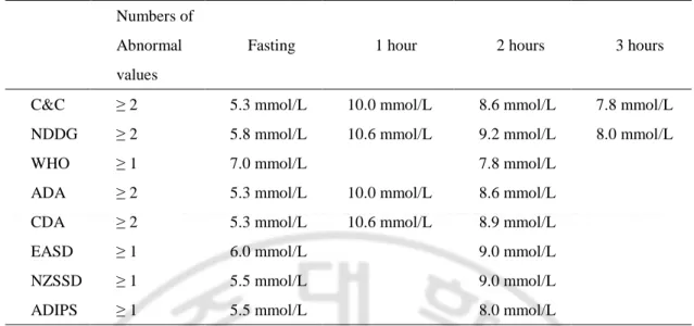
The gold standard for diagnosing GDM is the 3 hour 100g oral glucose tolerance test (OGTT). The patients should be expected to follow the strict conditions for this test: intake of at least 150 grams of carbohydrate per meal during the 3 days before the test, no other allowance of food and drink except for water between 8 and 14 hours before the test, prohibition of smoking for 12 hours before the test, rest for 30 minutes before the fasting glucose measurement, drinking of 100 grams glucose solution within 5 minutes of the test, and the restriction of smoking and walking during the test. Table 1 shows the various cutoff values for the diagnosis of GDM (Hanna and Peters, 2002).
There are some limitations in OGTT. These tests are not only time-consuming but also unpleasant, especially if administered during the first trimester. All the tests are performed under the conditions of supra-physiological glucose load that is irrespective of body weight. In addition, the limitation of age, various predictive values according to the ethnicity, and lack of reproducibility were considered to be limitations in OGTT (Hanna and Peters, 2002).
Table 1. Cutoff values for the different diagnostic criteria.
Numbers of Abnormal values
Fasting 1 hour 2 hours 3 hours C&C ≥ 2 5.3 mmol/L 10.0 mmol/L 8.6 mmol/L 7.8 mmol/L NDDG ≥ 2 5.8 mmol/L 10.6 mmol/L 9.2 mmol/L 8.0 mmol/L WHO ≥ 1 7.0 mmol/L 7.8 mmol/L
ADA ≥ 2 5.3 mmol/L 10.0 mmol/L 8.6 mmol/L CDA ≥ 2 5.3 mmol/L 10.6 mmol/L 8.9 mmol/L EASD ≥ 1 6.0 mmol/L 9.0 mmol/L NZSSD ≥ 1 5.5 mmol/L 9.0 mmol/L ADIPS ≥ 1 5.5 mmol/L 8.0 mmol/L
C&C, Carpenter and Coustan (Carpenter and Coustan, 1982); NDDG, National Diabetes Data Group (National Diabetes Data Group, 1979); WHO, World Health Organization (World Health Organization, 1999); ADA, American Diabetes Association (American Diabetes Association, 2004); CDA, Canadian Diabetes Association (Canadian Diabetes Association Clinical Practice Guidelines Expert Committee, 2003); EASD, European Association for the Study of Diabetes (Pregnancy and Neonatal Care Group of the European Association for the Study of Diabetes, 1996); NZSSD, New Zealand Society for the study of Diabetes (The Australasian Diabetes in Pregnancy Society, 1998); ADIPS, Australasian Diabetes in Pregnancy Society (The Australasian Diabetes in Pregnancy Society, 1998)
C. Implications for the offspring
Insulin resistance in pregnancy physiologically results from increased placental lactogen, growth hormone, progesterone, cortisol, and prolactin in late trimester and dissolves promptly postpartum (Ryan, 2003). Mothers with GDM have more elevated glucose, amino acids, and free fatty acids than those with normal glucose tolerance (NGT) in pregnancy (Catalano et al, 2003). Increased maternal nutrients are conveyed to the fetus and stimulate fetal insulin production (Pedersen, 1967). Since insulin plays an important role in the growth of fetus, macrosomia and is common in the offspring of GDM mothers (Jang et al, 1997). The frequent occurrence of perinatal morbidities such as injuries of the brachial plexus, neonatal hypoglycaemia, and foetal distress in offspring also results from glucose intolerance during gestation (Jang et al, 1997). Moreover, the significant increased fat mass in the infants of mothers with GDM is observed compared with the infants with NGT (Catalano et al, 2003). This elevated birth weight and body fat in infants is expected to be the precursor of obesity in adolescents as well as in later life.
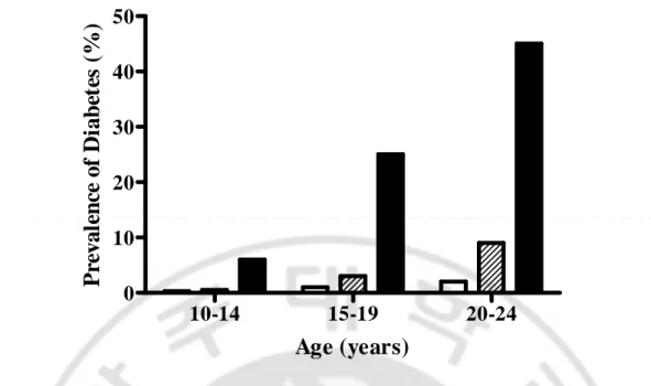
In Pima Indians study comparing the risk of obesity and diabetes in siblings born before and after the recognition of GDM in their mothers, the mean BMI was significantly higher in the offspring of GDM mothers than in the offspring of non diabetic mothers. In addition, the odds ratio for developing diabetes in the offspring of GDM mothers was 3.7 when compared to the offspring of non diabetic mother (Dabelea et al, 2000). Furthermore, a number of epidemiological studies have demonstrated that altered glucose metabolism in mothers during pregnancy has both short- and long-term adverse effects on their offspring
(Pettitt et al, 1983; Pettitt et al, 1988; Silverman et al, 1995; Cho et al, 2000; Boney et al, 2005; Krishnaveni et al, 2005) (Table2, Fig. 1). Pedersen and Freinkel established the theory of fuel-mediated teratogenesis, in which the intrauterine environment is influenced by excessive maternal fuels, which may have short- and long-term adverse effects on offspring (Pedersen, 1967; Freinkel, 1980).
Table 2. Mean percentage of desirable weight in offspring according to age and mother’’’’s diabetic status (Pettitt et al, 1983).
Age group of offspring
5-9 years 10-14 years 15-19 years Mother’s
Diabetic status
% desirable weight (95% confidence interval) no. in group
Nondiabetic 112 (111-114) 767 120 (118-122) 875 117 (114-119) 518 Prediabetic 114 (111-116) 296 123 (120-125) 430 125 (122-128) 336 Diabetic 132 (125-139) 48 149 (141-156) 51 145 (133-157) 24
10-14
15-19
20-24
0
10
20
30
40
50
Age (years)
P
re
v
a
le
n
ce
o
f
D
ia
b
et
es
(
%
)
Fig. 1. Prevalence of diabetes (2-h postload plasma glucose concentration ≥≥≥≥ 200mg/dl) according to age at examination and mother’’’’s diabetes status. White bars, nondiabetic; crosshatched bars,
D. Implications for mother at postpartum
Previous studies have indicated that mothers with GDM are more likely to present with pre-eclampsia, premature rupture of the membranes, shoulder dystocia, Caesarean section, and pre-term delivery than are those with NGT during pregnancy (Jang et al, 1997; Xiong et al, 2001; Kim et al, 2002).
Women with previous history of GDM had more elevated blood pressure and lipid profiles including total cholesterol, triglycerides, low density lipoprotein, and glucose (Meyers-Seifer and Vohr, 1996; Pallardo et al, 1999), and were at greater risk for developing overt type 2 diabetes mellitus (T2DM) at postpartum period (Jovanovic and Pettitt, 2001). The recent systematic review showed the conversion rate of diabetes in women with previous history of GDM was 3 to 65% resulted from the diversity of criterion on diabetes, length of follow-up, and ethnicity (Kim C et al, 2002). Our previous study showed 116 of 909 (12.8%) and 120 of 909 (13.2%) women were converted into either DM or impaired glucose tolerance (IGT), respectively during the 6 years postpartum follow-up (Cho et al, 2006). The incidence of diabetes in women with a previous history of GDM was almost
risk factors have been found: severity of glucose intolerance during pregnancy, insulin demand during pregnancy, earlier diagnosis of GDM, family history of diabetes, recurrence of GDM, increasing parity, maternal age, prepregnant body mass index, and weight gain during or postpartum (Dornhorst et al, 1990; Metzger et al, 1993; Kjos et al, 1995; Albareda et al, 2003). Pregnant women physiologically have decreased insulin sensitivity of maternal tissues (Agardh et al, 1996), which is considered to be caused by the changes in the endocrine and pancreas (Nieuwenhuizen et al, 1997). Nonetheless, women with GDM not only have usually more increased insulin resistance but also more impaired insulin secretion during pregnancy, independent of the development of diabetes at postpartum, than normal women (Kautzky-Willer A et al, 1997; Catalano et al, 1999; Homko et al, 2001). This metabolic aberration that is defective β-cell function is not capable of compensating increased insulin resistance, and it is likely to remain at postpartum and contribute to future diabetes (Buchanan TA, 2001).
Furthermore, among groups with a family history of diabetes, women with GDM have more metabolic syndrome and type 2 diabetes (T2DM) as well as higher prevalence of cardiovascular disease (CVD) than the ones without GDM (Carr DB et al, 2006). In nondiabetic African-American women with parental history of T2DM, women with prior GDM have been found to have inadequate β-cell function and decreased insulin sensitivity compared with age, waist-hip ratio, and body weight matched ones without prior GDM and healthy controls (Osei K et al, 1998). These results suggest that women with GDM might have an additional genetic predisposition distinct from that related to a family history of T2DM.
E. Purpose of the study
There have been limited studies to identify the early-stage manifestations and the relationship of CVD risk factors in the offspring of GDM mothers, especially in the Asian population. Therefore, in this prospective study, we hypothesized that the intrauterine environment of women with GDM is likely to influence the CVD risk factors of offspring. The objective of this study was to investigate the relationship between the maternal metabolism and the long-term adverse effects on the glucose metabolism and CVD risk factors in the offspring of diabetic mothers.
. SUBJECTS AND METHODS
Ⅱ
Ⅱ
Ⅱ
Ⅱ
A. Study design and subjects
This study was conducted at one major general hospital (Il-Shin Christian) and three university hospitals (Ajou, Seoul National, and Pochon Cha University) in Korea. During 24-28 weeks of gestation, a 50 g glucose challenge test was performed if the 1 h plasma glucose value was ≥ 130 mg/dL (7.2 mmol/L), a 3 h OGTT was performed during 28-32 weeks of gestation. GDM was diagnosed using the criteria of the National Diabetes Data Group (National Diabetes Data Group, 1979). Medical nutritional control was recommended to all women with either IGT or GDM. However some of the severe cases were treated with insulin (approximately 19.5%). The study subjects were recruited from August 1995 to May 1997 and were registered in the GDM registry. From 2300 entries in the GDM registry, we identified 1050 mothers who were available to be re-examined. Of the available subjects, 909 mothers were followed up more than once with post-partum evaluations. All the participants were followed up at six weeks and subsequent follow-ups were made annually. All the participants underwent a 2 h 75 g OGTT. The WHO criteria were used to diagnose DM (World Health Organization, 1985).
Of the 909 subjects, 298 offspring (3-5 years old) were recruited consecutively for the study and followed up every year. During the follow-up period, 160 (53.7%) of the 298 recruited offspring were visited twice or more: 91 were visited twice, 60 three times, and nine four times. The median follow-up period was 5.1 years (range, 3.8 - 8.9 years) for
offspring visited twice or more. Mothers of offspring visited only once had lower incomes, higher rates of GDM, and lower parity, and used more insulin relative to mothers of offspring visited twice or more. The area under the curve (AUC) for glucose in the mothers of offspring visited once was greater than that for mothers of offspring visited twice or more, whereas the AUC for insulin was lower, as calculated by a 3 h OGTT performed during 24-28 weeks of gestation. However, there were no significant differences in the neonatal characteristics, including gestational age at delivery, offspring's sex, and birth weight, between the two groups (i.e., those visited once and those visited twice or more) (Table 3). Of the 298 children, 202 were the offspring of GDM mothers and 96 were the offspring of mothers with IGT, with one abnormal value on the 3 h OGTT. All the participating women gave their written informed consent and also co-signed on behalf of their offspring. This study protocol was approved by the Ethical Committee of Ajou University School of Medicine Review Board.
Table 3. Antepartum maternal and neonatal characteristics in offspring of once and twice or more visited mothers.
Once visited (n = 138)
Twice or more visited (n = 160)
P
Maternal
Education level (> 12 years) 60 (43.8) 76 (47.5) NS Income level (> 2 million won) 83 (60.6) 125 (78.1) < 0.01 Family history of diabetes 54 (39.1) 65 (40.6) NS Age at delivery (years) 31.7 ± 4.4 31.4 ± 4.1 NS Parity 1.9 ± 0.7 2.0 ± 0.6 NS Pre-pregnant BMI (kg/m2) 23.0 ± 3.4 22.4 ± 3.5 NS HbA1c 5.4 ± 0.8 5.3 ± 1.2 NS
GDM 102 (73.9) 100 (62.5) < 0.05 AUC for glucose (mmol/L per 3 h) 28.1 ± 6.4 26.7 ± 4.5 < 0.05 AUC for insulin (µg/mL per 3 h) 175.7 ± 132.3 207.1 ± 141.8 NS Insulin used 185 (36.1) 58 (20.4) < 0.001
Neonatal
Gestational age at delivery (weeks) 38.5 ± 1.6 38.7 ± 1.9 NS Sex (male / female) 72 / 63 (53.3 / 46.7) 89 / 71 (55.3 / 44.4) NS Birth weight (g) 3347.4 ± 602.2 3308.5 ± 587.2 NS
B. Anthropometric measurements
Anthropometric data were collected by trained staff using standardized protocols. The height and weight of the mothers were measured while the patient was barefoot and wearing lightweight clothing. Each woman's BMI was calculated by dividing her weight (kg) by her height squared (m2). Pre-pregnant weight was reported at the gestational screening test for GDM. In offspring, waist circumference was measured at the level of the umbilicus and hip circumference was measured at the level of the greater trochanters. Skinfold thickness was measured using skinfold calipers. For blood pressure measurements, the patient sat quietly for 10 minutes before the measurements were made and a paediatric cuff was used. A five-minute rest period was allowed between each measurement. Total body fat content was measured as the percentage (%) of body fat using a bioelectric impedance analyser (Body Composition Analyzer, Gilwoo Co., Korea). The level of obesity was defined as a BMI ≥ 95th percentile specific for age and sex (Hong et al, 1999). AUC for glucose and insulin were calculated using the trapezoidal method.
Plasma glucose concentrations were measured immediately after the blood was drawn using a glucose oxidase method (YSI 2300-STAT; YSI Life Sciences, Yellow Springs, OH) and plasma insulin and C-peptide were measured using radioimmunoassay kits (Linco Research Inc., St Louis, MO). Total cholesterol and triglyceride concentrations were determined by enzymatic procedures with a Beckman analyser (Beckman Instruments, Brea, CA). High-density lipoprotein (HDL) cholesterol levels were determined using the direct Sigma EZ-HDL assay (Sigma Diagnostics, St Louis, MO).
D. Statistical analysis
Statistical analyses were performed using SPSS for Windows 12.0. All the results are expressed as means ± standard deviations (SD) and percentages, unless otherwise stated. Differences in the means of the continuous variables were tested with an independent t-test. Comparisons of proportions between the two groups were analyzed using the c2 test. To correct normality, log transformations were performed for those variables that were not normally distributed, such as triglyceride and insulin concentrations. However, to simplify the interpretation, all results are expressed as untransformed values. Comparisons of BMI between the offspring of IGT and GDM mothers were made using ANCOVA, adjusted for covariates. The covariates were:1) antepartum maternal variables, including age at delivery, pre-pregnant BMI, and insulin used; 2) neonatal variables, including gestational age at delivery, sex, age, and birth weight. The relationships between CVD risk factors and age were examined using Pearson's correlation test. To examine the differences in CVD risk
factors with age between the IGT and GDM mothers, the slopes of two straight lines using separate regression fits were compared (Kleinbaum et al). Statistical significance was defined as P < 0.05.
Ⅲ
Ⅲ
Ⅲ
Ⅲ. RESULTS
The 611 non-participating mothers had lower incomes, higher rates of GDM, and lowered parity, and used more insulin than the participating mothers. The AUC for insulin, calculated by a 3 h OGTT performed during 28-32 weeks of gestation, for non-participating mothers was less than that for the 298 participating mothers. However, there was no significant difference in the AUC for glucose at gestation among the mothers or in the neonatal characteristics of the two groups, such as gestational age at delivery, offspring's sex, and birth weight (Table 4).
Table 4. Antepartum maternal and neonatal characteristics in offspring of non-participated and participated mothers. Non-participated (n = 611) Participated (n = 298) P Maternal
Education level (> 12 years) 309 (50.7) 136 (45.8) NS Income level (> 2 million won) 233 (38.4) 208 (70.0) < 0.001 Family history of diabetes 209 (34.2) 119 (40.1) NS Age at delivery (years) 31.2 ± 4.2 31.2 ± 4.2 NS Parity 1.6 ± 0.7 1.9 ± 0.6 < 0.001 Pre-pregnant BMI (kg/m2) 22.9 ± 3.5 22.7 ± 3.4 NS HbA1c 5.2 ± 1.3 5.4 ± 1.1 NS
GDM 458 (74.8) 202 (68.0) < 0.05 AUC for glucose (mmol/L per 3 h) 27.7 ± 5.0 27.4 ± 5.5 NS AUC for insulin (µg/mL per 3 h) 109.0 ± 92.4 162.5 ± 133.2 < 0.001 Insulin used 185 (36.1) 58 (20.4) < 0.001
Neonatal
Gestational age at delivery (weeks) 38.7 ± 1.4 38.7 ± 1.7 NS Sex (male / female) 296 / 230 (56.3 / 43.7) 161 / 134 (54.6 / 45.4) NS Birth weight (g) 3386.8 ± 528.1 3326.2 ± 593.4 NS
Antepartum maternal and neonatal characteristics are summarized in Table 5. Compared with IGT mothers, GDM mothers were older at delivery, had a higher HbA1c level and a greater AUC for glucose on a 3 h OGTT during 28-32 weeks of gestation, and used more insulin. GDM mothers were slightly more obese than IGT mothers. There were no significant differences in the neonatal characteristics of the two groups. Fifty-nine GDM mothers (29.2%) and eight IGT mothers (8.4%) developed diabetes during the six years of the post-partum period. The GDM mothers showed a risk of developing diabetes that was 4.5 times (95% CI, 2.0-9.8) higher than that of IGT mothers. The risk of developing diabetes in GDM mothers remained 3.7 times (95% CI, 1.5-9.0) higher than that of IGT mothers after adjustment for potential confounding factors, such as age at delivery, education level, income level, family history of diabetes mellitus, parity, pre-pregnant BMI, and insulin used.
Table 5. Antepartum maternal and neonatal characteristics in offspring of IGT and GDM mothers.
IGT (n = 96) GDM (n = 202) P
Maternal
Education level (> 12 years) 40 (41.7) 96 (47.5) NS Income level (> 2 million won) 70 (71.9) 138 (68.3) NS Family history of diabetes 37 (38.5) 82 (40.6) NS Age at delivery (years) 30.3 ± 4.2 31.4 ± 4.2 < 0.05 Parity 2.0 ± 0.7 1.9 ± 0.6 NS Pre-pregnant BMI (kg/m2) 22.1 ± 3.1 22.9 ± 3.6 0.064 HbA1c 5.1 ± 1.4 5.5 ± 0.8 < 0.01
AUC for glucose (mmol/L per 3 h) 23.9 ± 2.0 29.0 ± 5.8 < 0.001 AUC for insulin (µg/mL per 3 h) 204.4 ± 154.0 192.6 ± 132.8 NS Insulin used 8 (8.3) 50 (24.8) 0.001
Neonatal
Gestational age at delivery (weeks) 38.7 ± 2.2 38.6 ± 1.5 NS Sex (male / female) 45 / 51 (46.9 / 53.1) 116 / 86 (57.4 / 42.6) NS Birth weight (g) 3286.6 ± 612.4 3344.6 ± 585.0 NS Data are presented as means ± SD or as numbers with percentages (%) in parentheses. BMI: body mass index; AUC: area under the curve; NS: not significant.
A total of 242 (82.4%) offspring were less than five years old at the first visit. The mean age of offspring at the first visit was 4.1 - 1.1 years (range, 2.4 - 8.8 years). There were no significant differences in CVD risk factors between the offspring in the two groups at the first visit (Table 6). BMI ≥ 95th percentile was observed in four (4.3%) offspring of IGT mothers and in 17 (8.5%) offspring of GDM mothers, but the differences between the groups was not significant.
Correlations between biochemical values of mothers during pregnancy and cardiovascular risk factors of offspring at first visit were analyzed using Pearson correlation test. There were no significant correlations between the groups, except for correlation between HbA1c in mothers and total cholesterol in offspring (Table 7).
Table 6. Cardiovascular disease risk factors of offspring of IGT mothers and GDM mothers at the first visit.
IGT GDM P Age (years) 4.0 ± 0.9 4.2 ± 1.1 NS Systolic blood pressure (mmHg) 92.3 ± 8.7 93.3 ± 9.3 NS Diastolic blood pressure (mmHg) 59.0 ± 7.5 59.6 ± 8.5 NS Skinfold thickness (mm) 9.3 ± 4.9 8.5 ± 4.4 NS Body fat (%) 20.2 ± 6.0 18.9 ± 5.3 NS Total cholesterol (mmol/L) 4.2 ± 0.8 4.4 ± 0.7 NS Triglyceride (mmol/L) 0.8 ± 0.3 0.9 ± 0.4 NS HDL-cholesterol (mmol/L) 1.4 ± 0.3 1.4 ± 0.3 NS Fasting plasma glucose (mmol/L) 4.7 ± 0.5 4.8 ± 0.5 NS 2 h plasma glucose (mmol/L) 5.3 ± 1.0 5.3 ± 0.9 NS Fasting plasma insulin (µg/mL) 6.8 ± 3.5 7.2 ± 3.9 NS 2 h plasma insulin (µg/mL) 17.3 ± 11.0 17.4 ± 15.1 NS Body mass index (kg/m2) 16.1 ± 1.7 16.1 ± 1.9 NS Data are presented as means ± SD. HDL: High Density Lipoprotein; NS: not significant.
Table 7. Correlation coefficients between the biochemical values of mothers during pregnancy and the cardiovascular risk factors of offspring at the first visit.
Mothers† Offspring FPG 1HPG 2HPG 3HPG HbA1c SBP 0.050 -0.012 0.025 0.043 0.010 DBP 0.097 0.006 0.082 0.091 0.022 SST 0.029 0.004 0.043 -0.019 0.097 BF% -0.060 -0.015 -0.057 -0.018 -0.009 TC -0.008 0.006 0.063 0.034 0.150* TG 0.050 -0.011 0.067 0.021 0.020 HDLC -0.026 -0.024 -0.057 0.004 0.008 FPG 0.013 -0.043 -0.038 -0.032 -0.026 2HPG 0.005 0.048 0.022 0.044 0.018 FPI 0.005 -0.003 -0.024 -0.044 -0.051 2HPI -0.031 -0.024 -0.004 -0.015 -0.004 BMI 0.036 0.052 0.029 -0.030 0.055 Pearson correlation was used to test the association among the variables. * P < 0.05.
†
Values derive from 3 hour oral glucose tolerance test during 28-32 weeks of gestation.
FPG, fasting plasma glucose; 1HPG, 1 hour plasma glucose; 2HPG, 2 hour plasma glucose; 3HPG, 3 hour plasma glucose; PBMI, prepregnant body mass index; SBP, systolic blood pressure; DBP, diastolic blood pressure; SST, suprailiac skinfold thickness; BF%, body fat %; TC, total cholesterol; TG, triglyceride; HDLC, high density lipoprotein cholesterol; 2HPG, 2 hour plasma glucose; FPI, fasting plasma insulin; 2HPI, 2 hour plasma insulin; BMI, body mass index.
The differences in CVD risk factors between the groups were analyzed according to the age at the first visit. Of these, the mean BMI of the offspring of GDM mothers at three years of age (15.9 ± 2.0 kg/m2, n = 100) was slightly lower than that of the offspring of IGT mothers (16.5 ± 2.0 kg/m2, n = 53). However, the BMI of offspring of GDM mothers who were five years old or more (16.9 ± 2.0 kg/m2, n = 35) was significantly higher than that of the offspring of IGT mothers (15.7 ± 1.5 kg/m2, n = 19) (P < 0.05) (Fig. 2). The BMI of the offspring of GDM mothers who were five years old or more (16.8 kg/m2 [95% CI, 16.2-17.4]) remained significantly higher than that of the offspring of IGT mothers (15.2 kg/m2 [95% CI, 14.4-16.1]) (P < 0.01), even after adjustments were made for the mother's age at delivery, pre-pregnant BMI, insulin used, and gestational age at delivery, and the offspring's age, sex, and birth weight.
Fig. 2. A comparison of BMI of the offspring of IGT and GDM mothers according to age. * P <
0.05. After adjustment for the mother’s age at delivery, pre-pregnant BMI, insulin used, and gestational age at delivery, and the offspring’s age, sex, and birth weight, the BMI of the offspring of GDM mothers, who were five years old or more (16.9 kg/m2 [95% CI, 16.2–17.4]) remained significantly higher than that of the offspring of IGT mothers (15.2 kg/m2, [95% CI, 14.3–16.1]) (P < 0.01).
All follow-up data were included in analyzing the correlation between age and CVD risk factors in both groups. The correlation coefficients between age and CVD risk factors for the offspring of IGT and GDM mothers are shown in Table 8. Positive relationships between age and systolic blood pressure, diastolic blood pressure, body fat, and 2 h plasma insulin levels were observed in both groups. In the offspring of GDM mothers, suprailiac skinfold thickness (r = 0.192, P < 0.001), fasting plasma glucose level (r = 0.233, P < 0.001), 2 h plasma glucose level (r= 0.112, P < 0.05), fasting plasma insulin level (r = 0.272, P < 0.001), and BMI (r = 0.174, P < 0.01) were positively correlated with age. However, a negative and significant relationship between age and BMI was observed in the offspring of IGT mothers (r = 0.230, P < 0.01). There was no significant correlation between age and the lipid profiles of either group.
Table 8. Correlation between age and cardiovascular disease risk factors in offspring of IGT and GDM mothers.
IGT GDM
r n P r n P Systolic blood pressure (mmHg) 0.328 181 < 0.001 0.305 336 < 0.001 Diastolic blood pressure (mmHg) 0.180 181 < 0.05 0.248 336 < 0.001 Suprailiac skinfold thickness (mm) –0.105 177 NS 0.192 335 < 0.001 Body fat (%) 0.182 181 < 0.05 0.341 324 < 0.001 Total cholesterol (mmol/L) 0.083 185 NS –0.043 338 NS Triglyceride (mmol/L) 0.006 185 NS –0.008 338 NS HDL cholesterol (mmol/L) 0.096 184 NS 0.020 338 NS Fasting plasma glucose (mmol/L) 0.057 186 NS 0.233 340 < 0.001 2 h plasma glucose (mmol/L) –0.020 173 NS 0.112 311 < 0.05 Fasting plasma insulin (µg/mL) 0.049 185 NS 0.272 337 < 0.001 2 h plasma insulin (µg/mL) 0.230 171 < 0.01 0.346 314 < 0.001 Body mass index (kg/m2) –0.230 187 < 0.01 0.174 339 < 0.01
The slopes of the linear regression lines for BMI and fasting plasma insulin levels with age were significantly steeper for the offspring of GDM mothers than for the offspring of IGT mothers (P < 0.001 and P < 0.05, respectively) (Figs 3, 4). The slope of the linear regression line for fasting plasma glucose levels with age was marginally steeper for the offspring of GDM mothers (P = 0.052).
2 3 4 5 6 7 8 9 10 15 20 25
Age (years)
B
o
d
y
m
a
s
s
i
n
d
e
x
(
k
g
/m
2)
2 3 4 5 6 7 8 9 10 15 20 25Age (years)
B
o
d
y
m
a
s
s
i
n
d
e
x
(
k
g
/m
2)
Fig. 3. The linear regression of fitted lines for BMI in the offspring of IGT (open squares) and GDM mothers (closed squares). The slope of the linear regression line for BMI with age is
significantly steeper for the offspring of GDM mothers than for the offspring of IGT mothers (P < 0.001).
2 3 4 5 6 7 8 9 0 5 10 15 20 25
Age (years)
F
a
s
ti
n
g
p
la
s
m
a
i
n
s
u
li
n
l
e
v
e
l
(
µ
g
/m
L
)
2 3 4 5 6 7 8 9 0 5 10 15 20 25F
a
s
ti
n
g
p
la
s
m
a
i
n
s
u
li
n
l
e
v
e
l
(
µ
g
/m
L
)
Ⅳ
Ⅳ
Ⅳ
Ⅳ. DISCUSSION
Women with normal pregnancies have elevated insulin resistance, induced by increasing adiposity and placental hormones, which reduce the action of insulin, and increasing insulin secretion by the pancreatic β-cells, which compensates for the insulin response (Buchanan and Xiang, 2005). This phenomenon of an inappropriate insulin supply to regulate normalglucose metabolism in pregnancy causes GDM (Buchanan et al, 1990; Catalano et al, 1993; Honk et al, 2001; Buchanan and Xiang, 2005). Moreover, GDM plays an important role in the post-partum development of diabetes (Jovanovic and Pettitt, 2001). A recent study that reviewed a long-term follow-up study of GDM subjects revealed that the cumulative incidence of diabetes after delivery ranged from 2.6% to 70%. These rate differences are due to ethnicity, different diagnostic criteria, and variations in the follow-up period (Kim et al, 2002). In our study, 59 GDM mothers (29.2%) and eight IGT mothers (8.4%) developed diabetes during the six-year post-partum period. The risk of developing diabetes in GDM mothers was 3.7 times (95% CI, 1.5-9.0) higher than that in IGT mothers after adjustments were made for potential confounding factors, such as age at delivery, education level, income level, family history of diabetes mellitus, parity, pre-pregnant BMI, and insulin used.
Numerous previous studies have indicated that maternal diabetes during pregnancy has both short- and long-term adverse effects on offspring (Pettitt et al, 1983; Pettitt et al, 1988; Silverman et al, 1995; Cho et al, 2000; Boney et al, 2005; Krishnaveni et al, 2005).
Metzger et al. (Silverman, 1993) reported that the frequency of IGT was increased in the adolescent offspring of diabetic mothers and established the hypothesis that "diabetes begets diabetes", indicating that the intrauterine environment and the altered glucose regulation in the second and third trimesters absolutely influenced the short- and long-term health of the foetus.
The results of our study are similar to those of previous studies. In this study, mothers were divided into two groups according to GDM status during pregnancy, because there were no significant correlations between the individual biochemical values of mothers during pregnancy and the cardiovascular risk factors of offspring at the first visit. Although there were no significant differences in the CVD risk factors between the offspring of IGT and GDM mothers at the first visit, the mean BMI of the offspring of GDM mothers increased at the first visit according to age. The BMI of the offspring of GDM mothers who were five years old or more was significantly higher than that of the offspring of IGT mothers, and this difference remained significant after adjustment for covariates. A significant positive correlation between age and BMI in the offspring of GDM mothers was observed, whereas a negative correlation was observed in the offspring of IGT mothers. The
plasma glucose levels between the two groups, childhood obesity as well as the altered glucose metabolism caused by the maternal uterine environment are expected to be more obvious in the offspring of GDM mothers than in the offspring of IGT mothers.
The lean body mass rather than the fat mass contributes the annual increment in the BMI of school-aged children and adolescents (Maynard et al, 2001). However, the BMI of children and adolescents is strongly associated with blood pressure (Falkner et al, 2006), as well as adiposity (measured by dual-energy X-ray absorptiometry) and metabolic risk factors, including glucose, insulin, and lipids (Lindsay et al, 2001). Furthermore, childhood BMI is related to BMI, adiposity, insulin, lipids, and systolic blood pressure in adulthood (Sinaiko et al, 1999; Li et al, 2003). It also predicts the carotid intima-media thickness, which is the best end point with which to predict carotid artery disease (Li et al, 2003). The Expert Committee on Clinical Guidelines for Overweight in Adolescent Preventive Services defined overweight as a BMI ≥ 95th percentile for age and sex, or greater than 30 (kg/m2) and defined the increased risk of obesity as a BMI between the 85th and 95th percentiles (Himes et al, 1994). In our study, 17 (8.5%) offspring of GDM mothers and four(4.3%) offspring of IGT mothers had BMI ≥ 95th percentile.
The BMI of the offspring of diabetic mothers was markedly reduced during the first two years after birth. A steep weight gain was observed subsequently until school-age (Silverman et al, 1998; Touger et al, 2005). In contrast, BMI is high in the first year of offspring, decreasing between 25 years, and then an upward tendency from school age to puberty in the normal paediatric population (Veldhuis et al, 2005). In this study, there was no significant difference inthe birth weight and the BMI of offspring up to three years of age
between IGT and GDM mothers. However, the BMI of the offspring of GDM mothers overtook those of IGT mothers and reached significant levels at five years of age. This difference between the offspring of IGT and GDM mothers could be due to the effect of maternal uterine environment during pregnancy. Another possible cause could be the influence of the lifestyle of GDM mothers versus IGT mothers on their offspring. Some studies (Mitchell et al, 2003; Davison et al, 2005) showed that parents' lifestyle did influence their children's lifestyle whereas other studies (Baughcum et al, 2001; Trost et al, 2003) showed no significant association between parents' and children's lifestyle in early age. However, these studies did not compare the difference of lifestyle between mothers with and without a history of GDM. Hence, further research is needed to explain the difference of BMI between the offspring of IGT and GDM mothers.
Our results are based on a study population that represented 32.8% (298 of 909) of eligible mothers. Of these, 53.7% (160 of 298) were followed up twice or more. Therefore, our study is susceptible to selection bias. However, there were no significant differences between the neonatal characteristics of the participating and non-participating group, or between the groups of participants who were followed up once or twice or more.
pregnancy when the study protocols were set up. However, women with IGT during pregnancy have higher elevated glucose levels and a higher risk of developing diabetes in the post-partum period than those with NGT (Bian et al, 2000). From the above mentioned study, we can infer that women with IGT had greater influences on CVD risk factors among offspring than those with NGT. The differences in CVD risk factors among the offspring of different mothers may have been more evident if the offspring of women with NGT during pregnancy were included in this study.
This prospective study identified early manifestations of CVD risk factors in the offspring of GDM mothers. Therefore, the implementation of early medical assessments for diabetes, obesity, and CVD in the offspring of diabetic mothers is warranted to prevent potential chronic diseases in later life.
Ⅴ
Ⅴ
Ⅴ
Ⅴ. CONCLUSIONS
Childhood obesity, as well as altered glucose metabolism influenced by the maternal uterine environment, is more likely with advancing years in the offspring of GDM mothers than in the offspring of IGT mothers.
REFERENCES
1. Agardh CD, Aberg A, Norden NE: Glucose levels and insulin secretion during a 75 g glucose challenge test in normal pregnancy. J Intern Med 240: 303-309, 1996
2. Albareda M, Caballero A, Badell G, Piquer S, Ortiz A, de Leiva A, Corcoy R: Diabetes and abnormal glucose tolerance in women with previous gestational diabetes. Diabetes
Care 26: 1199-1205, 2003
3. American Diabetes Association: Gestational diabetes mellitus. Position statement of the American Diabetes Association. Diabetes Care 27: S88-S90, 2004
4. Baughcum AE, Powers SW, Johnson SB, Chamberlin LA, Deeks CM, Jain A: Maternal feeding practices and beliefs and their relationships to overweight in early childhood. J
Dev Behav Pediatr 22: 391-408, 2001
5. Berger H, Crane J, Farine D: Screening for gestational diabetes mellitus. JOGC 24: 894-912, 2002
6. Bian X, Gao P, Xing X, Xu H, Qian M, Liu S: Risk factors for development of diabetes mellitus in women with a history of gestational diabetes mellitus. Chin Med J 113: 759-762, 2000
7. Boney CM, Verma A, Tucker R, Vohr BR: Metabolic syndrome in childhood: Association with birth weight, maternal obesity, and gestational diabetes mellitus.
8. Brody SC, Harris R, Lohr K: Screening for gestational diabetes: a summary of the evidence for the U.S. Preventive Services Task Force. Obstet Gynecol 101: 380-392, 2003
9. Buchanan TA, Metzger BE, Freinkel N, Bergman RN: Insulin sensitivity and b-cell responsiveness to glucose during late pregnancy in lean and moderately obese women with normal glucose tolerance or mild gestational diabetes. Am J Obstet Gynecol 162: 1008-1014, 1990
10. Buchanan TA: Pancreatic B-cell defects in gestational diabetes: implications for the pathogenesis and prevention of type 2 diabetes. J Clin Endocrinol Metab 86: 989-993, 2001
11. Buchanan TA, Xiang AH: Gestational diabetes mellitus. J Clin Invest 115: 485-491, 2005
12. Canadian Diabetes Association Clinical Practice Guidelines Expert Committee: Canadian Diabetes Association 2003 Clinical Practice Guidelines for the Prevention and Management of Diabetes in Canada. Can J Diabetes 27: S99-S105, 2003
increases the risk of cardiovascular disease in women with a family history of type 2 diabetes. Diabetes Care 29: 2078-2083, 2006
15. Catalano RM, Tyzbir ED, Wolfe RR, Calles J, Roman NM, Amini SB: Carbohydrate metabolism during pregnancy in control subjects and women with gestational diabetes.
Am J Physiol 264: E60-E67, 1993
16. Catalano PM, Huston L, Amini SB, Kalhan SC: Longitudinal changes in glucose metabolism during pregnancy in obese women with normal glucose tolerance and gestational diabetes mellitus. Am J Obstet Gynecol 180: 903-916, 1999
17. Catalano PM, Thomas A, Huston-Presley L, Amini SB: Increased fetal adiposity: a very sensitive marker of abnormal in utero development. Am J Obstet Gynecol 189: 1698-1704, 2003
18. Catalano PM, Kirwan JP, Haugel-de Mouzon S, King J: Gestational diabetes and insulin resistance: role in short- and long-term implications for mother and fetus. J Nutr 133 (Suppl 2): 1674S-1683S, 2003
19. Cheung NW, Byth K: Population health significance of gestational diabetes. Diabetes
Care 26: 2005-2009, 2003
20. Cho NH, Silverman BL, Rizzo TA, Metzger BE: Correlations between the intrauterine metabolic environment and blood pressure in adolescent offspring of diabetic mothers. J
21. Cho NH, Jang HC, Park HK, Cho YW: Waist circumference is the key risk factor for diabetes in Korean women with history of gestational diabetes. Diabetes Res Clin Pract 71: 177-183, 2006
22. Coustan DR, Nelson C, Carpenter MW, Carr SR, Rotondo L, Widness JA: Maternal age and screening for gestational diabetes: a population-based study. Obstet Gynecol 73: 557-561, 1989
23. Dabelea D, Hanson RL, Lindsay RS, Pettitt DJ, Imperatore G, Gabir MM, Roumain J, Bennett PH, Knowler WC: Intrauterine exposure to diabetes conveys risks for type 2 diabetes and obesity: a study of discordant sibships. Diabetes 49:2208-2211, 2000
24. Dabelea D, Snell-Bergeon JK, Hartsfield CL, Bischoff KJ, Hamman RF, McDuffie RS: Increasing prevalence of gestational diabetes mellitus (GDM) over time and by birth cohort: Kaiser Permanente of Colorado GDM Screening Program. Diabetes Care 28: 579-584, 2005
25. Davis CL, Gutt M, Llabre MM, Marks JB, O'Sullivan MJ, Potter JE: History of gestational diabetes, insulin resistance and coronary risk. J Diabetes Complications 13:
27. Dornhorst A, Bailey PC, Anyaoku V, Elkeles RS, Johnston DG, Beard RW: Abnormalities of glucose tolerance following gestational diabetes. Q J Med 77: 1219-1228, 1990
28. Egeland GM, Skjærven R, Irgens LM: Birth characteristics of women who develop gestational diabetes: population based study. BMJ 321: 545-547, 2000
29. Falkner B, Gidding SS, Ramirez-Garnica G, Wiltrout SA, West D, Rappaport EB: The relationship of body mass index and blood pressure in primary care pediatric patients. J
Pediatr 148: 195-200, 2006
30. Ferrara A, Kahn HS, Quesenberry CP, Riley C, Hedderson MM: An increase in the incidence of gestational diabetes mellitus: Northern California, 1991-2000. Obstet
Gynecol 103: 526-533, 2004
31. Freinkel N: Banting Lecture 1980. Of pregnancy and progeny. Diabetes 19: 1023-1035, 1980
32. Hanna FW, Peters JR: Screening for gestational diabetes; past, present and future.
Diabet Med 19: 351-358, 2002
33. Himes JH, Dietz WH: Guidelines for overweight in adolescent preventive services: recommendations from an expert committee. The Expert Committee on Clinical Guidelines for Overweight in Adolescent Preventive Services. Am J Clin Nutr 59: 307-316, 1994
34. Homko C, Sivan E, Chen X, Reece EA, Boden G: Insulin secretion during and after pregnancy in patients with gestational diabetes mellitus. J Clin Endocrinol Metab 86: 568-573, 2001
35. Hong YM, Moon KR, Seo JW, Sim JG, Yoo KW, Jeong BJ: Nation-wide study on body mass index, skinfold thickness and arm circumference in Korean children. J
Korean Pediatr Soc 42: 1186-1200, 1999
36. Honk C, Sivan E, Chen X, Reece EA, Borden G: Insulin secretion during and after pregnancy in patients with gestational diabetes mellitus. J Clin Endocrinol Metab 86: 568-573, 2001
37. Jang HC, Cho NH, Min YK, Han JK, Jung KB, Metzger BE: Increased macrosomia and perinatal morbidity independent of maternal obesity and advanced age in Korean women with GDM. Diabetes Care 20: 1582-1588, 1997
38. Jovanovic L, Pettitt DJ: Gestational diabetes mellitus. JAMA 286: 2516-2518, 2001
41. Kim C, Newton CM, R.H. Knopp: Gestational diabetes and the incidence of type 2 diabetes: a systematic review. Diabetes Care 25: 1862-1868, 2002
42. Kim HS, Chang KH, Yang JI, Yang SC, Lee HJ, Ryu HS: Clinical outcomes of pregnancy with one elevated glucose tolerance test value. Int J Gynaecol Obstet 78: 131-138, 2002
43. Kjos SL, Peters RK, Xiang A, Henry OA, Montoro M, Buchanan TA: Predicting future diabetes in Latino women with gestational diabetes. Utility of early postpartum glucose tolerance testing. Diabetes 44: 586-591, 1995
44. Kleinbaum DG, Kupper LL, Muller KE: Applied Regression Analysis and Other Multivariable Methods. 2nd ed. Boston, Massachusetts, PWS-KENT, pp.266-271
45. Krishnaveni GV, Hill JC, Leary SD, Veena SR, Saperia J, Saroja A: Anthropometry, glucose tolerance, and insulin concentrations in Indian children: relationships to maternal glucose and insulin concentrations during pregnancy. Diabetes Care 28: 2919-2925, 2005
46. Lauenborg J, Hansen T, Jensen DM, Vestergaard H, Molsted-Pedersen L, Hornnes P, Locht H, Pedersen O, Damm P: Increasing incidence of diabetes after gestational diabetes: a long-term follow-up in a Danish population. Diabetes Care 27: 1194-1199, 2004
47. Li S, Chen W, Srinivasan SR, Bond MG, Tang R, Urbina EM: Childhood cardiovascular risk factors and carotid vascular changes in adulthood: the Bogalusa Heart Study. JAMA 290: 2271-2276, 2003
48. Lindsay RS, Hanson RL, Roumain J, Ravussin E, Knowler WC, Tataranni PA: Body mass index as a measure of adiposity in children and adolescents: relationship to adiposity by dual energy x-ray absorptiometry and to cardiovascular risk factors. J
Clin Endocrinol Metab 86: 4061-4067, 2001
49. Maresh M: Screening for gestational diabetes mellitus. Semin Fetal Neonatal Med 10: 317-323, 2005
50. Maynard LM, Wisemandle W, Roche AF, Chumlea WC, Guo SS, Siervogel RM: Childhood body composition in relation to body mass index. Pediatrics 107: 344-350, 2001
51. Metzger BE, Bybee DE, Freinkel N, Phelps RL, Radvany RM, Vaisrub N: Gestational diabetes mellitus. Correlations between the phenotypic and genotypic characteristics of the mother and abnormal glucose tolerance during the first year postpartum. Diabetes
53. Metzger BE, Coustan DR: Summary and recommendations of the Fourth International Workshop-Conference on Gestational Diabetes Mellitus. Diabetes Care 37: B161-B167, 1998
54. Meyers-Seifer CH, Vohr BR: Lipid levels in former gestational diabetic mothers.
Diabetes Care 19: 1351-1356, 1996
55. Mitchell BD, Rainwater DL, Hsueh WC, Kennedy AJ, Stern MP, Maccluer JW: Familial aggregation of nutrient intake and physical activity: results from the San Antonio Family Heart Study. Ann Epidemiol 13: 128-135, 2003
56. Moses RG: The recurrence rate of gestational diabetes in subsequent pregnancies.
Diabetes Care 19: 1348-1350, 1996
57. National Diabetes Data Group: Classification and diagnosis of diabetes mellitus and other categories of glucose intolerance. Diabetes 28: 1039-1057, 1979
58. Nieuwenhuizen AG, Schuiling GA, Moes H, Koiter TR: Role of increased insulin demand in the adaptation of the endocrine pancreas to pregnancy. Acta Physiol Scand 159: 303-312, 1997
59. Osei K, Gaillard TR, Schuster DP: History of gestational diabetes leads to distinct metabolic alterations in nondiabetic African-American women with a parental history of type 2 diabetes. Diabetes Care 21: 1250-1257, 1998
60. O'Sullivan JB, Mahan CM: Prospective study of 352 young patients with chemical diabetes. N Engl J Med 278: 1038-1041, 1968
61. Pallardo F, Herranz L, Garcia-Ingelmo T, Grande C, Martin-Vaquero P, Janez M, Gonzalez A: Early postpartum metabolic assessment in women with prior gestational diabetes. Diabetes Care 22: 1053-1058, 1999
62. Pedersen JL: The Pregnant Diabetic and Her Newborn. Baltimore, Williams & Wilkins. 1967
63. Pettitt DJ, Baird HR, Aleck KA, Bennett PH, Knowler WC: Excessive obesity in offspring of Pima Indian woman with diabetes during pregnancy. N Engl J Med 308: 242-245, 1983
64. Pettitt DJ, Aleck KA, Baird HR, Carraher MJ, Bennett PH, Knowler WC: Congenital susceptibility to NIDDM - Role of intrauterine environment. Diabetes 37: 622-628, 1988
781-67. Ryan EA : Hormones and insulin resistance during pregnancy. Lancet 362: 1777-1778, 2003
68. Scott DA, Loveman E, McIntyre L, Waugh N: Screening for gestational diabetes: a systematic review and economic evaluation. Health Technol Assess 6: 1-161, 2002
69. Silverman BL, Cho NH, Metzger BE: Impaired glucose tolerance (IGT) in adolescent offspring of diabetic mothers (ODM): Does diabetes beget diabetes? Diabetes 42: 179A, 1993
70. Silverman BL, Metzger BE, Cho NH, Loeb CA: Impaired glucose tolerance in adolescent offspring of diabetic mothers: Relationship to fetal hyperinsulinism.
Diabetes Care 18: 611-617, 1995
71. Silverman BL, Rizzo TA, Cho NH, Metzger BE: Long-term effects of the intrauterine environment. Diabetes Care 21: B142-B149, 1998
72. Sinaiko AR, Donahue RP, Jacobs DR Jr, Prineas RJ: Relation of weight and rateof increase in weight during childhood and adolescence to body size, blood pressure, fasting insulin, and lipids in young adults. The Minneapolis Children's Blood Pressure Study. Circulation 99: 1471-1476, 1999
73. Solomon CG, Willett WC, Carey VJ, Rich-Edwards J, Hunter DJ, Colditz GA, Stampfer MJ, Speizer FE, Spiegelman D, Manson JE: A prospective study of pregravid determinants of gestational diabetes mellitus. JAMA 278: 1078-1083, 1997
74. Stone CA, McLachlan KA, Halliday JL, Wein P, Tippett C: Gestational diabetes in Victoria in 1996: incidence, risk factors and outcomes. Med J Aust 177: 486-491, 2002
75. The Australasian Diabetes in Pregnancy Society: Gestational diabetes mellitus guidelines. Med J Australia 169: 93-97, 1998
76. Touger L, Looker HC, Krakoff J, Lindsay RS, Cook V, Knowler WC: Early growth in offspring of diabetic mothers. Diabetes Care 28: 585-589, 2005
77. Trost SG, Sirard JR, Dowda M, Pfeiffer KA, Pate RR: Physical activity in overweight and nonoverweight preschool children. Int J Obes Relat Metab Disord 27: 834-839, 2003
78. Veldhuis JD, Roemmich JN, Richmond EJ, Rogol AD, Lovejoy JC, Sheffield-Moore M: Endocrine control of body composition in infancy, childhood, and puberty. Endo
Reviews 26: 114-146, 2005
79. Vidaeff AC, Yeomans ER, Ramin SM: Gestational diabetes: a field of controversy.
82. Xiong X, Saunders LD, Wang FL, Demianczuk NN: Gestational diabetes mellitus: prevalence, risk factors, maternal and infant outcomes. Int J Gynaecol Obstet 75: 221-228, 2001
- 국문요약 -
임신성
임신성
임신성
임신성 당뇨병을
당뇨병을
당뇨병을
당뇨병을 경험한
경험한
경험한 여성에서
경험한
여성에서
여성에서 태어난
여성에서
태어난
태어난
태어난 자녀들의
자녀들의
자녀들의
자녀들의
심혈관계
심혈관계
심혈관계
심혈관계 질환
질환
질환
질환 위험인자
위험인자
위험인자
위험인자
아주대학교 대학원의학과 이 훈 (지도교수: 조 남 한) 목적: 본 연구에서는 모체의 임신성 당뇨병이 자녀의 심혈관계 질환 위험인자에 장기적으로 미치는 악영향에 대해 조사하고자 하였다. 대상 및 방법 : 총 298 명의 자녀들이 본 연구에 참여하였는데, 이 중 202 명은 임신성 당뇨병을 경험한 여성의 자녀였고, 96 명은 임신 중 내당능장애를 경험한 여성들의 자녀였다. 심혈관계 위험인자에는 체질량지수, 피하지방두께, 체지방, 혈압, 지질대사와 2 시간 경구 당부하 검사를 통한 혈당치를 포함하였다.회귀 직선의 기울기를 비교한 결과 내당능장애를 경험한 여성들의 자녀에 비해 임신성 당뇨병을 경험한 여성들의 자녀의 선형 회귀 직선의 기울기가 유의하게 증가되어 있었다. 결론: 본 연구는 임신성 당뇨병을 경험한 여성들의 자녀에서 심혈관계 위험인자들의 조기 징후들이 나타남을 알 수 있었다. 그러므로 임신성 당뇨병을 경험한 여성들의 자녀들을 대상으로 당뇨병, 비만, 그리고 심혈관계 질환에 대한 의학적인 조기 평가를 시행하는 것이 이후 잠재적인 만성 질환의 예방에 도움이 될 것으로 생각된다. 핵심어: 소아, 임신성 당뇨병, 비만, 당불내인성