R E S E A R C H
Open Access
Three-dimensional computed tomography
evaluation of craniofacial characteristics
according to lateral deviation of chin
Hyo-Won CHOI, Bola KIM, Jae-Young KIM, Jong-Ki HUH and Kwang-Ho PARK
*Abstract
Background: The relationship between the lateral deviation of chin and the upper and middle facial third asymmetry is still controversial. The purpose of this study is to evaluate the correlation of upper and middle facial third asymmetry with lateral deviation of chin using 3-dimensional computed tomography. The study was conducted on patients who underwent orthognathic surgery from January 2016 to August 2017. A total of 40 patients were included in this retrospective study. A spiral scanner was used to obtain the 3-dimensional computed tomography scans. The landmarks were assigned on the reconstructed 3-dimensional images, and their locations were verified on the axial, midsagittal, and coronal slices. The Pearson correlation analysis was performed to evaluate the correlation between chin deviation and difference between the measurements of distances in paired craniofacial structures. Statistical analysis was performed at a significance level of 5%.
Results: In mandible, the degree of chin deviation was correlated with the mandibular length and mandibular body length. Mandibular length and mandibular body length are shorter on the deviated-chin side compared to that on the non-deviated side (mandibular length,r = −0.897, p value < 0.001; mandibular body length, r = −0.318, p value = 0.045). In the upper and middle facial thirds, the degree of chin deviation was correlated with the vertical asymmetry of the glenoid fossa and zygonion. Glenoid fossa and zygonion are superior on the deviated-chin side than on the non-deviated side (glenoid fossa,r = 0.317, p value = 0.046; zygonion, r = 0.357, p value = 0.024). Conclusion: Lateral deviation of chin is correlated with upper and middle facial third asymmetry as well as lower facial third asymmetry. As a result, treatment planning in patients with chin deviation should involve a careful evaluation of the asymmetry of the upper and middle facial thirds to ensure complete patient satisfaction. Keywords: Lateral deviation of chin, Upper and middle facial third asymmetry, Asymmetry of glenoid cavity
Background
Facial asymmetry is a relatively common feature with a prevalence rate of 21–85%. In majority of cases, facial asymmetry is mild and hardly recognizable, and hence, surgical intervention is not usually necessary [1–4]. However, patients with apparent facial asymmetry may not be satisfied with their appearance; such patients are more likely to opt for surgical intervention for esthetic and occlusal improvement [3].
The most common type of facial asymmetry is ob-served in the lower third of face with lateral deviation of
the chin (75%) [3]. The most common cause is unilateral mandibular hyperplasia, i.e., enlargement of the man-dible [5]. Functional disharmony of the masticatory mus-cles may be associated with lower facial third asymmetry with lateral deviation of chin [6].
Facial asymmetry often involves varying degrees of upper (5%) and middle (36%) facial third asymmetries [3]. In a previous study, asymmetry of the glenoid cavity, a type of upper and middle facial third asymmetry, was reported [7]. Asymmetry of the glenoid cavity is caused by defects in generation, proliferation, migration, and differentiation of cranial neural crest cells [8] or cranio-facial structure modeling from the cerebrum [9–11]. As a result, asymmetry of the glenoid cavity causes lateral deviation of the chin [7].
© The Author(s). 2019 Open Access This article is distributed under the terms of the Creative Commons Attribution 4.0 International License (http://creativecommons.org/licenses/by/4.0/), which permits unrestricted use, distribution, and reproduction in any medium, provided you give appropriate credit to the original author(s) and the source, provide a link to the Creative Commons license, and indicate if changes were made.
* Correspondence:[email protected]
Department of Oral and Maxillofacial Surgery, Gangnam Severance Hospital, Yonsei University College of Dentistry, Seoul, Korea
The relationship between the lateral deviation of chin and the upper and middle facial third asymmetry is still controversial. López Buitrago et al. reported lateral devi-ation of chin is associated with upper and middle facial third asymmetry, while Kwon et al. reported lateral devi-ation of chin is not closely related with upper and mid-dle facial third asymmetry [7, 12].The purpose of this study was to evaluate the correlation of upper and mid-dle facial third asymmetry with lateral deviation of chin using 3-dimensional computed tomography (3-D CT). Methods
Patients
The study was conducted on patients who underwent orthognathic surgery at the Department of Oral and Maxillofacial surgery, Gangnam Severance Hospital, Seoul, Korea, between January 2016 and August 2017. Patients with (1) history of trauma to the jaw and (2) congenital deformities, such as cleft lip and/or palate, were excluded from this study. Finally, 40 patients (18 males and 22 females; mean age, 25.50 years [range, 19 to 42]) were included this retrospective study. This study was approved by Gangnam Severance Hospital Institu-tional Review Board (Approval No. 3-2019-0119) Image acquisition and analysis
A spiral scanner was used for 3-D CT scans advised before orthognathic surgery for pre-surgical evaluation. (SOMA-TOM sensation 64; Siemens, Erlangen, Germany). During the process of CT scan, the patient’s teeth were maintained in centric occlusion, and the scan was obtained with follow-ing settfollow-ings: gantry angle of 0°, 1024 × 1024 matrix, 120 kV, 90 mA, 1.0 mm slice thickness, and 0.5 sec gantry rotation
time. The CT analysis software was used to reconstruct the digital imaging and communication in medicine (DICOM) images into 3-D images (Mimics version 23.0; Materalise Dental, Leuven, Belgium).
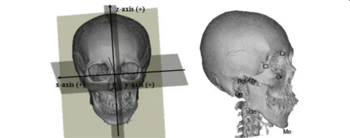
3-dimensional reference plane and craniofacial landmarks Landmarks were assigned on the reconstructed 3-D image, and their locations were verified on the axial, midsagittal, and coronal slices. The landmarks and mea-surements of the craniofacial structures to be performed were selected with reference to previous studies [12,13]. The various landmarks studies are summarized in Figs.
1,2and Table1.
To determine the standard orientation, 3-D reference planes were initially located. The axial plane (AxP) was defined as a plane including the porion (Po) on both sides and the left orbitale (OrL). The midsagittal plane (MSP) was defined as a plane perpendicular to the AxP, including the crista galli (Cr) and the midpoint of the anterior clinoid process (Cl). The coronal plane (CoP) was defined as a plane perpendicular to the AxP and the MSP passing through opisthion (Op).
Craniofacial measurements
The craniofacial measurements performed in the study are summarized in Table 2. Distance between the men-ton (Me) and MSP was defined as dMe, for convenience of comparison; (+) indicated right side deviation of men-ton. In the mandible, distance from the condylar super-ius (Con) to Me, distance from gonion (Go) to Me, and the distance from Con to Go were defined as mandibu-lar length (dML), mandibumandibu-lar body length (dMBL), and ramal height (dRH), respectively. Distance from glenoid
Fig. 1 Three-dimensional reference planes and craniofacial landmarks. Cr, crista galli; Cl, clinoid process; Op, opisthion; Po, porion; Me, menton; Go, gonion; Gf, glenoid fossa; Or, orbitale; Zy, zygonion
fossa (Gf) to MSP, CoP and AxP was defined as dGfx, dGfy, and dGfz, respectively. Distance from orbitale (Or) and zygonion (Zy) to each plane was defined as the same way.
Differences between the measurements of distances in the paired craniofacial structures are given in Table 3. (R-L) was the mean difference between the measure-ments of distances in the paired craniofacial structures. (R-L) from MSP, CoP, and AxP was defined as x(R-L), y(R-L), and z(R-L). A positive value of x(R-L) indicates that the right craniofacial structure is more lateral than the left craniofacial structure from MSP, positive value of y(R-L) indicates that the right craniofacial structure is more anterior than the left craniofacial structure from CoP, and positive value of z(R-L) indicates that the right
craniofacial structure is more superior than the left cra-niofacial structure from AxP. The relationship between the measurements of distances in the paired craniofacial structures and chin deviation was studied.
Statistical analysis
To avoid inter-observer errors in measurements, all the measurements were performed by a single observer. The Pearson’s correlation analysis was performed to evaluate the correlation between chin deviation and difference between the measurements of distances in paired cranio-facial structures. Statistical analysis was performed at a significance level of 5% with SPSS version 25.0 (IBM Corp, Armonk, NY, USA).
Table 1 Description of craniofacial landmarks and reference planes
Point Definition
Cr (crista galli) The superior-most edge of the crista galli Cl (clinoid process) Midpoint between the anterior clinoid processes
Con (condylar superius) The superior-most point of the condylar head; ConR: right, ConL: left Op (opisthion) Midpoint of the posterior arch of foramen magnum
Po (porion) The superior-most point of the external auditory meatus; PoR: right, PoL: left Me (menton) The inferior-most point on the symphysis of mandible
Go (gonion) The apex of the mandibular angle; GoR: right, GoL: left
Gf (glenoid fossa) The antero-superior-most point of the glenoid fossa; GfR: right, GfL: left Or (orbitale) The inferior-most point of the infraorbital rim; OrR: right, OrL: left Zy (zygonion) The lateral-most point of the zygomatic arch; ZyR: right, ZyL: left Axial plane (AxP) A plane passing through PoR, PoL, and OrL
Midsagittal plane (MSP) A plane perpendicular to the axial plane including Cr and Cl
Coronal plane (CoP) A plane perpendicular to the axial plane and sagittal planes passing through Op
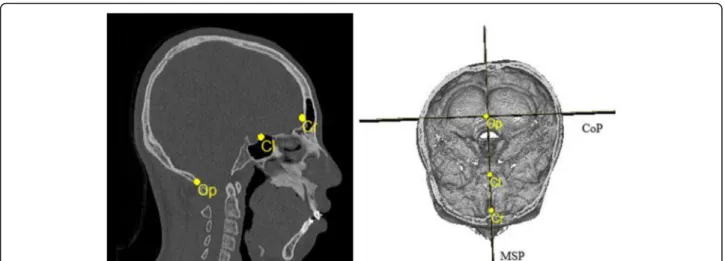
Fig. 2 Computed tomography (CT) scans of craniofacial landmarks. Cr, crista galli; Cl, clinoid process; Op, opisthion; MSP, midsagittal plane; CoP, coronal plane
The intraclass correlation coefficient was used to evaluate intra-observer error by the same observer 1 week apart. In this study, the second set of measure-ments was used.
Results
Study subjects
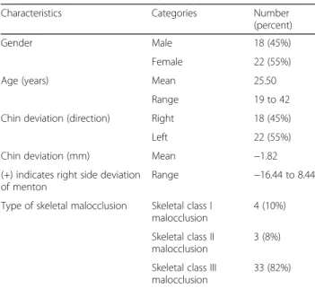
The characteristics of patients included in the study are summarized in Table4. A total of 40 patients (18 males and 22 females; mean age, 25.50 years [range, 19 to 42]) were included in this study. In our study, 18 patients
(45%) showed chin deviation to the right side, and 22 patients (55%) showed chin deviation to the left side (mean, −1.82 mm [range, −16.44 mm to 8.44 mm]), and (+) indicates right side deviation of menton. Of the 40 patients, skeletal class III, class II, and class I malocclu-sions were evident in 33, 3, and 4 patients, as deter-mined by lateral cephalograms.
The intraclass correlation coefficients of craniofacial dis-tance measurements are shown in Table5. The intraclass correlation coefficient ranged from 0.91 to 0.99, which showed that data from one observer were very reliable. Table 2 Description of craniofacial measurements of distances
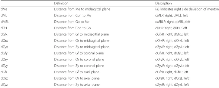
Definition Description
dMe Distance from Me to midsagittal plane (+) indicates right side deviation of menton
dML Distance from Con to Me dMLR: right, dMLL: left
dMBL Distance from Go to Me dMBLR: right, dMBLL:left
dRH Distance from Con to Go dRHR: right, dRHL: left
dGfx Distance from Gf to midsagittal plane dGfxR: right, dGfxL: left dOrx Distance from Or to midsagittal plane dOrxR: right, dOrxL: left dZyx Distance from Zy to midsagittal plane dZyxR: right, dZyxL: left dGfy Distance from Gf to coronal plane dGfyR: right, dGfyL: left dOry Distance from Or to coronal plane dOryR: right, dOryL: left dZyy Distance from Zy to coronal plane dZyyR: right, dZyyL: left dGfz Distance from Gf to axial plane dGfzR: right, dGfzL: left dOrz Distance from Or to axial plane dOrzR: right, dOrzL: left dZyz Distance from Zy to axial plane dZyzR: right, dZyzL: left
*Abbreviations: dML refers to mandibular length; dMBL refers to mandibular body length; dRH refers to ramal height; Me, menton; Con, condylar superius; Go, gonion; Gf, glenoid fossa; Or, orbitale; Zy, zygonion
Table 3 Description of difference between the measurements of distances in paired craniofacial structures.
Definition Description
dML(R-L) dMLR–dMLL (R-L) refers to the difference between the measurements of distances in paired craniofacial structures (right–left) dMBL(R-L) dMBLR–dMBLL
dRH(R-L) dRHR–dRHL
dGfx(R-L) dGfxR–dGfxL x(R-L) refers to the difference between the measurements of distances in paired craniofacial structures from MSP, and (+) indicates that the right craniofacial structure is more lateral than the left craniofacial structure from MSP
dZyx(R-L) dZyxR–dZyxL dOrx(R-L) dOrxR–dOrxL
dGfy(R-L) dGfyR–dGfyL y(R-L) refers to the difference between the measurements of distances in paired craniofacial structures from CoP, and (+) indicates that the right craniofacial structure is more anterior than left craniofacial structure form CoP
dZyy(R-L) dZyyR–dZyyL dOry(R-L) dOryR–dOryL
dGfz(R-L) dGfzR–dGfzL z(R-L) refers to the difference between the measurements of distances in paired craniofacial structures from AxP, and (+) indicates that the right craniofacial structure is more superior to the left craniofacial structure
dZyz(R-L) dZyzR–dZyzL dOrz(R-L) dOrzR–dOrzL
*Abbreviations: (R-L), difference between measurements of distances in paired craniofacial structures (right–left); dML refers to mandibular length; dMBL refers to mandibular body length; dRH refers to ramal height; dGfx, distance from glenoid fossa to midsagittal plane; dZyx, distance from zygonion to midsagittal plane; dOrx, distance from orbitale to midsagittal plane; dGfy, distance from glenoid fossa to coronal plane; dZyy, distance from zygonion to coronal plane; dOry, distance from orbitale to coronal plane; dGfz, distance from glenoid fossa to axial plane; dZyz, distance from zygonion to axial plane; dOrz, distance from orbitale to axial plane
The correlation between chin deviation and difference between the measurements of distances in paired lower facial third structures
The correlation between chin deviation and difference be-tween the measurements of distances in paired lower facial third structures is shown in Table 6. In the lower facial thirds, the degree of chin deviation was related to mandibu-lar length and mandibumandibu-lar body length. Mandibumandibu-lar length and mandibular body length are shorter on the deviated-chin side compared to that on the non-deviated side (man-dibular length,r = − 0.897, value < 0.001; mandibular body
length,r = − 0.318, p value = 0.045). However, no signifi-cant relation was observed between the degree of chin devi-ation and ramal height.
The correlation between chin deviation and difference between the measurements of distances in paired upper and middle facial third structures
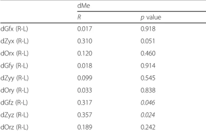
The correlation between chin deviation and difference between the measurements of distances in paired upper and middle facial third structures is shown in Table 7. In the upper and middle facial thirds, the degree of chin deviation was correlated with the vertical asymmetry of glenoid fossa and zygonion. Glenoid fossa and zygonion were superior on the deviated-chin side compared to that on the non-deviated side (glenoid fossa, r = 0.317, p value = 0.046; zygonion, r = 0.357, p value = 0.024). However, no significant relation was observed between the degree of chin deviation and position of orbitale. Discussion
The purpose of this study was to evaluate the correlation between upper and middle facial third asymmetry and lateral deviation of chin using 3-D CT.
Traditionally, posteroanterior cephalograms, submen-tovertex view radiographs, or frontal facial photos have been used for diagnosing facial asymmetry. Certainly, these diagnostic modalities have proven their worth over the years. However, they have limited diagnostic abilities due to problems related to magnification, distortion, and superimposition of craniofacial structures [14–17]. How-ever, 3-D CT reduces errors due to magnification and Table 4 Patients characteristics (N = 40).
Characteristics Categories Number (percent)
Gender Male 18 (45%)
Female 22 (55%)
Age (years) Mean 25.50
Range 19 to 42 Chin deviation (direction) Right 18 (45%)
Left 22 (55%)
Chin deviation (mm) Mean −1.82 (+) indicates right side deviation
of menton
Range −16.44 to 8.44 Type of skeletal malocclusion Skeletal class I
malocclusion
4 (10%) Skeletal class II
malocclusion
3 (8%) Skeletal class III
malocclusion
33 (82%)
Table 5 Intraclass correlation coefficient of craniofacial distance measurements (N = 40).
Intraclass correlation coefficient (single) 95% confidence interval (single)
dMe 0.947 0.903–0.972
dML dMLR: 0.994, dMLL: 0.994 dMLR: 0.988–0.997, dMLL: 0.988–0.997 dMBL dMBLR: 0.987, dMBLL: 0.988 dMBLR: 0.976–0.993, dMBLL: 0.977–0.994 dRH dRHR: 0.993, dRHL: 0.994 dRHR: 0.987–0.996, dRHL: 0.988–0.997 dGfx dGfxR: 0.972, dGfxL: 0.965 dGfxR: 0.948–0.985, dGfxL: 0.934–0.981 dOrx dOrxR: 0.971, dOrxL: 0.943 dOrxR: 0.945–0.984, dOrxL: 0.895–0.969 dZyx dZyxR: 0.983, dZyxL: 0.941 dZyxR: 0.968–0.991, dZyxL: 0.891–0.968 dGfy dGfyR: 0.973, dGfyL: 0.914 dGfyR: 0.949–0.986, dGfyL: 0.844–0.954 dOry dOryR: 0.982, dOryL: 0.989 dOryR: 0.967–0.991, dOryL: 0.980–0.994 dZyy dZyyR: 0.994, dZyyL: 0.987 dZyyR: 0.989–0.997, dZyyL: 0.976–0.993 dGfz dGfzR: 0.982, dGfzL: 0.910 dGfzR: 0.966–0.990, dGfzL: 0.836–0.911
dOrz dOrzR: 0.994 dOrzR: 0.988–0.997
dZyz dZyzR: 0.933, dZyzL: 0.981 dZyzR: 0.876–0.964, dZyzL: 0.964–0.990
*Abbreviations: dMe, distance from Me to midsagittal plane; dML, mandibular length; dMBL, mandibular body length; dRH, ramal height; dGfx, distance from glenoid fossa to midsagittal plane; dZyx, distance from zygonion to midsagittal plane; dOrx, distance from orbitale to midsagittal plane; dGfy, distance from glenoid fossa to coronal plane; dZyy, distance from zygonion to coronal plane; dOry, distance from orbitale to coronal plane; dGfz, distance from glenoid fossa to axial plane; dZyz, distance from zygonion to axial plane; dOrz, distance from orbitale to axial plane
distortion and allows the quantitative measurements of craniofacial structures [18–20].
Currently, the external auditory meatus is regarded as a reliable reference for the analysis of craniofacial char-acteristics because of its stable shape [21]. Previous 3-D studies use the Frankfort’s horizontal plane as the refer-ence axial plane [22–24]. For these reasons, in this study, the Frankfort’s horizontal plane passing through bilateral porion and left orbitale was used as the axial plane. Then, a plane perpendicular to the axial plane passing through the crita galli (Cr) and the midpoint between the anterior clinoid processes (Cl) was defined as mid-sagittal plane [12]. A plane perpendicular to axial and midsagittal plane with passing through opisthion (Op) was defined as a coronal plane based on the study of Kwon et al .[12].
The glenoid fossa is a depression in the temporal bone that articulates with the mandible to form the
temporomandibular joint [25]. Positional changes in the glenoid fossa during growth can lead to facial asymmetry and malocclusion [7]. The location of the orbit and zygomatic bone plays an important role in facial sym-metry and esthetics [26,27]. For these reasons, the glen-oid fossa, orbitale, and zygomatic arch were analyzed in this study. Mandibular length, mandibular body length, and ramal height were also analyzed to evaluate lower facial third asymmetry.
In the lower facial thirds, chin deviation is correlated with mandibular length and mandibular body length asymmetry, coincident with the findings of previous studies [12, 21, 28]. Moreover, in our study, chin devi-ation was also correlated with the upper and middle fa-cial third asymmetry, espefa-cially vertical asymmetry of the glenoid fossa and zygomatic arch, coincident with the findings of another stud y[7]. In a previous study, asymmetry of the glenoid cavity, a type of upper and middle facial third asymmetry, was reported [7]. The asymmetry of glenoid cavity is often caused by the de-fects in generation, proliferation, migration, and differen-tiation of cranial neural crest cells [8] or craniofacial structure modeling from the cerebrum [9–11]. As a re-sult, the glenoid cavity is located superiorly where devel-opmental defects occurred (affected side) [7]. Similarly, supraorbital arch, zygomatic bone, and external auditory meatus are also located superiorly on affected side (orbi-culo-zygomatic-meatal and articular asymmetry) [7]. Fi-nally, the asymmetry of glenoid cavity functionally affects condylar position, causing lateral deviation of chin to the affected side [7].
This study showed that lateral deviation of chin is corre-lated with upper and middle facial third asymmetry as well as lower facial third asymmetry, especially vertical asym-metry of the glenoid fossa and zygomatic arch. Correction of chin deviation by mandibular surgery alone will not cor-rect the asymmetry of the upper and middle facial thirds.
A limitation of this study is that a small number of craniofacial landmarks were analyzed for the correlation with the lateral deviation of chin, and further studies in-corporating more number of craniofacial landmarks should be conducted for a deeper understanding of the correlation between the lateral deviation of chin and cra-niofacial landmarks.
To be best of our knowledge, this is the first study to evaluate craniofacial characteristics associated with the lateral deviation of chin using 3-dimensional imaging mo-dalities. Considering the high prevalence and the impact of facial asymmetry on patient’s treatment outcome, this study is very relevant in the present scenario. Knowledge about the fact that facial symmetry is influenced by the upper and middle thirds of face will help clinicians around the world in proper treatment planning and hence, in pro-viding better treatment to such patients.
Table 6 The correlation between chin deviation and difference between the measurements of distances in paired lower facial third structures (N = 40). dMe R p value dML (R-L) − 0.897 < 0.001 dMBL (R-L) − 0.318 0.045 dRH (R-L) − 0.123 0.449
*Abbreviations: (R-L), difference between the measurements of distances in paired craniofacial structures (right–left); dML refers to mandibular length; dMBL refers to mandibular body length; dRH refers to ramal height
Table 7 The correlation between chin deviation and difference between the measurements of distances in paired upper and middle facial third structures (N = 40).
dMe R p value dGfx (R-L) 0.017 0.918 dZyx (R-L) 0.310 0.051 dOrx (R-L) 0.120 0.460 dGfy (R-L) 0.018 0.914 dZyy (R-L) 0.099 0.545 dOry (R-L) 0.033 0.838 dGfz (R-L) 0.317 0.046 dZyz (R-L) 0.357 0.024 dOrz (R-L) 0.189 0.242
*Abbreviations: (R-L), difference between the measurements of distances in paired craniofacial structures (right–left); dGfx, distance from glenoid fossa to midsagittal plane; dZyx, distance from zygonion to midsagittal plane; dOrx, distance from orbitale to midsagittal plane; dGfy, distance from glenoid fossa to coronal plane; dZyy, distance from zygonion to coronal plane; dOry, distance from orbitale to coronal plane; dGfz, distance from glenoid fossa to axial plane; dZyz, distance from zygonion to axial plane; dOrz, distance from orbitale to axial plane
Conclusions
Lateral deviation of chin is correlated with upper and mid-dle facial third asymmetry as well as lower facial third asymmetry. Correction of chin deviation by mandibular surgery alone will not correct the asymmetry of the upper and middle facial thirds. As a result, treatment planning in patients with chin deviation should involve a careful evalu-ation of the asymmetry of the upper and middle facial thirds to ensure complete patient satisfaction.
Abbreviations
(R-L):Difference between the measurements of distances in paired craniofacial structures (right–left); 3-D CT: 3-dimensional computed tomography; AxP: Axial plane; Cl: Clinoid process; Con: Condylar superius; ConL: Left condylar superius; ConR: Right condylar superius; CoP: Coronal plane; Cr: Crista galli; dGfx: Distance from Gf to midsagittal plane; dGfx(R-L): dGfxR–dGfxL; dGfxL: Left dGfx; dGfxR: Right dGfx; dGfy: Distance from Gf to coronal plane; dGfy(R-L): dGfyR–dGfyL; dGfyL: Left dGfy; dGfyR: Right dGfy; dGfz: Distance from Gf to axial plane; dGfz(R-L): dGfzR–dGfzL; dGfzL: Left dGfz; dGfzR: Right dGfz; DICOM: Digital imaging and communication in medicine; dMBL: Mandibular body length, distance from Go to Me; dMBL(R-L): dMBLR–dMBLL; dMe: Distance from Me to midsagittal plane;
dML: Mandibular length, distance from Con to Me; dML(R-L): dMLR–dMLL; dOrx: Distance from Or to midsagittal plane; dOrx(R-L): dOrxR–dOrxL; dOrxL: Left dOrx; dOrxR: Right dOrx; dOry: Distance from Or to coronal plane; dOry(R-L): dOryR–dOryL; dOryL: Left dOry; dOryR: Right dOry; dOrz: Distance from Or to axial plane; dOrz(R-L): dOrzR–dOrzL; dOrzL: Left dOrz; dOrzR: Right dOrz; dRH: Ramal height, distance from Con to Go; dRH(R-L): dRHR–dRHL; dZyx: Distance from Zy to midsagittal plane; dZyx(R-L): dZyxR–dZyxL; dZyxL: Left dZyx; dZyxR: Right dZyx; dZyy: Distance from Zy to coronal plane; dZyy(R-L): dZyyR–dZyyL; dZyyL: Left dZyy; dZyyR: Right dZyy; dZyz(R-L): dZyzR–dZyzL; dZyz: Distance from Zy to axial plane; dZyzL: Left dZyz; dZyzR: Right dZyz; Gf: Glenoid fossa; GfL: Left glenoid fossa; GfR: Right glenoid fossa; Go: Gonion; GoL: Left gonion; GoR: Right gonion; Me: Renton; MSP: Midsagittal plane; Op: Opithion; Or: Orbitale; OrL: Left orbitale; OrR: Right orbitale; Po: Porion; PoL: Left porion; PoR: Right porion; x(R-L): Difference between the measurements of distances in paired craniofacial structures from MSP; y(R-L): Difference between the measurements of distances in paired craniofacial structures from CoP; z(R-L): Difference between the measurements of distances in paired craniofacial structures from AxP; Zy: Zygonion; ZyL: Left zygonion; ZyR: Right zygonion Acknowledgements
We would like to thank Editage (www.editage.co.kr) for English language editing.
Authors’ contributions
HWC and BK were responsible for collecting and analyzing the data. JYK was responsible for statistical analysis and translation of the data. KHP and JKH contributed to the direction and design of the research and contributed to the review of the paper. All the authors have read the final version of the paper and have agreed to its publication.
Funding
This research received no specific grant from any funding agency in the public, commercial, or not-for profit sectors.
Availability of data and materials
The data sets used and/or analyzed during the current study are available from the corresponding author on reasonable request.
Ethics approval and consent to participate
This study was approved by Gangnam Severance Hospital Institutional Review Board (Approval No. 3-2019-0119).
Consent for publication Not applicable.
Competing interests
The authors declare that they have no competing interests. Received: 5 September 2019 Accepted: 20 November 2019
References
1. Haraguchi S, Takada K, Yasuda Y (2002) Facial asymmetry in subjects with skeletal class III deformity. Angle Orthod 72:28–35
2. Samman N, Tong AC, Cheung DL, Tideman H (1992) Analysis of 300 dentofacial deformities in Hong Kong. Int J Adult Orthodon Orthognath Surg 7:181–185
3. Severt TR, Proffit WR (1997) The prevalence of facial asymmetry in the dentofacial deformities population at the University of North Carolina. Int J Adult Orthodon Orthognath Surg 12:171–176
4. Burstone CJ (1998) Diagnosis and treatment planning of patients with asymmetries. Semin Orthod 4:153–164
5. Obwegeser HL, Makek MS (1986) Hemimandibular hyperplasia--hemimandibular elongation. J Maxillofac Surg 14:183–208
6. Poikela A, Kantomaa T, Pirttiniemi P (1997) Craniofacial growth after a period of unilateral masticatory function in young rabbits. Eur J Oral Sci 105: 331–337
7. López Buitrago DF, Ruiz Botero J (2017) Asymmetry of glenoid fossa as differential diagnosis for hemimandibular elongation. Revista Mexicana de Ortodoncia 5:e217–ee26
8. Minoux M, Rijli FM (2010) Molecular mechanisms of cranial neural crest cell migration and patterning in craniofacial development. Development 137: 2605–2621
9. de Cráneo AdlB, el Crecimiento D (2011) Asymmetry of human skull base during growth. Int J Morphol 29:1028-1032.
10. Basili C, Costa H, Sasaguri K, Akimoto S, Slavicek R, Sato S (2009) Comparison of the position of the mandibular fossa using 3D CBCT in different skeletal frames in human caucasic skulls. Int J Stomatol Occlusion Med 2:179–190
11. Slavicek R (2001) The dynamic functional anatomy of craniofacial complex and its relation to the articulation of the dentitions. Das Kauorgan Funktione und Dysfunktionen:482–514
12. Kwon TG, Park HS, Ryoo HM, Lee SH (2006) A comparison of craniofacial morphology in patients with and without facial asymmetry-a three-dimensional analysis with computed tomography. Int J Oral Maxillofac Surg 35:43–48
13. You KH, Lee KJ, Lee SH, Baik HS (2010) Three-dimensional computed tomography analysis of mandibular morphology in patients with facial asymmetry and mandibular prognathism. Am J Orthod Dentofacial Orthop 138:540.e1–540.e8 discussion -1
14. Ahlqvist J, Eliasson S, Welander U (1983) The cephalometric projection. part II. principles of image distortion in cephalography. Dentomaxillofac Radiol 12:101–108
15. Bergersen EO (1980) Enlargement and distortion in cephalometric radiography: compensation tables for linear measurements. Angle Orthod 50:230–244
16. Gravely JF, Benzies PM (1974) The clinical significance of tracing error in cephalometry. Br J Orthod 1:95–101
17. Savara BS, Tracy WE, Miller PA (1966) Analysis of errors in cephalometric measurements of three-dimensional distances on the mandible. Arch Oral Biol 11:209–217
18. Fuhrmann RA, Schnappauf A, Diedrich PR (1995) Three-dimensional imaging of craniomaxillofacial structures with a standard personal computer. Dentomaxillofac Radiol 24:260–263
19. Katsumata A, Fujishita M, Maeda M, Ariji Y, Ariji E, Langlais RP (2005) 3D-CT evaluation of facial asymmetry. Oral Surg Oral Med Oral Pathol Oral Radiol Endod 99:212–220
20. Xia J, Ip HH, Samman N, Wang D, Kot CS, Yeung RW et al (2000) Computer-assisted three-dimensional surgical planning and simulation: 3D virtual osteotomy. Int J Oral Maxillofac Surg 29:11–17
21. Kim YH, Sato K, Mitani H, Shimizu Y, Kikuchi M (2003) Asymmetry of the sphenoid bone and its suitability as a reference for analyzing craniofacial asymmetry. Am J Orthod Dentofacial Orthop 124:656–662
22. Captier G, Leboucq N, Bigorre M, Canovas F, Bonnel F, Bonnafe A et al (2003) Plagiocephaly: morphometry of skull base asymmetry. Surg Radiol Anat 25:226–233
23. Lo LJ, Marsh JL, Pilgram TK, Vannier MW (1996) Plagiocephaly: differential diagnosis based on endocranial morphology. Plast Reconstr Surg 97:282–291 24. Yu CC, Wong FH, Lo LJ, Chen YR (2004) Craniofacial deformity in patients
with uncorrected congenital muscular torticollis: an assessment from three-dimensional computed tomography imaging. Plast Reconstr Surg 113:24–33 25. Sato S, Takamoto K, Fushima K, Akimoto S, Suzuki Y (1989) A new
orthodontic approach to mandibular lateral displacement malocclusion-importance of occlusal plane reconstruction. Dent Jpn 26:81–85 26. Springer IN, Wannicke B, Warnke PH, Zernial O, Wiltfang J, Russo PA et al
(2007) Facial attractiveness: visual impact of symmetry increases significantly towards the midline. Ann Plast Surg 59:156–162
27. Khaqani MS, Tavosi F, Gholami M, Eftekharian HR, Khojastepour L (2018) Analysis of facial symmetry after zygomatic bone fracture management. J Oral Maxillofac Surg 76:595–604
28. Baek C, Paeng JY, Lee JS, Hong J (2012) Morphologic evaluation and classification of facial asymmetry using 3-dimensional computed tomography. J Oral Maxillofac Surg 70:1161–1169
Publisher’s Note
Springer Nature remains neutral with regard to jurisdictional claims in published maps and institutional affiliations.