Preparation and Characterization of Genetically Engineered Mesenchymal
Stem Cell Aggregates for Regenerative Medicine
Sun Hwa Kim1†, Hyung-Ho Moon1, Bong Genn Chung2 and Donghoon Choi1†
1Severance Integrative Research Institute for Cerebral & Cardiovascular Disease,
Yonsei University Health System, 250 Seongsanno, Seodaemun-gu, Seoul 120-752, South Korea
2Department of Bionano Engineering, Hanyang University ERICA Campus, 1271 Sa3-dong,
Sangnok-gu, Ansan-si, Gyeonggi-do 426-791, South Korea (Received July 20, 2010·Revised July 28, 2010·Accepted July 29, 2010)
ABSTRACT−Combining cell- and gene-based therapy is a promising therapeutic strategy in regenerative medicine. The aim of this study was to develop genetically modified mesenchymal stem cell (MSC) aggregates using a poly(ethylene gly-col) (PEG) hydrogel micro-well array technique. Stable PEG hydrogel micro-well arrays with diameters of 200 to 500µm were fabricated and used to generate genetically engineered MSC aggregates. Rat bone marrow-derived MSCs were trans-fected with a green fluorescent protein (GFP) plasmid as a reporter gene, and aggregated by culturing in the PEG hydrogel micro-well arrays. The resultant cell aggregates had a mean diameter of less than 200µm, and maintained the mesenchymal phenotype even after genetic modification and cell aggregation. Transplantation of MSC aggregates that are genetically modified to express therapeutic or cell-survival genes may be a potential therapeutic approach for regenerative medicine. Key words−Cell and gene therapy, Mesenchymal stem cells, Micro-well array, Cell aggregates
Transplantation of progenitor/stem cells has been studied over the last decade as a promising treatment for the regen-eration of damaged tissues (Humes et al., 2003). Bone mar-row-derived mesenchymal stem cells (MSCs), in particular, have received recent attention in the cell therapy field, because of their numerous beneficial properties such as self-renewal ability, multipotential nature, regenerative and immunomod-ulatory potential, and genetic stability (Tae et al., 2006). Despite increasing experimental and clinical interest in using MSCs in regenerative medicine, clinical MSC-based thera-peutic approaches have not been well established because poor cell viability and low engraftment limit the therapeutic efficacy of MSC transplantation (Pittenger et al., 2004). To overcome the lack of long-term MSC engraftment and low-level engraft-ment efficiency, the genetic modification to promote MSC via-bility and therapeutic efficacy has been suggested as a potentially effective approach to tissue repair and regeneration (Cheng et al., 2008; Noiseux et al., 2006). For instance, genet-ically modifying MSCs to produce growth factors, such as VEGF and IGF-I, improved their retention and growth prop-erties after cell transplantation into ischemic myocardium in rats (Pons et al., 2008; Sadat et al., 2007).
In recent years, microfabricated cell clusters or aggregates have been used in biomedical applications, such as cell ther-apy, tissue engineering, developmental biology, and drug dis-covery (Gallego-Perez et al., 2010). In particular, transplantation of different types of assembled cell clusters including cell aggregates and cellular sheets has been suggested for tissue repair and regeneration. Transplantation of a group of cells markedly improves cell survival rates and transplantation effi-ciencies compared to injection of single cells (Kato et al., 1993). In this study, genetically engineered MSC aggregates prepared by a microfabrication technique were developed for a combination cell- and gene- therapy approach. To generate genetically modified MSC aggregates, micro-well arrays were made using PEG hydrogels. Green fluorescent protein (GFP) plasmid was used as a reporter gene to assess the possibility of the fabrication of MSC aggregates expressing exogenous tar-get genes without causing phenotypic changes. When used with therapeutic target genes such as cell survival and angio-genic factors, this technique to generate genetically engineered MSC aggregates may provide a promising strategy for regen-erative therapy applications.
†Co-corresponding Author :
Tel : +82-2-2228-0375, E-mail : shkim77@yuhs.ac Tel : +82-2-2228-8460, E-mail : cdhlyj@yuhs.ac DOI : 10.4333/KPS.2010.40.6.333
Materials and Methods
Materials
SU-8 photoresist was purchased from Microchem Corp. (Newton, MA, USA). Poly(dimethylsiloxane) (PDMS) was obtained from Dow Corning Corp. (Midland, MI, USA). Poly(ethylene glycol)-diacrylate (PEG-diacrylate, 1,000 Da) was supplied by PolySciences Inc. (UK). Irgacure 2959 was obtained from Ciba Specialty Chemicals Corp. (Tarrytown, NY, USA). 3-(Trimethoxysilyl)propylmethacrylate (TMSPMA), linear poly(ethylenimine) (LPEI, mw = 25 kDa), and penicillin/ streptomycin solution were purchased from Sigma-Aldrich (St. Louis, MO, USA). Ficoll-Paque Plus was supplied by Amer-sham Biosciences (Uppsala, Sweden). The peGFP-C1 vector was obtained from BD Bioscience Clontech (Palo Alto, CA, USA). Fetal bovine serum (FBS), Dulbecco’s modified Eagle’s medium (DMEM), and Dulbecco's phosphate-buffered saline (PBS) were supplied by Gibco BRL (Grand Island, NY, USA). All other chemicals and reagents were used as received unless otherwise noted.
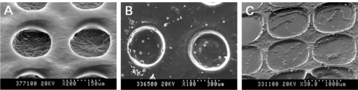
Fabrication of PEG hydrogel micro-well arrays PEG hydrogel micro-well arrays were generated by pho-tolithography and micro-molding techniques. Briefly, SU-8 photoresist was spin-coated on a silicon wafer and exposed to UV light, resulting in micropatterns with 200 µm to 500 µm in diameter. After the photolithography process, PDMS mixed silicone elastomer with curing agent (10:1) was poured into a SU-8 photoresist-patterned silicon wafer, and baked at 70oC for 2 h. To generate PEG micro-well arrays, PEG-diacrylate was mixed with 1% Irgacure 2959. PDMS stamps were gently placed on PEG prepolymer solutions on a glass substrate and exposed to UV light (EXFO Photonic Solutions Inc., Omn-iCure, Canada, 50 mW/cm2) for 50 sec. To enhance the bond-ing between PEG micro-well arrays and glass substrates, glass substrates were treated with TMSPMA for 30 min and baked at 70oC for 2 hours. After removing the PDMS stamps, PEG micro-well arrays of 200µm, 300 µm, and 500 µm in diameter were generated on the glass substrates. Morphological obser-vation of the PEG hydrogel micro-well arrays was carried out with scanning electron microscopy (SEM, Philips 535 M, Netherlands) and optical images were generated under light microscopy (100 × magnification).
MSC preparation, culture, and characterization MSCs were isolated from the bone marrow of four-week-old male Sprague-Dawley rats (Samtako Bio Co., Osan, Korea). The use of animals was in accordance with the International
Guide for the Care and Use of Laboratory Animals. Rat MSCs were harvested and propagated as described previously (Song et al., 2007; Chang et al., 2009). Briefly, the rat bone marrow-derived cells were flushed out from femurs and tibias with cul-ture medium (DMEM with 10% FBS). Mononuclear cells were isolated by Ficoll-Paque density gradient centrifugation. After 48 h incubation, non-adherent cells were discarded, and adherent cells were expanded until confluent. MSCs were cul-tured in DMEM supplemented with 10% FBS and 1% pen-icillin-streptomycin up to 80% confluency for three or four passages before use.
In vitro transfection
For in vitro transfection, MSCs were plated at a density of 1.0 × 10⁶ cells in a 100 mm tissue culture dish. After 48 h of incubation, the culture medium was exchanged with fresh serum-free medium. Cells were transfected with 6µg of peGFP prepared with LPEI at a N/P ratio (nitrogen of PEI/ phosphate of DNA) of 10:1. After 4 h for transfection, the medium was replaced with fresh 10% serum medium. Trans-fected cells were incubated at 37oC for 24 h and used for the formation of MSC aggregates using the PEG hydrogel micro-well arrays. GFP expression in MSC aggregates was visualized by a confocal laser scanning microscope (Olympus Fluoview FV300, Melville, NY) using an argon-krypton mixed gas laser (Ex. 494 nm). The nuclei of MSCs were stained with 4',6-dia-midino-2-phenylindole (DAPI, 1µg/mL, Molecular Probes Inc., Eugene, OR, USA) for 30 min one day before MSC aggregates formation, according to the manufacturer’s sug-gested protocol.
Preparation of MSC aggregates
PEG hydrogel micro-well arrays were washed three times with PBS and exposed to UV for 30 min for sterilization. After the fourth passage, rat MSCs grown in monolayer cultures were trypsinized, resuspended in media, counted with a hema-cytometer, and plated onto the PEG micro-wells at 1.0 × 10⁶ cells/plate with 0.2 mL culture medium. Cells were allowed to settle into the micro-wells for 4 h before the PEG micro-well arrays were washed with a gentle flow of culture medium to remove undocked cells. Seeded micro-wells were incubated at 37oC and 5% CO2 for 2 days. The diameters of the resultant
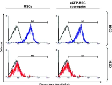
MSC aggregates were directly measured from optical images taken for a total of 50 cell aggregates at three different regions of the specimen. To assess the MSC phenotype, cultured single MSCs and peGFP-modified MSC aggregates were labeled against positive and negative cell-surface markers, CD90-flu-orescein isothiocyanate (FITC) and CD34-phycoerythrin (PE)
(BD Biosciences, San Diego, CA), and analyzed by flow cytometry. In brief, harvested cells were incubated with anti-bodies (0.5 mg/mL) for 15 min at room temperature and washed twice with PBS. After staining with fluorescence-labeled monoclonal antibodies, fluorescence intensity was detected using fluorescence-activated cell sorting analysis (Becton Dickinson, San Jose, CA).
Results and Discussion
To develop a novel cell- and gene-based therapy strategy for efficient tissue regeneration, genetically engineered MSC aggregates were prepared using a micro-well array technique. The fabrication process for genetically modified MSC aggre-gates is schematically described in Figure 1. Isolated MSCs were transfected with LPEI (25 kDa) as a polymeric gene car-rier and an eGFP plasmid as a reporter gene. The genetically modified MSCs were loaded into the microfabricated wells to
generate small cell clusters. After two days of culture, the final MSC aggregates were harvested from the micro-well plates. For the fabrication of the PEG hydrogel microstructures on the glass substrates, a photolithographic method based on PDMS stamps was used (Xue et al., 2010). As shown in Figure 2, microfabricated arrays of PEG hydrogel micro-wells with different pore diameters of 200µm, 300 µm, and 500 µm were produced. To investigate the influence of micro-well array pore size on the extent of cell cluster formation, unmodified MSC aggregates were prepared using micro-wells of different sizes. MSC aggregate sizes were determined by median diameters measured from optical images selected at random. Although the pore sizes of the micro-well arrays were not directly reflected in the results of MSC aggregate formation, cell aggre-gate size increased with increasing pore size in the PEG hydro-gel micro-well arrays from 200µm to 500 µm (Figure 3). The cell mass of the aggregates, however, did not expanded indef-initely with increasing micro-well pore size. The MSC
aggre-Figure 1. Schematic representation of the preparation of genetically engineered MSC aggregates using PEG hydrogel micro-well array technology. The inset is a representative optical image of a PEG hydrogel micro-well array with a 200µm diameter.
gates generated by the micro-well array technique generally reached a maximum of around 200µm. Specifically, cell aggregate sizes obtained from micro-wells had diameters of 100.2 ± 12.56 µm for 200 µm pores, 152.4 ± 14.27 µm for 300µm pores, and 206.7 ± 27.92 µm for 500 µm pores. Since low gauge needles cause a lot of tissue damage and scar for-mation with repeated injections, needles with a gauge over 30 (an inner diameter of ca. 150 µm) are commonly used in ani-mal experiments to avoid the tissue damage and pain asso-ciated with cell therapy injections (Ratcliff et al., 2008). Therefore, MSC aggregates with a size of less than 150 µm could be efficiently used as injectable cell aggregates for ther-apeutic applications in the field of tissue engineering and regenerative medicine.
After in vitro cell transfection with an interesting gene, MSC aggregates were prepared with the micro-well-based fabrica-tion method (Figure 4). Figures 4 A and B show the successful formation of micro-scale spherical MSC aggregates using the
PEG hydrogel micro-well technique. As shown in Figures 4 C and D, eGFP-modified MSC aggregates exhibited GFP expression in their constituent cells, although the observed gene expression level was low. Induction of in vitro gene expression is highly dependent upon the selection of appro-priate gene carriers, and dependent on cell-related factors such as cell type and cell conditions. Since primary cells including adult bone marrow-derived MSCs show very low in vitro transfection efficiencies of generally no more than a few per-cent using common transfection methodologies (Haleem-Smith et al, 2005; Otani et al., 2009), the development of effi-cient transfection reagents for MSCs is needed for successful therapeutic approaches using genetically engineered MSC aggregates. To exclude the possibility that the genetic mod-ification and cell aggregation processes caused phenotypic changes of the MSCs, phenotypic analysis of intact MSCs and eGFP-modified MSC aggregates was conducted by flow cytometry after staining for positive (CD90) and negative (CD34) MSC membrane surface markers. As shown in Figure 5, no phenotypic changes were observed in MSCs after eGFP modification and cell aggregate formation, possibly due to the genetic stability of MSCs, which is one of their advantages mentioned above (Pittenger et al., 2004; Mosca et al., 2000). Thus, the transplantation of genetically engineered MSC aggregates prepared through a micro-well array method could be suggested as a potential therapeutic toll in regenerative medicine strategies.
In conclusion, this work provides a platform technology for Figure 3. Sizes of MSC aggregates produced by PEG hydrogel
micro-well arrays with different pore sizes. ns = not significant.
Figure 4. (A,B) Phase contrast images of MSC aggregates and (C,D) fluorescence images of eGFP-modified MSC aggregates stained with DAPI (blue). Scale bar is 200 µm.
Figure 5. Phenotypic analysis of MSCs surface markers. MSCs and eGFP-modified MSC aggregates were stained for different selectable markers (positive: CD90, blue; negative: CD34, red). An unstained MSC sample was used as a negative control (black). M refers to the gated area (fluorescence intensity in arbitrary units: 101-104).
the production of genetically engineered MSC aggregates, that can be applied for combined cell and gene therapy. Using a PEG hydrogel micro-well array technique, we successfully generated genetically modified MSC aggregates without changing phenotype. With efficient MSC transfection meth-odologies and potential therapeutic target genes, this novel fab-rication approach has potential for use in regenerative technology.
Acknowledgements
This work was supported by grants from the Korea Health-care Technology R&D Project, Ministry for Health, Welfare & Family Affairs, Republic of Korea (A085136), Basic Science Research Program through the National Research Foundation of Korea (NRF) funded by the Ministry of Education, Science and Technology (Grant Number R11-2008-044-01002-0 and 20100022471), and Korea Industrial Technology Foundation (KOTEF) through the Human Resource Training Project for Strategic Technology.
References
Chang, W., Song, B., Lim, S., Song, H., Shim, C.Y., Cha, M., Ahn, D.H., Jung, Y., Lee, D., Chung, J.H., Choi, K., Lee, S., Chung, N., Lee, S., Jang, Y., Hwang, K., 2009. Mesenchymal stem cells pretreated with delivered Hph-1Hsp70 protein are pro-tected from hypoxia-mediated cell death and rescue heart func-tions form myocardial injury. Stem Cells 27, 2283-2292. Cheng, Z., Ou, L., Zhou, X., Li, F., Jia, X., Zhang, Y., Liu, X., Li,
Y., Ward, C.A., Melo, L.G., Kong, D., 2008. Targeted migra-tion of mesenchymal stem cells modified with CXCR4 gene to infarcted myocardium improves cardiac performance. Mol. Ther. 16, 571-579.
Gallego-Perez, D., Higuita-Castro, N., Sharma, S., Reen, R.K., Palmer, A.F., Gooch, K.J., Lee, L.J., Lannutti, J.J., Hansford, D.J., 2010. High throughput assembly of spatially controlled 3D cell clusters on a micro/nanoplatfom. Lab Chip 10, 775-782.
Haleem-Smith, H., Derfoul, A., Okafor, C., Tuli, R., Olsen, D., Hall, D.J., Tuan, R.S., 2005. Optimization of high-efficiency transfection of adult human mesenchymal stem cells in vitro. Mol. Biotechnol. 30, 9-19.
Humes, H.D., 2003. Cell therapy: leveraging nature’s therapeutic potential. J. Am. Soc. Nephrol. 14, 2211-2213.
Kato, K., Gurdon, J.B., 1993, Single-cell transplantation deter-mines the time when Xenopus muscle precursor cells acquire a capacity for autonomous differentiation. Proc. Natl. Acad. Sci. USA 90, 1310-1314.
Mosca, J.D., Hendricks, J.K., Buyaner, D., Davis-Sproul, J., Chuang, L.C., Majumdar, M.K., Chopra, R., Barry, F., Mur-phy, M., Thiede, M.a., Junker, U., Rigg, R.J, Forestell, S.P., Bohnlein, E., Storb, R., Sandmaier, B.M., 2000. Mesenchymal stem cells as vehicles for gene delivery. Clin. Orthop. Rel. Res, 379, S71-90.
Noiseux, N., Gnecchi, M., Lopez-Ilasaca, M., Zhang, L., Solomon, S.D., Deb A., Dzau, V.J., Pratt, R.E., 2006. Mesenchymal stem cells overexpressing Akt dramatically repair infarcted myo-cardium and improve cardiac function despite infrequent cel-lular fusion or differentiation. Mol. Ther. 14, 840-850. Otani, K., Yamahara, K., Ohnishi, S., Obata, H., Kitamura, S.,
Nagaya, N., 2009. Nonviral delivery of siRNA into mesen-chymal stem cells by a combination of ultrasound and microbubbles. J. Control. Release 133, 146-153.
Pittenger, M.F., Martin, B.J., 2004. Mesenchymal stem cells and their potential as cardiac therapeutics. Circ. Res. 95, 9-20. Pons, J., Huang, Y., Arakawa-Hoyt, J., Washko, D., Takagawa, J.,
Ye, J., Grossman, W., Su, H., 2008. VEGF improves survival of mesenchymal stem cells in infarcted hearts. Biochem. Bio-phys. Res. Commun. 376, 419-422.
Ratcliff, A., 2008. Choosing the correct needle for neuroscience injections. American Biotechnology Laboratory (Newsletter) 26, 18-19.
Sadat, S., Gehmert, S., Song, Y.H., Yen, Y., Bai, X., Gaiser, S., Klein, H., Alt, E., 2007. The cardioprotective effect of mes-enchymal stem cells is mediated by IGF-I and VEGF. Bio-chem. Biophys. Res. Commun. 363, 674-679.
Song, H., Chang, W., Lim, S., Seo, H.S., Shim, C.Y., Park, S., Yoo, K.J., Kim, B.S., Min, B.H., Lee, H., Jang, Y., Chung, N., Hwang, K.C., 2007. Tissue transglutaminase is essential for integrin-mediated survival of bone marrow-derived mesen-chymal stem cells. Stem Cells 25, 1431-1438.
Tae, S.K., Lee, S.H., Park, J.S., Im, G.I., 2006. Mesenchymal stem cells for tissue engineering and regenerative medicine. Biomed. Mater. 1, 63-71.
Xue, C., Chin, S.Y., Khan, S.A., Yang, K., 2010. UV-defined flat PDMS stamps suitable for microcontact printing. Langmuir 26, 3739-3743.