INTRODUCTION
Glioblastoma represents 15-20% of brain tumors and 50% of all gliomas (1). A characteristic histopathologic hallmark of glioblastoma is marked cytologic heterogeneity of astrocytic tumor cells associated with necrosis and/or vas-cular endothelial proliferation (2, 3). Molevas-cular biologic studies indicate that glioblastoma is the common end point in the progression of grade 2 (low-grade) or grade 3 (anaplastic) astrocytomas (4, 5). In the revised WHO classi-fication of tumors of the central nervous system, glioblas-toma is classified under the group of astrocytic tumors, grade 4, as opposed to its prior classification under embry-onal tumors (6).
Recent clinical and molecular studies indicate the two different genetic pathways leading to the development of glioblastomas: (a) final progression of lower grade astrocy-toma, which arise due to p53 mutation, loss of heterozygosi-ty (LOH) 17p, amplification of MDM2, CDK4, Rb
muta-tion, LOH 10, and epidermal growth factor receptor (EGFR) amplification (7-10); and (b) de novo from rapid malignant transformation of a normal glial cell due to LOH 10 and EGFR amplification (11, 12). Another possible pathway of glioblastoma development is progression from other types of gliomas, such as oligodendrogliomas and oligoastrocytomas by way of genetic lesions of LOH 19q and 1p (13-15), 9p and 10 (16). Common association of EGFR amplification and p53 mutation is very rare in the first pathway (17, 18).
Attempts have been made to link these different molecu-lar genetic pathways in the development of glioblastomas to clinical features (17-20). Secondary glioblastomas derived from lower grade astrocytomas occurs in younger patients (mean age, 40.5 yr), while de novo glioblastomas tend to occur in older patients (mean age, 56.3 yr). However, these studies are primarily based on histopathologic diagnosis of glioblastomas without clinical distinction between primary and secondary etiologies.
Kyung Sik Yoon, Min Cheol Lee*, Sam Suk Kang, Jae-Hyoo Kim, Shin Jung, Yoon Ji Kim*, Jae Hyuk Lee*, Kyu Yoon Ahn�
, Ji Shin Lee�
, Jong Yoon Cheon� Department of Neurosurgery, Pathology* and Anatomy�
, Chonnam National University Medical School and Research Institute of Medical Sciences, Kwangju; Department of Pathology�
, College of Medicine, Seonam University, Kwangju; Department of Pathology, College of Natural Science�
, Ewha Womans University, Seoul, Korea
Received : 30 October 2000
Accepted : 16 March 2001
Address for correspondence Min Cheol Lee, M.D.
Department of Pathology, Chonnam National University Medical School and Hospital, 5 Hak-dong, Dong-gu, Kwangju 501-190, Korea Tel : +82.62-220-4300, Fax : +82.62-227-3429 E-mail : mclee@chonnam.ac.kr
*This study was partly supported by the Research Grants of Chonnam National University in 1997.
481
p53
Mutation and Epidermal Growth Factor Receptor Overexpression
in Glioblastoma
Recent molecular studies indicate two different genetic pathways leading to the development of glioblastoma; final progression of astrocytoma and de novo for-mation. To define the mutual relationships of cytogenetic changes in the patho-genesis of glioblastoma, molecular histopathologic alterations of p53 and epi-dermal growth factor receptor (EGFR) were evaluated by single stranded con-formational polymorphion, reverse transcriptase-polymerase chain reaction and immunohistochemical stains in 15 primary and 21 secondary glioblastomas. Mutations in p53 gene and positive immunoreactivity to p53 protein (DO1) were more prevalent in secondary glioblastomas than in primary glioblastomas. A cor-relation between p53 mutations and p53 immunopositivities in glioblastomas was observed in 83.3% of the cases. All cases with positive p53 immunoreactiv-ities showed p53 mutations; however, 13.9% of glioblastomas with p53 immuno-positivities lacked the relevant mutations. EGFR amplifications were detected in 73.3% of primary glioblastomas and 9.5% of secondary glioblastomas (p<0.001). The concurrence of p53 mutation and EGFR amplification was revealed in only 2 out of 15 primary glioblastomas and none among the secondary glioblastomas. Immunoreactivities for EGFR were noted in 66.7% of primary glioblastomas and in 9.5% of secondary glioblastomas (p <0.001). A correlation between EGFR amplification and EGFR immunopositivity in glioblastomas was observed in 91.7% of the cases. These data indicate that EGFR amplification and p53 mutations are two independent genetic events in the development of glioblastomas.
The aims of this study are to investigate molecular genet-ic abnormalities and their correlation to clingenet-ical criteria of primary and secondary glioblastomas and to define the mutual relationships between p53 mutation and EGFR amplification.
MATERIALS AND METHODS Tumor samples
All tumor specimens were obtained from patients under-going therapeutic operation for brain tumors at Chonnam National University Hospital from 1993 to 1998. Samples consisted of fresh-frozen tissues and paraffin-embedded tis-sue blocks, which were selected based on the adequacy for the molecular biologic and immunohistochemical studies.
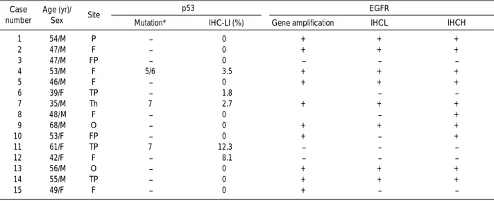
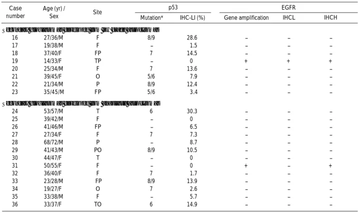
Of the 36 cases, 15 patients had a clinical history of less than 3 months and were designated as primary glioblas-tomas. Twenty-one patients had secondary glioblasglioblas-tomas. Previous operations with initial histopathologic diagnosis of low grade astrocytomas and anaplastic astrocytomas were performed in 8 and 13 patients, respectively. The details of the clinical histories are shown in Table 1 and 2.
Immunohistochemistry for p53 and EGFR proteins Paraffin-embedded tissue slices, 6 m in thickness, were immunostained by the avidin-biotin peroxidase (ABC) method (21). Monoclonal antibodies for DO1 (Santa Cruz, U.S.A.) and EGFR-NCL (Novocastra, UK) were used at a titer of 1:100. DO1 binds to both mutant and wild type p53 proteins, and EGFR-NCL attaches to the extracellular
ligand-binding domain of EGFR (22). The tissue slices were deparaffinated with xylene. Endogenous peroxidase activity was quenched by incubation in 0.3% hydrogen peroxide in 10% methanol. The tissue slices were hydrated with graded alcohol, treated with 10% normal goat serum for 30 min, and then incubated with primary antibodies overnight at a temperature of 4℃They were incubated with biotinylated anti-mouse IgG for 30 min at room tem-perature. The tissue slices for p53 protein immunostaining were incubated with streptavidin-alkaline phosphatase for 1 hr and developed with fast red TR salt (FRT). EGFR-labelled tissue slices were incubated with 1% avidin-biotinylated horseradish peroxidase in 10% normal goat serum for 1 hr at room temperature and developed in a mixture of 0.4 mg/mL diaminobenzidine (DAB) in 0.1% hydrogen peroxide solution for 40 sec. For negative con-trols, the primary antibodies were omitted in the process of immunostaining. All immunostained tissues were counter-stained with hematoxylin solution for 5 min. After dehy-dration, the tissue was sealed with a universal mount (Research Genetics, U.S.A.) and examined under a light microscope. Nuclear staining of p53 was scored semi-quan-titatively by counting 1,000 cells in the most prominently stained area of the tissue slides (23). Staining of cell mem-brane for EGFR was marked as+or - in both low grade and high grade areas of the tissue slides (24).
SSCP analysis for p53 mutations
DNA was extracted from paraffin sections. For samples with positive immunostaining with DO1 antibody, the same areas were chosen for DNA extraction. For samples with negative immunostaining, DNA was extracted from
Case number
Age (yr)/
Sex Site Mutation* IHC-LI (%) Gene amplification IHCL IHCH
1 54/M P - 0 + + + 2 47/M F - 0 + + + 3 47/M FP - 0 - - -4 53/M F 5/6 3.5 + + + 5 46/M F - 0 + + + 6 39/F TP - 1.8 ±� - -7 35/M Th 7 2.7 + + + 8 48/M F - 0 ±� - + 9 68/M O - 0 + + + 10 53/F FP - 0 + - + 11 61/F TP 7 12.3 - - -12 42/F F - 8.1 - - -13 56/M O - 0 + + + 14 55/M TP - 0 + + + 15 49/F F - 0 + -
-Table 1. Clinical data and molecular genetic alterations of cases with primary glioblastomas
Sex: M (male), F (female); Site: F (frontal), P (parietal), T (temporal), O (occipital), Th (thalamic) *: exons, �
: equivocal amplification interpreted as-(negative) in case 6, + (positive) in case 8
IHC-LI, labeling index by immunohistochemistry; IHCL, immunohistochemistry in low-grade area; IHCH, immunohistochemistry in high-grade area
the lesion which was histologically typical of a glioblas-tomas, avoiding the peripheral infiltration zone. Primers for exons 4-9 of p53 were synthesized (Life Tec, Korea) accord-ing to published sequences (25). Details of primers are shown in Table 3. Polymerase chain reaction (PCR)-single stranded conformational polymorphism (SSCP) analysis was performed on 50 L of PCR mixture containing PCR reac-tion buffer (10 mM Tris-HCl, pH 8.8 at 25℃, 1.5 mM MgCl2, 50 mM KCl, 0.1% Triton X-100), 10 pmol of each primer, 50 M dNTPs, 3 L template DNA, 1 U DNA polymerase, and 0.1 Ci -32P dCTP (NEN, U.S.A.).
After adding 20 L of mineral oil (Sigma, U.S.A.), 35 cycles were executed. Each cycle consisted of denaturation (94℃) for 1 min, annealing for 60 sec (58℃for exons 5/6 and 8/9, and 64℃for exons 4 and 7), and extension for 2 min (72℃), and was carried out in an automated DNA Thermal Cycler (Perkin-Elmer, France). Forty microliter of
stop buffer (95% formamide, 10 mM EDTA, 0.05% bro-mophenol blue and 0.05% xylene cyanol) was added to the PCR product, and 2 L of the final mixture was sampled.
Samples were heated at 95℃for 5 min and immediately loaded onto a 6% non-denaturating polyacrylamide gel containing 6% glycerol. Gels were run at 30 W for 3 hr, cooled by fan at room temperature, dried at 60℃, and autoradiographed for 12-48 hr.
RT-PCR for EGFR gene amplification
To investigate the expression level of EGFR in human glioblastoma, we employed a reverse transcriptase (RT)-PCR assay using EGFR primers. Total RNA was extracted from the tumor tissues of each glioblastoma patient. After the reverse transcription, EGFR primers were used for cDNA amplification. RNA integrity was confirmed with Case
number
Age (yr)� /
Sex Site Mutation* IHC-LI (%) Gene amplification IHCL IHCH
Secondary glioblastomas developed from low grade astrocytomas
16 27/36/M F 8/9 28.6 - - -17 19/38/M F - 1.5 - - -18 37/40/F FP 7 14.5 - - -19 14/33/F TP - 0 + + + 20 25/34/M F 7 13.6 - - -21 39/45/F O 5/6 7.9 - - -22 21/34/M P 8/9 12.4 - - -23 35/45/M FP 5/6 3.4 - -
-Secondary glioblastomas developed from anaplastic astrocytomas
24 53/57/M T 6 30.3 - - -25 39/42/M F - 0 - - -26 41/46/M FP - 6.5 - - -27 27/34/F F 7 7.3 - - -28 68/72/M P - 8.7 - - -29 41/43/M PO 8/9 10.5 - - -30 44/47/F T - 0 - - -31 50/55/F F - 0 + - + 32 36/40/F F 7 1.7 - - -33 23/28/M FP 8/9 13.9 - - -34 19/27/F O 7 2.6 - - -35 33/38/M F - 5.7 - - -36 33/37/F TO 6 14.9 - -
-Table 2. Clinical data and molecular genetic alterations of cases with secondary glioblastomas
*, exons; �
, age at the first biopsy of astrocytomas
Sex: M (male), F (female); Site: F (frontal), P (parietal), T (temporal), O (occipital)
IHC-LI, labeling index by immunohistochemistry; IHCL, immunohistochemistry in low-grade area; IHCH, immunohistochemistry in high-grade area
p53 EGFR
4 ATC-TAC-AGT-CCC-CCT-TGC-CG GCA-ACT-GAC-CGT-GCA-AGT-CA
5/6 TC-CTC-TTC-CTG-CAG-TAC-TC AGT-TGC-AAA-CCA-GAC-CTC-AG
7 GTG-TTG-CCT-CCT-AGG-TTG-GC CAA-GTG-GCT-CCT-GAC-CTG-GA
8/9 CCT-ATC-CTG-AGT-AGT-GGT-AT CCA-AGA-CTT-AGT-ACC-TGA-AG
Table 3. The oligonucleotide primers used to amplify exons 4-9 of p53 gene in polymerase chain reaction (5'-3')25
parallel RT-PCR amplification using -actin primers. PCR products were electrophoresed on agarose gels containing ethidium bromide and visualized by UV photography. The detailed procedure was as follows.
Total RNA was extracted from frozen tissues using the LiCl/Urea method (26). For cDNA synthesis, 3 g of total RNA was annealed to oligo (dT) 15 in 20 L total volume containing 4 L of 5 XM-MLV RT buffer (50 mM Tris-HCl, pH 8.3, 7.5 mM KCl, 3 mM MgCl2, and 10 mM DTT), 0.5 mM dNTP, 20 U RNase and 200 U M-MLV RT (Promega, Germany) and was incubated at 37℃for 1.5 hr. PCR mixture contained 1X PCR buffer (50 mM KCl, 10 mM Tris-HCl, pH 9.0, and 0.1% Triton X-100), 1 mM MgCl2, 0.2 M of each primer, 0.2 mM dNTP, 2.5 U of Taq DNA polymerase (Promega, Germany) and 1.5 g of cDNA in a final volume of 50 L. The amplification was performed in the DNA Thermal Cycler. Twenty-five PCR cycles were performed which consisted of 1 min at each temperature (94℃, 53℃, 72℃), except for the initial cycle (5 min at 94℃) and the final extension step (5 min at 72℃). Primer sequences for EGFR and -actin (reference gene) were as follows: 5′ -AGCCATGCCCGCATTAGCTC-3′(5′primer) and 5′-AAAGGAATGCAACTTCCCAA-3′ (3′primer) for EGFR (24) resulting in a 110-bp PCR prod-uct, 5′-GACTATGACTTAGTTGCGTTA-3′(5′primer) and 5′- GCCTTCATACATCTCAAGTTG-3′(3′primer) for -actin resulting in a 501-bp PCR product.
Statistical Analysis
All statistical analysis was performed using Statistical Package for the Social Sciences (SPSS/PC
+
7.5, Chicago, IL, U.S.A.). A p value of less than 0.05 was accepted as sta-tistically significant. The Student t-test was used to evalu-ate differences in the age of patients and p53 labeling index (LI) between primary and secondary glioblastomas. The chi-square and Fisher’s exact tests were used to analyze differ-ences in sex ratio, p53 mutations, p53 expression, EGFR amplification, and EGFR expression.RESULTS
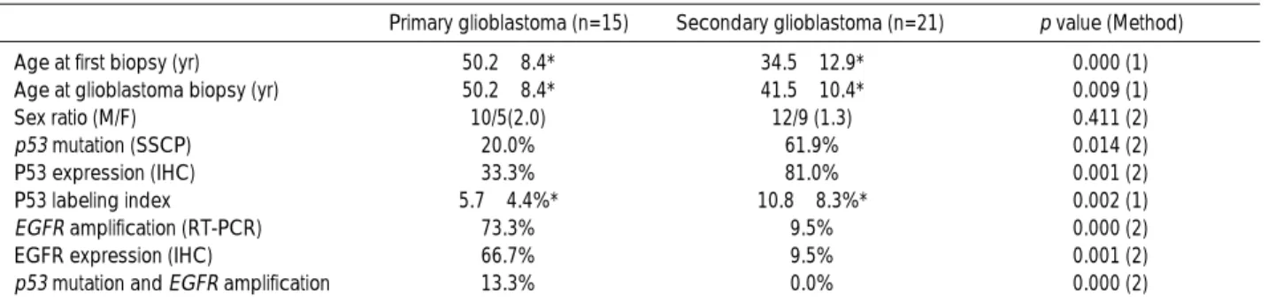
The results of this study are summarized in Table 4. The mean ages of the patients with primary and secondary glioblastomas at the time of surgery were 50.2±8.4 yr and 41.5±10.4 yr respectively. The mean age of the patients with primary glioblastomas was significantly higher than that with secondary glioblastomas. The frequencies of both primary and secondary glioblastoma were higher in males than in females but the difference was not statistically sig-nificant.
p53 mutations were detected in 3 out of 15 (20.0%) pri-mary glioblastomas (Fig. 1) and in 13 out of 21 (61.9%) secondary glioblastomas (p=0.014). All mutations were located between exons 5 to 9. Immunoreactivity to the p53 gene product was expressed on nuclei of glioblastoma cells. The distribution of p53-positive cells was very heteroge-nous in many cases (Fig. 2), and there was no specific
rela-1353
310
1 2 3 4 5 6 7 8 9 10 11
* * *
Fig. 1. Presence of exon 7 DNA of the p53 gene in glioblastomas by PCR (A), and mutations (*) revealed by single stranded con-formational polymorphism (Lane 1-11; secondary glioblastoma cases).
Age at first biopsy (yr) 50.2±8.4* 34.5±12.9* 0.000 (1)
Age at glioblastoma biopsy (yr) 50.2±8.4* 41.5±10.4* 0.009 (1)
Sex ratio (M/F) 10/5(2.0) 12/9 (1.3) 0.411 (2)
p53 mutation (SSCP) 20.0% 61.9% 0.014 (2)
P53 expression (IHC) 33.3% 81.0% 0.001 (2)
P53 labeling index� 5.7±4.4%* 10.8±8.3%* 0.002 (1)
EGFR amplification (RT-PCR) 73.3% 9.5% 0.000 (2)
EGFR expression (IHC) 66.7% 9.5% 0.001 (2)
p53 mutation and EGFR amplification 13.3% 0.0% 0.000 (2) Table 4. Summary of clinical data and molecular genetic alterations in 36 patients with glioblastomas
Primary glioblastoma (n=15) Secondary glioblastoma (n=21) p value (Method)
n, number of cases; *, Mean±Standard deviation (S.D.); �
, Mean±S.D in P53-positive cases
SSCP, single stranded conformational polymorphiom; RT-PCR, reverse transcriptase polymerase chain reaction; IHC, immunohistochemistry; EGFR, epidermal growth factor receptor; (1), Student t-test; (2), chi-square test and Fisher’s exact test
tionship between p53 immunoreactivity and nuclear mor-phology. Immunoreactivity to DO1 was observed in 5 out of 15 (33.3%) primary glioblastomas, and in 17 out of 21 (81.0%) secondary glioblastomas (p=0.001). The mean p53 labeling index (LI) was 5.7±4.4 % and 10.8±8.3 % in primary and secondary glioblastomas, respectively (p=0.002). The correlative results between p53 mutations and p53 immunoreactivities were observed in 30 out of 36 (83.3%) glioblastomas (both positive, 44.4%; both negative, 38.9%). p53 labeling index mutations were observed in all p53-posi-tive glioblastomas; however, 5 out of 36 (13.9%) glioblas-tomas revealed p53-immunopositivities without p53 muta-tions.
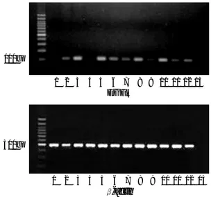
There were two cases with equivocal amplifications of EGFR by RT-PCR. One showed EGFR-positive immuno-reactivity and the other was negative. Among the two, the one with positive immunoreactivity to EGFR was consid-ered as positive EGFR amplification. EGFR amplifications were detected in 11 out of 15 (73.3%) primary glioblas-tomas and 2 out of 21 (9.5%) secondary glioblasglioblas-tomas (p<0.001). The concurrence of p53 mutation and EGFR amplification was observed only in 2 among 15 primary glio-blastomas (Table 1), and even absent in secondary glioblas-tomas (Table 2). EGFR immunoreactivity was noted along the cell membrane of glioblastoma cells (Fig. 3). Immuno-reactivity for EGFR was noted in 10 out of 15 (66.7%) pri-mary glioblastomas, and in 2 out of 21 (9.5%) in secondary glioblastomas (p<0.001). The correlative results between EGFR amplification by RT-PCR and EGFR immunoreac-tivity was observed in 33 out of 36 (91.7%) glioblastomas (both positive, 30.6%; both negative, 61.1%). Only one case revealed EGFR amplification without EGFR-positive
glioblastoma cells. Among the 12 immunopositive cases, nine (75.0%) showed immunoreactivities in both low and high grade areas of glioblastomas while three (25.0%) showed only in high grade areas.
DISCUSSION
The recent molecular biologic studies of astrocytic tumors suggest that both activation of oncogenes and inactivation of tumor suppressor genes are involved in the development of glioblastomas. Von Deimling et al. indicated that there were two types of glioblastomas based on their molecular biologic pathogenesis (27). The type 1 (secondary) glioblas-toma occurs with LOH on chromosome 17p closely associ-ated with p53 mutations and is more common in younger patients. The type 2 (primary) glioblastoma is characterized by higher frequencies of EGFR amplification and LOH on chromosome 10, and is common in elderly patients. The molecular features of primary and secondary glioblastomas were shown to be strongly correlated with the patients’ clinical history in the recent studies (19, 27, 28).
Although some studies support the classification of glioblastomas into two subtypes, only a few studies have analyzed molecular genetic alterations of EGFR and p53 in association with immunohistochemical findings on tumor samples. According to our results, the mutations of the p53 gene were found in 20.0% of primary glioblastomas and 61.9% of secondary glioblastomas. This observation is con-sistent with the high rate of p53 mutations reported by Watanabe et al. (29). The high rate of p53 mutation in sec-ondary glioblastomas might be due to the selection of biop-sies according to the definite evidence of clinical progres-sion from low grade or anaplastic astrocytomas. p53 protein immunoreactivity was noted in 33.3% and 80.1% of pri-mary and secondary glioblastomas, respectively. In the pres-ence of p53 gene mutations, positive staining of tumor cells for p53 protein might be expected; however, correlative studies of p53 protein immunohistochemistry and p53 gene mutation in glioblastomas reported a higher rate of protein expression than the gene mutation (30, 31). The results of this study suggest that an alternative mechanism of p53 gene mutation can result in p53 protein accumulation. In this study, p53 LI was scored 5.7±4.4% and 10.8±8.3% in primary and secondary glioblastomas, respectively. There has been reported a wide range of nuclear staining of p53 in glioblastomas (23, 29), and the difference might be attributed to the divergent monoclonal antibodies or to the scoring system used in each study.
EGFR is a member of the tyrosine kinase family of cell surface receptors and demonstrates various levels of expres-sion throughout the cellular development and in a variety of different cell types. The receptor can transduce signals into the cells upon: 1) interaction with ligands such as EGF, 1 2 3 4 5 6 7 8 9 10 11 12 13
1 2 3 4 5 6 7 8 9 10 11 12 13
Fig. 2. EGFR amplification detected by reverse transcriptase polymerase chain reaction in a control brain (lane 1), glioblas-tomas (lanes 2-12), and negative control (lane 13). Note regular 500bp bands of -actin mRNA as a positive control.
110bp
500bp
EGFR
transforming growth factor (TGF- ), amphiregulin or hep-arin-binding EGF; 2) truncation or mutation of extracellu-lar and/or intracelluextracellu-lar domains; and 3) amplification of a basal receptor activity in the absence of ligand through cooperation with other cellular signaling pathways or nucle-ar events, such as an expression of v-erbB (32, 33). EGFR has been implicated in human cancers, where it may con-tribute both to the initiation (glioblastoma) and progression (epithelial tumors) of the disease. It is frequently present in an amplified or over-expressed form in up to 30-40% of malignant gliomas (34, 35).
According to the present study, the incidence of EGFR amplification was 73.3% and 9.5% in primary and sec-ondary glioblastomas, respectively. Recent studies on EGFR amplification in glioblastoma were carried out by sub-grouping as primary and secondary forms. Lang et al. (36) reported 5 cases out of 34 glioblastomas had EGFR amplifi-cation and LOH on chromosome 10 without p53 muta-tions. Watanabe et al. (29) demonstrated a 63% incidence of EGFR amplification in primary glioblastomas compared to that of only 10% in secondary glioblastomas. According to the present study, positive immunohistochemical
stain-C D
A B
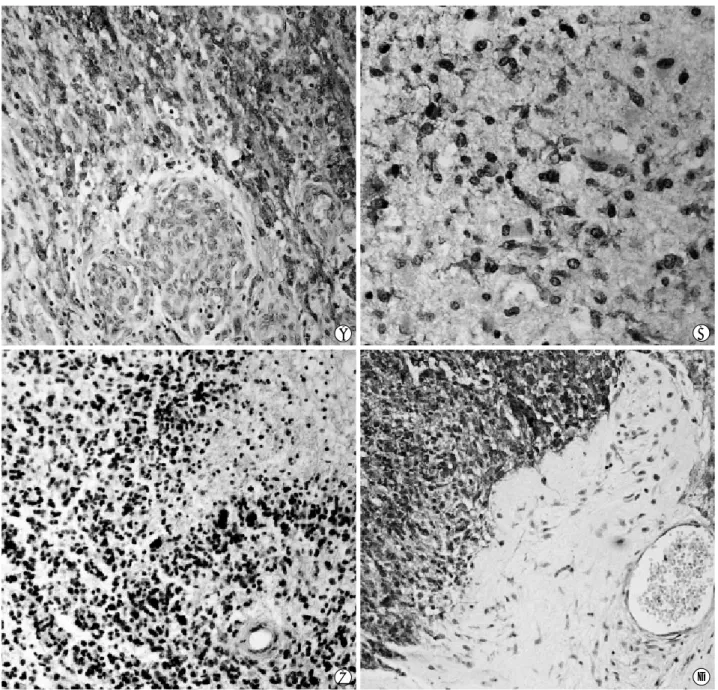
Fig. 3. EGFR immunohistochemistry revealed strong positivity on cell membrane of primary glioblastomas (A; high grade area, B; low grade area) and no immunoreactivity in high grade area (C) in a single case. Similar immunoreactivities are noted in a few cases of sec-ondary glioblastoma (D) (A,B,D×100, C×90).
ings for EGFR were observed in 66.7% and 9.5% of prima-ry and secondaprima-ry glioblastomas, respectively. Correlative studies between EGFR gene amplification and immunohis-tochemical finding are rare. A comparative study by Rieske et al. (37) reported 13 cases (45%) with immunopositivity for EGFR in a series of 28 glioblastomas, while amplifica-tion of EGFR gene was observed in 12 cases. In this study, only one case showed no EGFR immunoreactivity in the presence of EGFR gene amplification, and two cases demon-strated weak EGFR immunoreactivity without EGFR gene amplification. There seems to be no strict correlations bet-ween EGFR gene amplification and the protein overexpres-sion. EGFR was relatively well expressed in both low grade and high grade areas of glioblastomas, despite a few excep-tional cases. Three out of 12 EGFR immunopositive cases were not stained in low grade areas. Concerning with the morphological heterogeneity of glioblastoma, the other study (38) reported that genetic abnormalities seen in the low grade areas were conserved in the high grade areas sug-gesting that these morphologically different cellular subsets were derived from a common transformed clone. These fin-dings might be helpful to detect the presence or absence of tumor cell infiltrations in the peripheral margin or edema-tous area of glioblastomas.
In this study, the p53 mutation with concomitant EGFR gene amplification was observed in 13.3% of primary glioblastoma. There were no such cases among secondary glioblastomas. In line with this observation, Watanabe et al. reported only one case with such profile out of 20 sec-ondary glioblastomas and none in 19 primary glioblastomas (29). These data indicate that EGFR amplification and p53 mutation are two different genetic events in the develop-ment of glioblastomas.
REFERENCES
1. Russell DS, Rubinstein LJ. Pathology of tumors of the central ner-vous system. London, Arnold, 1989.
2. Burger PC, Green SB. Patient age, histologic features, and length of survival in patients with glioblastoma multiforme. Cancer 1987; 59: 1617-25.
3. Daumas-Duport C, Scheithauer B, O'Fallon J, Kelly P. Grading of astrocytomas. A simple and reproducible method. Cancer 1988; 62: 2152-65.
4. James CD, Carlbom E, Dumanski JP, Hansen M, Nordenskjold M, Collins VP, Cavenee WK. Clonal genomic alterations in glioma malignancy stages. Cancer Res 1988; 48: 5546-51.
5. von Deimling A, von Ammon K, Schoenfeld D, Wiestler OD, Seizinger BR, Louis DN. Subsets of glioblastoma multiforme defined by molecular genetic analysis. Brain Pathol 1993; 3: 19-26. 6. Kleihues P, Burger PC, Scheithauer BW. The new WHO
classifica-tion of brain tumours. Brain Pathol 1993; 3: 255-68.
7. Giani C, Finocchiaro G. Mutation rate of the CDKN2 gene in
malignant gliomas. Cancer Res 1994; 54: 6338-9.
8. Louis DN. The p53 gene and protein in human brain tumors. J Neuropathol Exp Neurol 1994; 53: 11-21.
9. Biernat W, Tohma Y, Yonekawa Y, Kleihues P, Ohgaki H. Alter-ations of cell cycle regulatory genes in primary (de novo) and sec-ondary glioblastomas. Acta Neuropathol Berl 1997; 94: 303-9. 10. Biernat W, Debiec-Rychter M, Libeski PP. Mutations of TP53,
amplification of EGFR, MDM2 and CDK4, and deletions of CDKN2A in malignant astrocytomas. Pol J Pathol 1998; 49: 267-71. 11. Hurtt MR, Moossy J, Donovan-Peluso M, Locker J. Amplification
of epidermal growth factor receptor gene in gliomas: Histopatholo-gy and prognosis. J Neuropathol Exp Neurol 1992; 51: 84-90. 12. Ekstrand AJ, James CD, Cavenee WK, Seliger B, Pettersson RF,
Collins VP. Genes for epidermal growth factor receptor, transform-ing growth factor alpha, and epidermal growth factor and their expression in human gliomas in vivo. Cancer Res 1991; 51: 2164-72. 13. von Deimling A, Louis DN, von Ammon K, Petersen I, Wiestler
OD, Seizinger BR. Evidence for a tumor suppressor gene on chro-mosome 19q associated with human astrocytomas, oligoden-drogliomas, and mixed gliomas. Cancer Res 1992; 52: 4277-9. 14. Bello MJ, Vaquero J, de Campos JM, Kusak ME, Sarasa JL,
Saez-Castresana J, Pestana A, Rey JA. Molecular analysis of chromo-some 1 abnormalities in human gliomas reveals frequent loss of 1p in oligodendroglial tumors. Int J Cancer 1994; 57: 172-5. 15. Kraus JA, Koopmann J, Kaskel P, Maintz D, Brandner S, Schramm
J, Louis DN, Wiestler OD, von Deimling A. Shared allelic losses on chromosomes 1p and 19q suggest a common origin of oligoden-droglioma and oligoastrocytoma. J Neuropathol Exp Neurol 1995; 54: 91-5.
16. Reifenberger J, Reifenberger G, Liu L, James CD, Wechsler W, Collins VP. Molecular genetic analysis of oligodendroglial tumors shows preferential allelic deletions on 19q and 1p. Am J Pathol 1994; 145: 1175-90.
17. Chung R, Whaley J, Kley N, Anderson K, Louis D, Menon A, Het-tlich C, Freiman R, Hedley-Whyte ET, Martuza R. TP53 gene mutations and 17p deletions in human astrocytomas. Genes Chro-mosomes Cancer 1991; 3: 323-31.
18. Rasheed BK, McLendon RE, Herndon JE, Friedman HS, Friedman AH, Bigner DD, Bigner SH. Alterations of the TP53 gene in human gliomas. Cancer Res 1994; 54: 1324-30.
19. von Deimling A, von Ammon K, Schoenfeld D, Wiestler OD, Seizinger BR, Louis DN. Subsets of glioblastoma multiforme defined by molecular genetic analysis. Brain Pathol 1993; 3: 19-26. 20. von Deimling A, Louis DN, von Ammon K, Petersen I, Hoell T, Chung RY, Martuza RL, Schoenfeld DA, Yasargil MG, Wiestler OD. Association of epidermal growth factor receptor gene amplifi-cation with loss of chromosome 10 in human glioblastoma multi-forme. J Neurosurg 1992; 77: 295-301.
21. Reed JA, Manahan LJ, Park CS, Brigati DJ. Complete one-hour immunohistochemistry based on capillary action. Biotechniques 1993; 13: 434-43.
22. Cheng Y, Ng HK, Ding M, Zhang SF, Pang C, Lo KW. Molecular analysis of microdissected de novo glioblastomas and paired astro-cytic tumors. J Neuropathol Exp Neurol 1999; 58: 120-8.
23. Ng HK, Lo SY, Huang DP, Poon WS. Paraffin section p53 protein immunohistochemistry in neuroectodermal tumors. Pathology 1994; 26: 1-5.
24. Reifenberger J, Ring GU, Gies U, Cobbers L, Oberstrass J, An HX, Niederacher D, Wechsler W, Reifenberger G. Analysis of p53 muta-tion and epidermal growth factor receptor amplificamuta-tion in recur-rent gliomas with malignant progression. J Neuropathol Exp Neu-rol 1996; 55: 822-31.
25. Brustle O, Ohgaki H, Schmitt HP, Walter GF, Ostertag H, Kleihues P. Primitive neuroectodermal tumors after prophylactic central ner-vous system irradiation in children. Association with an activated K-ras gene. Cancer 1992; 69: 2385-92.
26. Neubauer A, Neubauer B, He M, Effert P, Iglehart D, Frye RA, Liu E. Analysis of gene amplification in archival tissue by differential polymerase chain reaction. Oncogene 1992; 7: 1019-25.
27. von Deimling A, Louis DN, Wiestier OD. Molecular pathways in the formation of gliomas. Glia 1995; 15: 328-38.
28. Nakamura M, Yang F, Fujisawa H, Yonekawa Y, Kleihues P, Ohgaki H. Loss of heterozygosity on chromosome 19 in secondary glioblastoma. J Neuropathol Exp Neurol 2000; 59: 539-43. 29. Watanabe K, Tachibana O, Sato K, Yonekawa Y, Kleihues P,
Ohgaki H. Overexpression of the EGF receptor and p53 mutations are mutually exclusive in the evolution of primary and secondary glioblastomas. Brain Pathol 1996; 6: 217-23.
30. Newcomb EW, Madonia WJ, Pisharody S, Lang FF, Koslow M, Miller DC. A correlative study of p53 protein alteration and p53 gene mutation in glioblastoma multiforme. Brain Pathol 1993; 3:
229-35.
31. Lang FF, Miller DC, Pisharody S, Koslow M, Newcomb EW. High frequency of p53 protein accumulation without p53 gene mutation in human juvenile pilocytic, low grade and anaplastic astrocy-tomas. Oncogene 1994; 9: 949-54.
32. Khazaie K, Schirrmacher V, Lichtner RB. EGF receptor in neopla-sia and metastasis. Cancer Metast Rev 1993; 12: 255-74.
33. Downward J, Yarden Y, Mayes E, Scrace G, Totty N, Stockwell P, Ullrich A, Schlessinger J, Waterfield MD. Close similarity of the epidermal growth factor receptor and the v-erb-B oncogene protein sequences. Nature 1984; 307: 521-7.
34. Collins VP. Amplified genes in human gliomas. Semin Cancer Biol 1993; 4: 27-32.
35. Ohgaki H, Schauble B, zur Hausen A, von Ammon K, Kleihues P. Genetic alterations associated with the evolution and progression of astrocytic brain tumours. Virchows Arch 1995; 427: 113-8. 36. Lang FF, Miller DC, Koslow M, Newcomb EW. Pathways leading
to glioblastoma multiforme: a molecular analysis of genetic alter-ations in 65 astrocytic tumors. J Neurosurg 1994; 81: 427-36. 37. Rieske P, Kordek R, Bartkowiak J, Debiec-Rychter M, Bienhat W,
Liberski PP. A comparative study of epidermal growth factor recep-tor (EGFR) and MDM2 gene amplification and protein immunore-activity in human glioblastomas. Pol J Pathol 1998; 49: 145-9. 38. Cheng Y, Ng HK, Ding M, Zhang SF, Pang JC, Lo KW. Molecular
analysis of microdissected de nove glioblastomas and paired astro-cytic tumors. J Neuropathol Exp Neurol 1999; 58: 120-8.