Clinical Implication of Optical Coherence Tomography-Based
Neoatherosclerosis
Recent research has indicated neoatherosclerosis (NA), the de novo development of atherosclerosis within the neointimal region of the stented segment after coronary stent implantation, as a mechanism of late/very late stent thrombosis (VLST) and restenosis. This research is based on histologic and intravascular imaging studies. Optical coherence tomography (OCT) is an imaging tool that is superior with regard to resolution capacity, and can be used to visualize detailed information about distinct morphological
characteristics of the restenotic tissue. Thus, OCT is a valuable imaging tool for examining NA, such as macrophage infiltration, lipid accumulation, in-stent calcification, or neointimal rupture. This article discusses the prevalence, predictors, and clinical implications of NA that can be observed by OCT.
Keywords: Atherosclerosis; Drug-Eluting Stent; Optical Coherence Tomography
Sung-Jin Hong,1 Seung-Yul Lee,2
and Myeong-Ki Hong1,3,4
1Division of Cardiology, Severance Cardiovascular
Hospital, Yonsei University College of Medicine, Seoul, Korea; 2Department of Internal Medicine,
Sanbon Hospital, Wonkwang University College of Medicine, Gunpo, Korea; 3Severance Biomedical
Science Institute, Yonsei University College of Medicine, Seoul, Korea; 4Cardiovascular Research
Institute, Yonsei University College of Medicine, Seoul, Korea
Received: 10 February 2017 Accepted: 2 April 2017 Address for Correspondence: Myeong-Ki Hong, MD, PhD
Division of Cardiology, Severance Cardiovascular Hospital, Yonsei University College of Medicine, 50-1 Yonsei-ro, Seodaemun-gu, Seoul 03722, Korea
E-mail: [email protected]
https://doi.org/10.3346/jkms.2017.32.7.1056 • J Korean Med Sci 2017; 32: 1056-1061
INTRODUCTION
Drug-eluting stents (DESs) have markedly reduced in-stent re-stenosis (ISR) and repeat revascularizations (1,2). However, in spite of the use of DESs, percutaneous coronary intervention (PCI) is still associated with stent failure and is related to stent thrombosis or restenosis (3,4), which in turn may be related to fatal clinical events. Thus, it is important to investigate the path-ological mechanisms of stent failure. Although several factors for stent failure have been reported, one of the main mechanisms is neoatherosclerosis (NA), i.e., the de novo development of ath-erosclerosis within the neointimal region (5).
This article discusses the prevalence, predictors, and clinical implications of NA observed by optical coherence tomography (OCT).
OCT AND NA OCT-defined NA
NA is defined as atherosclerotic changes in neointimal tissue. It was firstly described in pathologic specimens with bare metal stents (BMSs), and also recently reported in DES (5). It is char-acterized by the presence of clusters of lipid-laden foamy mac-rophages with or without necrotic core formation and/or calci-fication within the neointimal tissue of stented segments.
His-tological examination can be used for diagnosis; however, it can be difficult to obtain pathologic specimens. Therefore, several intra-coronary imaging modalities, including intravascular ul-trasound (IVUS) and OCT, can be used to evaluate coronary stents, particularly the mechanisms of stent failure. Current guidelines recommend IVUS or OCT to assess the mechanisms of stent failure (6,7). However, compared to IVUS, OCT is preferred over IVUS due to the resolution capacity (80–120 vs. 10–20 µm), and OCT is better for evaluating neointimal tissue within the stent-ed segment. OCT can provide detailstent-ed information regarding restenotic tissue (tissue structure, backscatter, microvessels, and composition) (8-10). Therefore, it can be used to visualize dis-tinct morphological characteristics of NA, such as macrophage infiltration, lipid accumulation, in-stent calcification, or neointi-mal rupture.
On OCT images, OCT-defined NA is mainly confined to li-pidic or calcific neointima (11,12). Lili-pidic neointima is shown as diffusely bordered, poor regions with overlying rich bands. Calcific neointima shows well-delineated, signal-poor regions with sharp borders (13,14). Fig. 1 presents the li-pidic and calcific neointima defined by OCT evaluation. Prevalence and predictors of NA
Several studies used OCT to examine the prevalence and risk factors of NA. Table 1 summarizes the prevalence of OCT-based Cardiovascular Disorders
2017-03-16 https://crossmark-cdn.crossref.org/widget/v2.0/logos/CROSSMARK_Color_square.svg
NA. The prevalence varies, according to the stent type, the fol-low-up duration, and clinical indications. Takano et al. (9)
eval-uated neointimal characteristics after BMS implantation at ear-ly (< 6 months, n = 20) and late (≥ 5 years, n = 21) phases, us-Table 1. Prevalence, predictors, and clinical implication of OCT-based NA
Authors Year Subjects number of lesionsStent type and Duration Prevalence Predictors of NA Clinical implication Takano et al.
(9) 2009 Patients with OCT follow-up BMS, n = 21 ≥ 5 yr 67% -
-Kang et al.
(17) 2011 Symptomatic patients with ISR lesions and intimal hyperplasia > 50% of stent area
1st and 2nd DES,
n = 50 Median, 32 mon 90% - Patients with unstable angina (vs. stable angina) had more unstable OCT findings. Kim et al.
(16) 2012 Patients with serial OCT follow-up at 9 mon and 2 yr 1st DES, n = 43; and 2nd DES, n = 33
9 mon; and 2 yr 15%; and
28% -
-Yonetsu et al.
(12) 2012 Patients with OCT follow-up and mean neointimal thickness > 100 μm
BMS, n = 73; DES,
n = 106 Mean, 26.9 mon 47% Stent age, DES vs. BMS, smoking, CKD, no use of ACEi/ARB
-Ko et al.
(31) 2012 Patients with VLST after DES implantation 1st and 2nd DES, n = 18 42 mon 22% Time to OCT study -Lee et al.
(34) 2013 Patients with OCT follow-up and > 50% CSA neointimal stenotic lesions
BMS, n = 24; DES,
n = 128 Median, 70.7 mon 35.5% Stent age, use of first-gen-eration DES, and hyper-tension
NA was associated with higher TLR (93% vs. 78%) and higher stent thrombosis (15% vs. 0%). Kim et al.
(26) 2015 Patients with OCT follow-up ( ≤ 12 mon) and mean neointi-mal thickness > 100 μm
1st and 2nd DES,
n = 482 neoatherosclero-≤ 12 mon, early sis
6% Hypertension, pre-stent LDL-cholesterol ≥ 130 mg/dL
NA was associated with higher clini-cal symptoms (13% vs. 57%) and higher TLR (9% vs. 55%). Lee et al.
(18) 2015 Patients with > 50% neointimal CSA stenosis 1st DES, n = 101; and 2nd DES, n = 111
55 mon; and
12 mon 46%; and 11% CKD, LDL-cholesterol at follow-up more than 70 mg/dL, stent age
NA was associated with a higher acute coronary syndrome (19.0% vs. 3.9%).
Kuroda et al.
(27) 2016 Patients with OCT follow-up > 1 yr after stent implantation BMS, n = 37; DES, n = 277 > 1 yr 17% LDL-cholesterol, CRP levels at follow-up NA was associated with a higher MACE (composite of death, myocardial infarction, and TLR) (37% vs. 9%).
OCT = optical coherence tomography, BMS = bare metal stent, ISR = in-stent restenosis, DES = drug-eluting stent, CKD = chronic kidney disease, ACEi = angiotensin con-verting enzyme inhibitor, ARB = angiotensin-II receptor blocker, VLST = very late stent thrombosis, CSA = cross-sectional area, NA = neoatherosclerosis, TLR = target-lesion revascularization, LDL = low-density lipoprotein, CRP = C-reactive protein, MACE = major adverse cardiovascular event.
Fig. 1. OCT images of neointima. (A) Lipidic change. (B) Calcific change. Arrows indicate lipid and calcification within neointima, respectively. OCT = optical coherence tomography.
ing OCT. Lipid-laden neointima was not observed in the early phase, whereas it was observed at the late phase (67% of the pa-tients). Habara et al. (15) compared neointimal characteristics between early (≤ 1 year) and late restenosis within BMS (> 5 years), and showed that the heterogeneous appearance of neo-intima was more frequent in late restenosis compared with ear-ly restenosis (61% vs. 6%). Thus, after BMS implantation, the prev-alence of NA increased with the stent age. Similarly, after DES implantation, stent age is a major predictor of NA. We previous-ly reported on the serial changes (9 months and 2-year follow-up after DES implantation) of neointima characteristics in DES-treated lesions using OCT (16). Lipid-laden neointima (27.6% vs. 14.5%, P = 0.009) and thin-cap neoatheroma (13.2% vs. 3.9%, P = 0.070) were more frequently detected at 2-year follow-up compared with 9 months of follow-up. In addition, the change of neointimal morphology from homogeneous to heterogeneous or a lipid-laden pattern was observed in 30% of cases. Kang et al. (17) investigated 50 patients who presented with stable (n = 30) or unstable angina (n = 20) with DES restenosis using OCT, and found lipid-containing neointima in 90% of lesions. Twenty-six lesions (52%) had thin-cap fibroatheroma (TCFA)-containing neointima and 29 lesions (58%) had at least one in-stent neointi-mal rupture. In addition, when we analyzed a total of 212 DES-treated patients (second-generation DES, 52.4%) with > 50% neointimal cross-sectional area (CSA) stenosis that were retro-spectively enrolled from the Korean multicenter OCT registry, the incidence of NA increased with stent age (18). NA was found in 1.6% (1/64) of the lesions identified in less than 1 year. In com-parison, NA was observed in 73.9% (17/23) of the lesions over 7 years (18).
With regard to stent type, DES might be associated with a high-er risk of NA, compared with BMS, and Yonetsu et al. (12) eval-uated NA predictors, where all subtypes of DES were found to be independent predictors of NA compared with BMS. Similar-ly, Ali et al. (19) reported that prior DES vs. BMS (odds ratio [OR], 7.0; P = 0.006) was a predictor of NA. However, among the DES cases, second-generation DES was not found to be more pro-tective against NA compared with the first-generation DES (18). Additionally, NA was observed earlier in DES-treated lesions compared with BMS-treated lesions based on the results of a pathologic study (5). A possible explanation for the higher risk of NA with DES vs. BMS could be that the inhibition of neointi-mal hyperplasia by local drug delivery causes delayed coverage and dysfunction of endothelial cells. Although endothelial cells could be barriers that prevent lipid infiltration and inflamma-tory cell migration, incomplete maturation of the regenerated endothelium was more frequently observed in DES vs. BMS (20, 21). Considering these suggested mechanisms, although these metallic stents have been related to NA, bioresorbable vascular scaffolds could be expected to reduce NA. The recovery of va-somotion in the scaffolded segment was noted after
bioresorb-able vascular scaffold implantation, and a signal-rich layer sep-arated the potentially thrombogenic plaque components from the lumen; this suggests a favorable long-term healing response with potential plaque sealing, which was observed in a recent OCT follow-up study (22,23). Furthermore, no cases of necrotic core accumulation of adluminal origin were observed (24). This study did not evaluate whether the initial OCT findings at the index PCI can predict the development of NA at follow-up OCT, and the presence of unstable underlying lesion mor-phology was found to be a significant risk factor of NA based on autopsy cases (5). Nakazawa et al. (5) reported that stent struts embedded in the necrotic core, where the effect of the drug like-ly persists for a long period of time, potentiallike-ly causes dysfunc-tional and/or incompetent endothelium, which can lead to the development of NA. Similarly, Tian et al. (25) found that NA was more frequently associated with adjacent lipid plaque at follow-up OCT. Therefore, the plaque character can be determined by pre-procedural OCT and can be used as a predictor of NA. Although one of the strongest risk factors of NA is a longer time interval to follow-up, when we used OCT to evaluate the incidence of NA that developed after short post-DES implanta-tion duraimplanta-tions, early NA was also observed in 6.4% of 482 DES-treated lesions within 12 months after stent implantation (at a median post-implantation follow-up of 9.1 months) (26). Inde-pendent predictors of early NA were hypertension (OR, 3.20; P = 0.010) and pre-stent low-density lipoprotein (LDL) choles-terol ≥ 130 mg/dL at the time of index procedure (OR, 3.89; P = 0.002). Other clinical predictors of NA are also summarized in Table 1. Yonetsu et al. (12) assessed 179 stents (mean duration, 26.9 months; DES, 59%) and reported OCT-detected NA (lipid-laden neointimal and/or calcification within the neointima) in 84 lesions (47%). In this study, independent determinants of OCT-detected NA were current smoking, chronic kidney disease (CKD), and absence of angiotensin-converting enzyme inhibi-tors or angiotensin II receptor blockade usage. We also analyzed the restenotic lesions with first-generation DES (n = 101) and second-generation DES (n = 111), and found CKD and LDL cho-lesterol at follow-up with values that were more than 70 mg/dL, which were both independent predictors of NA. From the Kobe University Hospital OCT registry (175 patients, 314 lesions), Ku-roda et al. (27) found that C-reactive protein (CRP) level (OR, 1.022; P = 0.001) and LDL cholesterol (OR, 1.022; P = 0.008) at follow-up were independently associated with the presence of NA. Although the exact mechanisms need to be investigated further, these results additionally support the postulated mech-anism of NA; that is to say, oxidative stress and inflammation lead to atherosclerotic changes inside neointima (12,26). These findings support the importance of secondary prevention after stent implantation.
CLINICAL IMPLICATION OF NA: ISR AND STENT THROMBOSIS
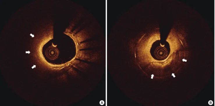
Recent studies with OCT suggest that NA may play an impor-tant role in late stent failure, both for ISR and stent thrombosis. Vergallo et al. (28) reported that the mean neointimal thickness was independently associated with the presence of NA (OR, 2.53; P < 0.001). This association was independent from stent type and time from implantation, suggesting a possible pathologic link between the 2 processes, neointimal thickness and NA. Sim-ilarly, after DES implantation, late ISR and very late ISR were associated with NA, suggesting that these could contribute to the late catch-up phenomenon after DES (29). Additionally, re-cent OCT findings in patients with late and very late stent throm-bosis (VLST) reported a high prevalence of plaque rupture and impaired healing (30-33). According to a prospective multicenter registry, ruptured neoatherosclerotic lesions were more frequent with BMS than with DES (36% vs. 14%, P = 0.005), and late/VLST were mainly related to malapposition (31%) and NA (28%) (32). Therefore, NA may contribute to both ISR and stent thrombo-sis. Neoatherosclerotic OCT images in patients that presented with ISR and stent thrombosis are shown in Fig. 2.
Patients with NA had higher chances of symptoms or need of repeat vascularization (Table 1) (34). We evaluated the neointi-mal characteristics of 152 lesions (128 DESs and 24 BMSs) with > 50% CSA neointimal stenosis. NA was observed in 54 lesions (35.5%, 35 DESs and 19 BMSs), and patients with NA vs.
with-out NA had a higher rate of target lesion revascularization (92.6% vs. 77.6%, P = 0.018) and stent thrombosis (14.8% vs. 0%, P < 0.001) (34). Similarly, from the Korean multicenter OCT registry, pati-ents with NA showed a higher rate of acute coronary syndrome at follow-up OCT (19.0% vs. 3.9%, P = 0.001) (18). In addition, based on data from the Kobe University Hospital OCT registry, the incidence of major adverse cardiovascular events (MACEs; e.g., composite of death, myocardial infarction, and target-lesion revascularization [TLR]) was significantly higher in patients with NA vs. those without NA (36.9% vs. 9.3%, P < 0.001). This was mainly driven by an increase in the rate of cardiac death (6.5% vs. 0%, P = 0.017) and TLR (30.4% vs. 7.7%, P < 0.001). Moreover, the incidence of stent thrombosis (definite/probable) was sig-nificantly higher in patients with NA (6.5% vs. 0%, P = 0.017) (27). Patients with early NA (within 12 months after stent implanta-tion) also had a higher incidence of clinical symptoms (13% vs. 57%, P < 0.001) and had undergone a higher frequency of TLR (9% vs. 55%, P < 0.001) at the time of OCT follow-up (26). Clinical presentation of NA can vary from asymptomatic or stable angina to life-threatening acute coronary syndrome be-cause of stent thrombosis. Compared to patients with stable an-gina, patients with unstable angina show higher incidences of unstable OCT findings, including TCFA-containing neointima, neointima rupture, and thrombus (P = 0.027) (17). OCT-defined rupture of lipidic neointima was observed in patients with VLST (31).
In addition, when treating ISR, the neointimal characteristic
Fig. 2. OCT images of neoatherosclerotic neointima in patients with ISR (A) and stent thrombosis (B). (A) Lumen is narrow for NA in a patient with ISR. (B) Disrupted neointima (arrows) with thrombi (arrowheads) is observed in a patient with stent thrombosis.
ISR = in-stent restenosis, NA = neoatherosclerosis.
of ISR lesions, particularly NA, could impact the periprocedural myocardial injury. We analyzed a total of 125 patients with ISR lesions (35). Post-PCI creatine kinase-myocardial band (CK-MB) elevation was observed in 20 (16.0%) patients, and multivariate analysis revealed that the maximum length of segments with NA (OR, 1.463; P = 0.011) and TCFA (OR, 14.328; P = 0.041) were independent predictors for post-PCI CK-MB elevation. Thus, NA may underlie the vulnerability of neointima to PCI, with a distal microembolization of neointimal debris.
Although routine OCT examination is not necessary for all patients who undergo follow-up angiography, the data from these studies indicates that OCT examination can be valuable for patients with symptoms and angiographically-significant stenosis. For these patients, OCT examination can determine the presence of NA for future risk stratification and can be use-ful for evaluating the mechanism of stent failure for targeted therapeutic approaches. Additional studies are needed to de-termine whether these results can be translated to clinical im-provements.
CONCLUSION
Recent evidence has indicated that NA is a considerable mech-anism of late/VLST and restenosis. In addition, it is an impor-tant substrate for future adverse events. Thus, detection of NA by OCT and identification of morphological characteristics, in-cluding in-stent lipidic or calcific neointima, is helpful for pre-dicting and preventing greater future adverse events.
ACKNOWLEDGMENT
This is an invited review article for Professor Myeong-Ki Hong who is the laureate of the 14th Pfizer Medical Award, The Nation-al Academy of Medicine Korea, 2016.
DISCLOSURE
The authors have no potential conflicts of interest to disclose. AUTHOR CONTRIBUTION
Conceptualization: Hong SJ, Lee SY, Hong MK. Writing - origi-nal draft: Hong SJ, Lee SY, Hong MK. Writing - review & editing: Hong SJ, Lee SY, Hong MK.
ORCID
Sung-Jin Hong https://orcid.org/0000-0003-4893-039X Seung-Yul Lee https://orcid.org/0000-0002-9039-9806 Myeong-Ki Hong https://orcid.org/0000-0002-2090-2031
REFERENCES
1. Stone GW, Ellis SG, Cannon L, Mann JT, Greenberg JD, Spriggs D, O’Shau ghnessy CD, DeMaio S, Hall P, Popma JJ, et al. Comparison of a polymer based paclitaxeleluting stent with a bare metal stent in patients with com plex coronary artery disease: a randomized controlled trial. JAMA 2005;
294: 121523.
2. Stone GW, Ellis SG, Cox DA, Hermiller J, O’Shaughnessy C, Mann JT, Tur co M, Caputo R, Bergin P, Greenberg J, et al. A polymerbased, paclitaxel eluting stent in patients with coronary artery disease. N Engl J Med 2004;
350: 22131.
3. Räber L, Magro M, Stefanini GG, Kalesan B, van Domburg RT, Onuma Y, Wenaweser P, Daemen J, Meier B, Jüni P, et al. Very late coronary stent thrombosis of a newergeneration everolimuseluting stent compared with earlygeneration drugeluting stents: a prospective cohort study. Cir-culation 2012; 125: 111021.
4. Yamaji K, Kimura T, Morimoto T, Nakagawa Y, Inoue K, Soga Y, Arita T, Shirai S, Ando K, Kondo K, et al. Very longterm (15 to 20 years) clinical and angiographic outcome after coronary bare metal stent implantation.
Circ Cardiovasc Interv 2010; 3: 46875.
5. Nakazawa G, Otsuka F, Nakano M, Vorpahl M, Yazdani SK, Ladich E, Kolo dgie FD, Finn AV, Virmani R. The pathology of neoatherosclerosis in hu man coronary implants baremetal and drugeluting stents. J Am Coll Cardiol 2011; 57: 131422.
6. Levine GN, Bates ER, Blankenship JC, Bailey SR, Bittl JA, Cercek B, Cham bers CE, Ellis SG, Guyton RA, Hollenberg SM, et al. 2011 ACCF/AHA/SCAI Guideline for Percutaneous Coronary Intervention. A report of the Amer ican College of Cardiology Foundation/American Heart Association Task Force on Practice Guidelines and the Society for Cardiovascular Angiog raphy and Interventions. J Am Coll Cardiol 2011; 58: e44122.
7. Windecker S, Kolh P, Alfonso F, Collet JP, Cremer J, Falk V, Filippatos G, Hamm C, Head SJ, Jüni P, et al. 2014 ESC/EACTS guidelines on myocar dial revascularization: the task force on myocardial revascularization of the European Society of Cardiology (ESC) and the European Association for CardioThoracic Surgery (EACTS) developed with the special contri bution of the European Association of Percutaneous Cardiovascular In terventions (EAPCI). Eur Heart J 2014; 35: 2541619.
8. Gonzalo N, Serruys PW, Okamura T, van Beusekom HM, GarciaGarcia HM, van Soest G, van der Giessen W, Regar E. Optical coherence tomog raphy patterns of stent restenosis. Am Heart J 2009; 158: 28493.
9. Takano M, Yamamoto M, Inami S, Murakami D, Ohba T, Seino Y, Mizuno K. Appearance of lipidladen intima and neovascularization after implan tation of baremetal stents extended latephase observation by intracoro nary optical coherence tomography. J Am Coll Cardiol 2009; 55: 2632.
10. Lee SJ, Kim BK, Kim JS, Ko YG, Choi D, Jang Y, Hong MK. Evaluation of neointimal morphology of lesions with or without instent restenosis: an optical coherence tomography study. Clin Cardiol 2011; 34: 6339.
11. Yonetsu T, Kim JS, Kato K, Kim SJ, Xing L, Yeh RW, Sakhuja R, McNulty I, Lee H, Zhang S, et al. Comparison of incidence and time course of neo atherosclerosis between bare metal stents and drugeluting stents using optical coherence tomography. Am J Cardiol 2012; 110: 9339.
12. Yonetsu T, Kato K, Kim SJ, Xing L, Jia H, McNulty I, Lee H, Zhang S, Uemu ra S, Jang Y, et al. Predictors for neoatherosclerosis: a retrospective obser vational study from the optical coherence tomography registry. Circ
Car-diovasc Imaging 2012; 5: 6606.
13. Tearney GJ, Regar E, Akasaka T, Adriaenssens T, Barlis P, Bezerra HG, Bou ma B, Bruining N, Cho JM, Chowdhary S, et al. Consensus standards for acquisition, measurement, and reporting of intravascular optical coher ence tomography studies: a report from the International Working Group for Intravascular Optical Coherence Tomography Standardization and Validation. J Am Coll Cardiol 2012; 59: 105872.
14. Prati F, Regar E, Mintz GS, Arbustini E, Di Mario C, Jang IK, Akasaka T, Cos ta M, Guagliumi G, Grube E, et al. Expert review document on methodol ogy, terminology, and clinical applications of optical coherence tomogra phy: physical principles, methodology of image acquisition, and clinical application for assessment of coronary arteries and atherosclerosis. Eur Heart J 2010; 31: 40115.
15. Habara M, Terashima M, Nasu K, Kaneda H, Inoue K, Ito T, Kamikawa S, Kurita T, Tanaka N, Kimura M, et al. Difference of tissue characteristics between early and very late restenosis lesions after baremetal stent im plantation: an optical coherence tomography study. Circ Cardiovasc In-terv 2011; 4: 2328.
16. Kim JS, Hong MK, Shin DH, Kim BK, Ko YG, Choi D, Jang Y. Quantitative and qualitative changes in DESrelated neointimal tissue based on serial OCT. JACC Cardiovasc Imaging 2012; 5: 114755.
17. Kang SJ, Mintz GS, Akasaka T, Park DW, Lee JY, Kim WJ, Lee SW, Kim YH, Whan Lee C, Park SW, et al. Optical coherence tomographic analysis of instent neoatherosclerosis after drugeluting stent implantation. Circu-lation 2011; 123: 295463.
18. Lee SY, Hur SH, Lee SG, Kim SW, Shin DH, Kim JS, Kim BK, Ko YG, Choi D, Jang Y, et al. Optical coherence tomographic observation of instent neoatherosclerosis in lesions with more than 50% neointimal area steno sis after secondgeneration drugeluting stent implantation. Circ Cardio-vasc Interv 2015; 8: e001878.
19. Ali ZA, Roleder T, Narula J, Mohanty BD, Baber U, Kovacic JC, Mintz GS, Otsuka F, Pan S, Virmani R, et al. Increased thincap neoatheroma and periprocedural myocardial infarction in drugeluting stent restenosis: mul timodality intravascular imaging of drugeluting and baremetal stents.
Circ Cardiovasc Interv 2013; 6: 50717.
20. Joner M, Nakazawa G, Finn AV, Quee SC, Coleman L, Acampado E, Wil son PS, Skorija K, Cheng Q, Xu X, et al. Endothelial cell recovery between comparator polymerbased drugeluting stents. J Am Coll Cardiol 2008;
52: 33342.
21. Nakazawa G, Nakano M, Otsuka F, Wilcox JN, Melder R, Pruitt S, Kolodgie FD, Virmani R. Evaluation of polymerbased comparator drugeluting stents using a rabbit model of iliac artery atherosclerosis. Circ Cardiovasc Interv 2011; 4: 3846.
22. Simsek C, Karanasos A, Magro M, GarciaGarcia HM, Onuma Y, Regar E, Boersma E, Serruys PW, van Geuns RJ. Longterm invasive followup of the everolimuseluting bioresorbable vascular scaffold: fiveyear results of multiple invasive imaging modalities. EuroIntervention 2016; 11: 996
1003.
23. Karanasos A, Simsek C, Serruys P, Ligthart J, Witberg K, van Geuns RJ, Sia nos G, Zijlstra F, Regar E. Fiveyear optical coherence tomography follow up of an everolimuseluting bioresorbable vascular scaffold: changing the paradigm of coronary stenting? Circulation 2012; 126: e8991.
24. Karanasos A, Simsek C, Gnanadesigan M, van Ditzhuijzen NS, Freire R, Dijkstra J, Tu S, Van Mieghem N, van Soest G, de Jaegere P, et al. OCT as sessment of the longterm vascular healing response 5 years after evero limuseluting bioresorbable vascular scaffold. J Am Coll Cardiol 2014; 64:
234356.
25. Tian J, Ren X, Uemura S, Dauerman H, Prasad A, Toma C, Jia H, Abtahian F, Vergallo R, Hu S, et al. Spatial heterogeneity of neoatherosclerosis and its relationship with neovascularization and adjacent plaque characteris tics: optical coherence tomography study. Am Heart J 2014; 167: 884892.
e2.
26. Kim C, Kim BK, Lee SY, Shin DH, Kim JS, Ko YG, Choi D, Jang Y, Hong MK. Incidence, clinical presentation, and predictors of early neoatherosclero sis after drugeluting stent implantation. Am Heart J 2015; 170: 5917.
27. Kuroda M, Otake H, Shinke T, Takaya T, Nakagawa M, Osue T, Taniguchi Y, Iwasaki M, Nishio R, Kinutani H, et al. The impact of instent neoathero sclerosis on longterm clinical outcomes: an observational study from the Kobe University Hospital optical coherence tomography registry. Eu-roIntervention 2016; 12: e136674.
28. Vergallo R, Yonetsu T, Uemura S, Park SJ, Lee S, Kato K, Jia H, Abtahian F, Tian J, Hu S, et al. Correlation between degree of neointimal hyperplasia and incidence and characteristics of neoatherosclerosis as assessed by optical coherence tomography. Am J Cardiol 2013; 112: 131521.
29. Habara M, Terashima M, Nasu K, Kaneda H, Yokota D, Ito T, Kurita T, Tera moto T, Kimura M, Kinoshita Y, et al. Morphological differences of tissue characteristics between early, late, and very late restenosis lesions after first generation drugeluting stent implantation: an optical coherence to mography study. Eur Heart J Cardiovasc Imaging 2013; 14: 27684.
30. Amabile N, Souteyrand G, Ghostine S, Combaret N, Slama MS, Barber Chamoux N, Motreff P, Caussin C. Very late stent thrombosis related to incomplete neointimal coverage or neoatherosclerotic plaque rupture identified by optical coherence tomography imaging. Eur Heart J Car-diovasc Imaging 2014; 15: 2431.
31. Ko YG, Kim DM, Cho JM, Choi SY, Yoon JH, Kim JS, Kim BK, Choi D, Jang Y, Hong MK. Optical coherence tomography findings of very late stent thrombosis after drugeluting stent implantation. Int J Cardiovasc Imag-ing 2012; 28: 71523.
32. Souteyrand G, Amabile N, Mangin L, Chabin X, Meneveau N, Cayla G, Vanzetto G, Barnay P, Trouillet C, Rioufol G, et al. Mechanisms of stent thrombosis analysed by optical coherence tomography: insights from the national PESTO French registry. Eur Heart J 2016; 37: 120816.
33. Taniwaki M, Radu MD, Zaugg S, Amabile N, GarciaGarcia HM, Yamaji K, Jørgensen E, Kelbæk H, Pilgrim T, Caussin C, et al. Mechanisms of very late drugeluting stent thrombosis assessed by optical coherence tomog raphy. Circulation 2016; 133: 65060.
34. Lee SY, Shin DH, Mintz GS, Kim JS, Kim BK, Ko YG, Choi D, Jang Y, Hong MK. Optical coherence tomographybased evaluation of instent neoath erosclerosis in lesions with more than 50% neointimal crosssectional area stenosis. EuroIntervention 2013; 9: 94551.
35. Lee SY, Hong MK, Shin DH, Kim JS, Kim BK, Ko YG, Choi D, Jang Y. Opti cal coherence tomographybased predictors for creatine kinasemyocar dial band elevation after elective percutaneous coronary intervention for instent restenosis. Catheter Cardiovasc Interv 2015; 85: 56472.