Factors Affecting Postural Reduction in
Posterior Surgery for Thoracolumbar Burst
Fracture
by
Sang-Jin Youn
Major in Medicine
Department of Medical Sciences
The Graduate School, Ajou University
Factors Affecting Postural Reduction in
Posterior Surgery for Thoracolumbar Burst
Fracture
by
Sang-Jin Youn
A Dissertation Submitted to The Graduate School of
Ajou University in Partial Fulfillment of the Requirements
for the Degree of
Master of Science in Medicine
Supervised by
Chang-Hoon Jeon, M.D., Ph.D.
Major in Medicine
Department of Medical Sciences
The Graduate School, Ajou University
This certifies that the dissertation
of Sang-Jin Youn is approved.
SUPERVISORY COMMITTEE
Chang-Hoon Jeon
Kyeong-Jin Han
Jae- Ho Cho
The Graduate School, Ajou University
December, 5th, 2014
i
- ABSTRACT -
Factors Affecting Postural Reduction in Posterior Surgery for
Thoracolumbar Burst Fracture
Study Design: Retrospective analysis of a prospectively collected patient database.
Objective: To investigate the significance and relevant factors of postural kyphosis reduction during posterior surgical treatment of thoracolumbar burst fracture.
Summary of Background Data: Optimal reduction of kyphosis is a goal in the surgical treatment of thoracolumbar burst fracture. Several factors are known to limit the amount of posterior surgical reduction. However, few comprehensive assessments of intraoperative postural reduction have been reported.
Methods: Seventy-two consecutive patients who underwent posterior surgical treatment for thoracolumbar (T11–L2) burst fracture were included. Postural reduction was evaluated using C-arm fluoroscopic images and regarded as insufficient when the lateral Cobb angle was ≥20 degrees or AP Cobb angle ≥10 degrees. Clinical characteristics
including sex, age, body mass index, time to operation, injury level, and neurological injury, as well as radiologic characteristics including fracture morphology, fracture deformity, canal stenosis, and ligament injuries were investigated to determine the relevant factors.
ii
Results: The mean lateral Cobb angle was 22.2±11.0 degrees preoperatively, 16.4±7.7 degrees after postural reduction (P<0.001), and 13.4±6.9 degrees after instrumental
reduction (P<0.001). Insufficient postural reduction was found in 25 (34.7%) patients, all of which were lateral. The relevant factors for insufficient reduction, as identified by
multivariate analysis, were time to operation <72 hours (OR, 6.453; 95% CI,1.283–32.553), burst-split type injury (OR, 4.689; 95% CI,1.314–25.225), and anterior compression ratio >0.5 (OR, 2.284;95% CI, 1.151–19.811).
Conclusions: Postural reduction plays an important role in the reduction of kyphosis and compression deformity after thoracolumbar burst fracture. However, it was affected by delayed operation time, burst-split type injury, and severe anterior vertebral compression. Keyword : Thoracolumbar, Burst fracture, Postural reduction, Instrumental reduction, Risk factor
iii
TABLE OF CONTENTS
ABSTRACT ··· ⅰ TABLE OF CONTENTS ··· ⅲ LIST OF FIGURES ··· ⅳ LIST OF TABLES ··· ⅴ . Ⅰ INTRODUCTION ··· 1 .Ⅱ MATERIALS AND METHODS ··· 3 . Ⅲ RESULTS ··· 7 . Ⅳ DISCUSSION ··· 11 . Ⅴ CONCLUSION ··· 14 REFERENCES ··· 15 국문요약 ··· 19
iv
LIST OF FIGURES
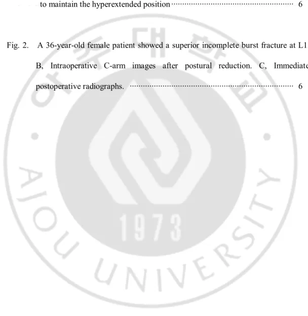
Fig. 1.
After postural kyphosis reduction, high bolsters were placed under the iliac crests to maintain the hyperextended position ··· 6
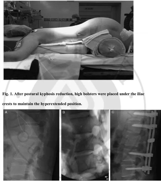
Fig. 2. A 36-year-old female patient showed a superior incomplete burst fracture at L1. B, Intraoperative C-arm images after postural reduction. C, Immediate postoperative radiographs. ··· 6
v
LIST OF TABLES
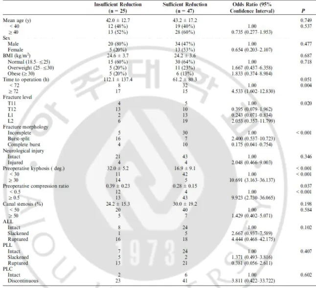
Table 1. Univariate analysis of insufficient postural reduction ··· 9
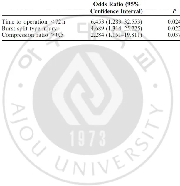
Table 2. Significant factors in insufficient postural reduction, determined by multivariate logistic regression analysis··· 10
1
I. INTRODUCTION
Thoracolumbar burst fracture results from axial compression with an associated flexion moment, creating kyphotic deformity. The posterior vertebral body is injured by definition, and in many cases it is retropulsed into the spinal canal.(Denis F, 1983; Cotterill PC et al, 1987; Hashimoto T et al, 1988) Thus, management is determined by the degree of mechanical and neurological instability. A posterior approach is generally suggested for most unstable thoracolumbar burst fractures, unless incomplete neurological injury or severe vertebral comminution has occurred.(Vaccaro AR et al, 2005; Vaccaro AR et al, 2006) In the posterior approach, ligamentotaxis can indirectly achieve correction of kyphosis, restoration of vertebral height, and clearing of canal compromise.(Zou D et al, 1993) Extension and distraction forces generate an anteriorly directed reduction load to be applied through the bony attachments along the vertebral body, annulus, and paravertebral ligaments.(Zou D et al, 1993; Xu Y, et al, 2008) The mechanism and efficacy of ligamentotaxis in thoracolumbar burst fractures have been well described.(Gertzbein SD et al, 1992; Kuner EH et al, 1994; Fredrickson BE et al, 1998; Mueller LA et al, 2006) Positional reduction and instrumental distraction can generate ligamentotaxis.(Kuner EH et al, 1994; Mueller LA et al, 2006; Whang PG and Vaccaro AR, 2007; Xu Y et al, 2008) Tensing of the posterior longitudinal ligament (PLL) and, to a lesser extent, the anterior longitudinal ligament (ALL) reduces retropulsed fragments that are still attached to a ligamentous structure or the outermost annular layer of the intervertebral disk.(Kuner EH et al, 1994) Several radiologic and clinical conditions, including destruction of the PLL or the outermost annular layer of the
2
intervertebral disk, severe canal compromise, and delayed surgical timing are known to limit the amount of canal clearance.(Fredrickson BE et al, 1998; Gertzbein SD et al, 1992; Chang KW, 1992; Katonis PG et al, 1999) However, few comprehensive assessments of the effects of radiologic and clinical conditions on the amount of postural kyphosis reduction have been reported. Moreover, the significance of postural reduction is not fully understood. The aim of this study was to investigate the significance of postural reduction in the posterior surgical treatment of thoracolumbar burst fracture and to determine the relevant factors affecting the optimal restoration of postural reduction.
3
Ⅱ
. MATERIALS AND METHODS
This study involved a retrospective analysis of 72 consecutive patients who underwent surgical treatment with a posterior approach for thoracolumbar (T11–L2) burst fracture at a department of orthopedic surgery in a tertiary hospital between March 2009 and June 2012. Burst fractures with preexisting spinal deformity, no spinal canal violation, and involving >2 levels were excluded. Burst fractures treated with additional anterior surgery after a posterior approach were included. All included patients were evaluated with plain radiographs, computed tomography (CT), magnetic resonance imaging, and neurological examination at the time of presentation. Surgical treatment was determined by calculating the
thoracolumbar injury severity score (TLISS; TLISS>5 or TLISS=4 with the presence of clinical, local, or systemic qualifiers). The ethical committee of the hospital reviewed and approved the design of this study. Information on each patient's age, sex, body mass index, time from injury to operation, neurological status, and other medical conditions were obtained from the medical records. The morphology of burst fractures was categorized as a superior incomplete burst fracture, inferior incomplete burst fracture, burst-split fracture, or complete burst fracture using preoperative CT images according to the classification
described by Magerl et al(Magerl F, et al, 1994) with modification. Angular deformity of the burst fractures was measured on preoperative supine anteroposterior (AP) and lateral plain radiographs using the Cobb technique(Kuklo TR, et al, 2001), taken from the superior endplate of the vertebra 1 level above the fractured vertebra to the inferior endplate of the vertebra 1 level below the fractured vertebra. Compression deformity of the burst fractures
4
was also calculated by measuring the anterior and posterior vertebral compression ratios (VCRs) at the level of injury to the estimated normal height using the vertebra above and below the injury.(Keynan O et al, 2006) The degree of canal stenosis was calculated by measuring the ratio of cross-sectional area at the level of injury to the estimated normal canal dimensions at that level on axial CT images.(Keynan O et al, 2006) The integrity of the ALL and PLL was described as intact, slackened, or ruptured using preoperative
magnetic resonance images, as described by Oner et al.(Oner FC et al, 1999) The integrity of the posterior ligamentous complex was described as intact or discontinuous by intraoperative gross morphology. All radiologic parameters were measured by 3 orthopedic surgeons not involved with the care of the study subjects and averaged. Following the induction of general anesthesia, each patient was placed in a prone position supported by a chest roll or a Jackson spinal surgery table (OSI, Union City, CA). Rocuronium was used for muscle relaxation to ensure zero train-of-four. Postural reduction was performed by 1 spinal surgeon (N.-S.C.) using manual forces and bolsters under intermittent C-arm fluoroscopic guidance. For kyphosis reduction, the patient's pelvis was gradually pulled and elevated to a
hyperextended position. The kyphosis vertex was gently pushed down using manual force. High bolsters were then placed under the iliac crests to maintain the hyperextended position (Fig. 1). When needed, scoliosis reduction was also performed using manual force and counterforce. AP and lateral C-arm fluoroscopic images demonstrating the superior endplate of the level above the injury and the inferior endplate of the level below the injury were obtained to evaluate the amount of postural reduction. Instrumental reduction was also performed by the same surgeon (N.-S.C.) using a pedicle screw instrument. After preparing and draping the patient, a standard midline incision was performed to allow for meticulous
5
exposure of the posterior elements. Pedicle screws were placed using standard landmarks and techniques. In most cases, long-segment instrumentation was performed 2 levels above and below the fractured vertebra. Bilateral rods were adequately precontoured and
positioned into the pedicle screws. Longitudinal distraction was then applied under C-arm fluoroscopic guidance. Posterior fusion with autoiliac corticocancellous bone graft was subsequently performed in all patients. Each radiologic parameter on preoperative plain radiographs was compared with those on the C-arm images after postural reduction and on immediately postoperative supine radiographs (Fig. 2). A paired t test was used to compare the change in each parameter. On the basis of the degree of postural reduction, patients were grouped as having insufficient reduction (lateral Cobb angle≥20 degrees or AP Cobb angle≥10 degrees) or sufficient reduction (lateral Cobb angle <20 degrees or AP Cobb angle <10 degrees). Associations between the dichotomous outcome variables (insufficient or sufficient reduction) and the independent variables were examined using univariate and multivariate logistic regression analysis, and adjusted odds ratios (ORs) with 95% confidence intervals (CIs) were calculated. Variables with a P-value <0.10 on univariate analysis were included in the multivariate logistic regression analysis. Reliability of the measurement of radiologic parameters among the 3 orthopedic surgeons was evaluated by calculating the interclass correlation coefficients for numeric variables and k-values for categorical variables. Statistical analysis was carried out using SPSS software (version 12.0; SPSS Inc.,Chicago, IL).
6
Fig. 1. After postural kyphosis reduction, high bolsters were placed under the iliac crests to maintain the hyperextended position.
Fig. 2. A, A 36-year-old female patient showed a superior incomplete burst fracture at L1. B, Intraoperative C-arm images after postural reduction. C, Immediate
7
Ⅲ
. RESULTS
The study sample comprised 54 men (75%) and 18 women with a mean age of
42.8±15.7 (range, 14–72) years. The mean body mass index was 24.3±3.6 (18.9–32.1) m/kg2. The mean time from injury to operation was 78.9±105.6 (8–672) hours. The most frequently injured level was L2 (25 cases) followed by T12 (23 cases), L1 (15 cases), and T11 (9 cases). The morphologies of burst fracture included 31 (43.1%) superior incomplete burst fractures, 4 (5.6%) inferior incomplete burst fractures, 23 (31.9%) burst-split fractures, and 14 (19.4%) complete burst fractures. Two (3.4%) patients had complete spinal cord injury, 2 (3.4%) patients had incomplete spinal cord injury, and 4 (6.9%) patients had root injury. At presentation, the mean AP Cobb angle was 3.8±3.6 degrees (0–20.0 degrees) and the mean lateral Cobb angle was 22.2±11.0 degrees (1.1–41.2 degrees). The mean anterior and
posterior VCRs were 0.32±0.19 (0.05–0.69) and 0.08±0.10 (0–0.34), respectively. The mean degree of canal stenosis was 28.0%±18.1% (9.8%–98.2%). The ALL was intact in 32 (44.4%) patients, slackened in 6 (8.3%) patients, and ruptured in 34 (45.9%) patients. The PLL was intact in 34 (47.2%) patients, slackened in 5 (6.8%) patients, and ruptured in 33 (45.8%) patients. Discontinuity of the posterior ligamentous complex was found in 64 (88.9%) patients. The mean AP Cobb angle decreased to 3.5±2.3 degrees (0–13.0 degrees) after postural reduction (P=0.058), and to 1.8±1.4 degrees (0–7.0 degrees) after instrumental reduction (P<0.001). The mean lateral Cobb angle decreased to 16.4±7.7 degrees (4–33.8 degrees) after postural reduction (P<0.001), and to 13.4±6.9 degrees (0–7.0 degrees) after instrumental reduction (P<0.001). The mean anterior VCR decreased to 0.24±0.13 (0.05–
8
0.55) after postural reduction (P<0.001), and to 0.14±0.11 (0–0.57) after instrumental reduction (P<0.001). The mean posterior VCR decreased to 0.08±0.09 (0–0.44) after postural reduction (P=0.409), and to 0.05±0.07 (0–0.38) after instrumental reduction
(P=0.001). Our series included 25 (34.7%) insufficient postural reductions, all of which were lateral (Cobb angle Z20 degrees). The results of our univariate analysis suggested that time to operation, fracture level, fracture morphology, preoperative kyphosis, and preoperative anterior VCR were significant risk factors for insufficient postural reduction (Table 1). The multivariate analysis results demonstrated that time to operation >72 hours (OR, 6.453; 95% CI, 1.283–32.553), burst-split morphology (OR, 4.689; 95% CI, 1.314–25.225), and anterior compression ratio >0.5 (OR, 2.284; 95% CI, 1.151–19.811) were significant risk factors for insufficient postural reduction. Fracture level was a significant risk factor in the univariate analysis, but not in the multivariate analysis (Table 2). The interclass correlation
coefficients of interobserver agreement for radiologic measurements were 0.69–0.72 (fair to good) for measurement of the Cobb angle, 0.73–0.75 (fair to good) for measurement of the VCR, and 0.56–0.71 (fair to good) for measurement of the cross-sectional area of canal encroachment. The k-values of interobserver agreement were 0.63–0.71 (substantial agreement) for ALL integrity, and 0.62–0.68 (substantial agreement) for PLL integrity.
9
Table 1. Univariate analysis of insufficient postural reduction
ALL indicates anterior AQ4 longitudinal ligament; BMI, body mass index; PLC, posterior ligamentous complex; PLL, posterior longitudinal ligament.
10
Table 2. Significant factors in insufficient postural reduction, determined by multivariate logistic regression analysis
11
.
Ⅳ
DISCUSSION
Many different techniques involve anterior, posterior, or combined approaches in the surgical treatment of thoracolumbar burst fractures, indicating that controversy remains regarding the optimal surgical technique. Because thoracolumbar burst fractures are created by axial compression and flexion forces, reduction can be achieved by extension and distraction forces.(Dai LY et al, 2007) Although the amount of kyphosis reduction is gradually lost and does not correlate with the clinical results,(Benson DR et al, 1992; Stephens GC et al, 1992) the surgical goal is arguably optimal restoration of spinal anatomy with stability.(Been HD and Bouma GJ, 1999; Leferink VJ et al, 2003; Verlaan JJ et al, 2004; Tezeren G and Kuru I, 2005) Posterior indirect reduction using pedicle screw
instrumentation has become a common procedure because of the advantages of familiarity to spine surgeons, avoidance of vital structures, and allowance of safe surgical
reexploration.(Vaccaro AR et al, 2006) In the posterior approach, indirect reduction is generated by postural and instrumental traction.(Zou D et al, 1993; Mueller LA et al, 2006; Xu Y et al, 2008) Few reports have examined the effectiveness of postural reduction. Xu et al(Xu Y et al, 2008) analyzed the effectiveness of postural and instrumental reduction. They found that the restored prevertebral height after postural reduction did not significantly increase after additional reduction using pedicle instrument distraction. However, recovery of the intervertebral spaces was more significant in instrumental reduction. Cho et al(Cho KJ et al, 2000) measured changes of the sagittal index and anterior vertebral height after
12
preoperatively, 9.2±6.1 degrees after postural reduction, and 0.9±6.1 degrees after instrumental reduction. The mean anterior vertebral height was 0.57±0.11 preoperatively, 0.78±0.11 after postural reduction, and 0.85±0.11 after instrumental reduction. In our study, the mean AP Cobb angle did not significantly correct after postural reduction (P=0.058), but corrected after additional instrumental reduction (P<0.001). The mean lateral Cobb angle significantly decreased after postural reduction (P<0.001), and decreased further after additional instrumental reduction (P<0.001). The mean amount of kyphosis reduction was 66% in postural reduction and 34% in instrumental reduction. In addition, the mean anterior VCR significantly decreased after postural reduction (P<0.001), and decreased further after additional instrumental reduction (P<0.001). The mean amount of anterior VCR restoration was 44% in postural reduction and 56% in instrumental reduction. Our results imply that postural reduction is more important in sagittal reduction and that instrumental reduction is more important in coronal reduction. Another aim of this study was to comprehensively assess the effects of radiologic and clinical conditions on the amount of postural reduction. Several conditions are known to limit the amount of indirect canal decompression.
Disruption of the PLL and, to a lesser extent, the ALL compromises its effectiveness.(Kuner EH et al, 1994) A delay between the time of injury and that of operation is thought to limit indirect reduction.(Gertzbein SD et al, 1992) Sjostrom et al(Sjostrom L et al, 1996) reported that indirect canal decompression was effective in moderate (<50%) canal stenosis when performed within the first 48 hours. Few studies have reported the relevant factors of
postural kyphosis reduction during posterior surgery. Xu et al(Xu Y et al, 2008) reported that neither postural nor instrumental reductions were effective when the compression was more than two thirds of the normal condition. We found that a time to operation >72 hours,
13
burstsplit type injury, and anterior compression ratio >0.5 were significantly relevant factors to determine insufficient postural reduction. Severe compression and delayed operation are known to limit canal clearance.(Chang KW, 1992; Fredrickson BE et al, 1998; Katonis PG et al, 1999) These factors were shown to affect postural kyphosis as well. In burst-split type fractures, one half of the vertebra (usually the upper half) has the burst, whereas the other half is split sagittally. The lamina or spinous process is split vertically, creating a more kyphotic and unstable injury.(Lindahl S et al, 1983; Magerl F et al, 1994) These factors may explain the difficulty of postural reduction for burst-split type fractures. In this study, sufficient postural kyphosis reduction was not affected by the degree of canal stenosis or PLL integrity. This finding is explained by the different mechanisms of kyphosis reduction and indirect canal decompression. Extension and distraction forces generated from postural reduction may produce a sufficient reduction load through the vertebral body, with the exception of ligamentotaxis.(Kuner EH et al, 1994) The strengths of this study include the comprehensive assessment of the effects of radiologic and clinical conditions on the amount of postural kyphosis reduction. Nevertheless, this study has several limitations. The
retrospective nature of this study precluded complete analysis of some potentially important relevant factors for insufficient postural reduction, such as the reduction technique. The reduction maneuver, height of the elevated iliac crest, gross posture of the patients, distracted length of the rod instrument, and other findings during postural and instrumental reduction could be evaluated quantitatively or qualitatively. The clinical and radiologic outcomes were not provided. Additional studies are warranted to determine whether these technical factors are associated with postural kyphosis reduction during posterior surgical treatment of thoracolumbar burst fractures.
14
Ⅴ
. CONCLUSION
In the present study, kyphosis and compression deformity of thoracolumbar burst fractures were significantly decreased after postural reduction. A time to operation >72 hours, burst-split type injury, and anterior compression ratio >0.5 were significantly relevant factors to determine insufficient postural reduction.
15
REFERENCES
1. Been HD, Bouma GJ. Comparison of two types of surgery for thoraco-lumbar burst fractures: combined anterior and posterior stabilisation vs. posterior instrumentation only.
Acta Neurochir (Wien) 141:349–357, 1999
2. Benson DR, Burkus JK, Montesano PX, et al. Unstable thoracolumbar and lumbar burst fractures treated with the AO fixateur interne. J Spinal Disord 5:335–343, 1992
3. Chang KW. A reduction-fixation system for unstable thoracolumbar burst fractures. Spine
(Phila Pa 1976) 17:879–886, 1992
4. Cho KJ, Kim RS, Kim MG, et al. The significance of postural reduction for kyphotic deformity in the posterior instrumentation of unstable burst fracture. J Korean Spine Surg 7:632–638, 2000
5. Cotterill PC, Kostuik JP, Wilson JA, et al. Production of a reproducible spinal burst fracture for use in biomechanical testing. J Orthop Res 5:462–465, 1987
6. Dai LY, Jiang SD, Wang XY, et al. A review of the management of thoracolumbar burst fractures. Surg Neurol 67:221–231; discussion 31, 2007
7. Denis F. The three column spine and its significance in the classification of acute thoracolumbar spinal injuries. Spine (Phila Pa 1976) 8:817–831, 1983
8. Fredrickson BE, Mann KA, Yuan HA, et al. Reduction of the intracanal fragment in experimental burst fractures. Spine (Phila Pa 1976) 13:267–271, 1998
9. Gertzbein SD, Crowe PJ, Fazl M, et al. Canal clearance in burst fractures using the AO internal fixator. Spine (Phila Pa 1976) 17:558–560, 1992
16
10. Hashimoto T, Kaneda K, Abumi K. Relationship between traumatic spinal canal stenosis and neurologic deficits in thoracolumbar burst fractures. Spine (Phila Pa 1976) 13:1268– 1272, 1988
11. Katonis PG, Kontakis GM, Loupasis GA, et al. Treatment of unstable thoracolumbar and lumbar spine injuries using Cotrel-Dubousset instrumentation. Spine (Phila Pa 1976) 24:2352–2357, 1999
12. Keynan O, Fisher CG, Vaccaro A, et al. Radiographic measurement parameters in thoracolumbar fractures: a systematic review and consensus statement of the spine trauma study group. Spine (Phila Pa 1976) 31:E156–E165, 2006
13. Kuklo TR, Polly DW, Owens BD, et al. Measurement of thoracic and lumbar fracture kyphosis: evaluation of intraobserver, interobserver, and technique variability. Spine (Phila
Pa 1976) 26:61–65; discussion 6, 2001
14. Kuner EH, Kuner A, Schlickewei W, et al. Ligamentotaxis with an internal spinal fixator for thoracolumbar fractures. J Bone Joint Surg Br 76:107–112, 1994
15. Leferink VJ, Nijboer JM, Zimmerman KW, et al. Burst fractures of the thoracolumbar spine: changes of the spinal canal during operative treatment and follow-up. Eur Spine J 12:255–260, 2003
16. Lindahl S, Willen J, Nordwall A, et al. The crush-cleavage fracture. A “new” thoracolumbar unstable fracture. Spine (Phila Pa 1976) 8:559–569, 1983
17. Magerl F, Aebi M, Gertzbein SD, et al. A comprehensive classification of thoracic and lumbar injuries. Eur Spine J 3:184–201, 1994
17
18. Mueller LA, Mueller LP, Schmidt R, et al. The phenomenon and efficiency of ligamentotaxis after dorsal stabilization of thoracolumbar burst fractures. Arch Orthop
Trauma Surg 126:364–368, 2006
19. Oner FC, van Gils AP, Dhert WJ, et al. MRI findings of thoracolumbar spine fractures: a categorisation based on MRI examinations of 100 fractures. Skeletal Radiol 28: 433–443, 1999
20. Sjostrom L, Karlstrom G, Pech P, et al. Indirect spinal canal decompression in burst fractures treated with pedicle screw instrumentation. Spine (Phila Pa 1976) 21:113–123, 1996
21. Stephens GC, Devito DP, McNamara MJ. Segmental fixation of lumbar burst fractures with Cotrel-Dubousset instrumentation. J Spinal Disord 5:344–348, 1992
22. Tezeren G, Kuru I. Posterior fixation of thoracolumbar burst fracture: short-segment pedicle fixation versus long-segment instrumentation. J Spinal Disord Tech 18:485–488, 2005
23. Vaccaro AR, Lehman RA Jr, Hurlbert RJ, et al. A new classification of thoracolumbar injuries: the importance of injury morphology, the integrity of the posterior ligamentous complex, and neurologic status. Spine (Phila Pa 1976) 30:2325–2333, 2005
24. Vaccaro AR, Lim MR, Hurlbert RJ, et al. Surgical decision making for unstable
thoracolumbar spine injuries: results of a consensus panel review by the Spine Trauma Study Group. J Spinal Disord Tech 19:1–10, 2006
25. Verlaan JJ, Diekerhof CH, Buskens E, et al. Surgical treatment of traumatic fractures of the thoracic and lumbar spine: a systematic review of the literature on techniques,
18
26. Whang PG, Vaccaro AR. Thoracolumbar fracture: posterior instrumentation using distraction and ligamentotaxis reduction. J Am Acad Orthop Surg 15:695–701, 2007
27. Xu Y, Zhou X, Yu C, et al. Effectiveness of postural and instrumental reduction in the treatment of thoracolumbar vertebra fracture. Int Orthop 32:361–365, 2008
28. Zou D, Yoo JU, Edwards WT, et al. Mechanics of anatomic reduction of thoracolumbar burst fractures. Comparison of distraction versus distraction plus lordosis, in the anatomic reduction of the thoracolumbar burst fracture. Spine (Phila Pa 1976) 18:195–203, 1993
19 - 국문요약 -
흉요추부
방출 골절의 수술적 치료에서의 척추 후만 정복에
영향을
미치는 요인
아주대학교 대학원 의학과 윤 상 진 (지도교수: 전 창 훈) 연구 계획 및 목적: 흉요추부 방출 골절의 수술적 치료에서의 척추 후만 정복에 영향을 미치는 요인을 후향적 연구 방법으로 분석하였다. 서론: 흉요추부 방출 골절 치료에 있어서 척추 후만의 정복은 수술의 중요한 목표이다. 몇몇 요인들이 척추 수술적 정복에 영향을 주는 것으로 알려져 있으나, 아직 많은 비교 연구가 시행되지 않았다. 연구 방법: 흉요추부 방출 골절(T11-L2)에 대하여 수술적 치료를 시행한 72 명의 환자를 대상으로 연구를 시행하였다. 체위 정복은 C-arm 을 이용하여 측정하였으며, 측면 Cobb angle ≥20 도 혹은 전후면 Cobb angle ≥10 도인 경우 불충분한 정복으로 판단하였다. 성별, 나이, 체 질량 지수, 수상 후 수술까지의 시간, 수상 부위, 신경 손상 여부 뿐만 아니라 방사선학적으로 골절의 형태, 골절의 변형, 척추관 협착 정도, 인대 손상 여부까지 모두 포함하여 영향을 미치는 요인을 분석하였다. 결과: 평균 측면 Cobb angle 은 수술 전 22.2±11.0 도, 체위 정복 후 16.4±7.7 도였으며, 기기를 이용한 정복 후 13.4±6.9 도 였다(P<0.001). 불충분한20
후만 정복은 25(34.7%)명 환자의 측면 측정상 나타났다. 다변량 기법을 이용하여 불충분한 후만 정복에 영향을 미치는 요인을 분석한 결과, 수술까지 걸린 시간이 72 시간이 초과된 경우(OR, 6.453; 95% CI,1.283–32.553), Burst-split type 으로 손상된 경우(OR, 4.689; 95% CI,1.314–25.225), 전방 추체 압박이 50%이상 일어난 경우(OR, 2.284;95% CI, 1.151–19.811)에 불충분한 후만 정복이 일어나는 것으로 나타났다. 결론: 체위 정복은 흉요추부 방출 골절의 후만 정복과 압박 변형 수복에 있어 중요한 역할을 한다. 후만의 정복은 지연된 수술 시간, Burst-split type 손상, 심한 전방 추체 압박에 영향을 받는 것으로 사료된다. 핵심어 : 흉요추부 골절, 방출 골절, 체위 정복, 기기 정복